final biology quiz
1/43
There's no tags or description
Looks like no tags are added yet.
Name | Mastery | Learn | Test | Matching | Spaced | Call with Kai |
|---|
No analytics yet
Send a link to your students to track their progress
44 Terms
6.5 Neurons and synapsis
Central nervous system
CNS
Brain and spinal cord
Peripheral nervous system
PNS
every other aspect of the body
neurons
cells that transmit electrical impulses
they connect to other cells at a Synapse
nerve impulses are carried out by temporary shifts in electrical charge
Post synaptic cells may be…..
another nueron
effector cell (glands, muscles etc.)
dendrites
short branched nerve fibers
many in the brain
axons
transmit impulses over a long distance
tips of toes to the spinal cord
inside of the axon is Negatively charged with respect to the outside
resting potential
the neuron is not transmitting an impulse
around -70 mV
Action potential
rapid change is membrane potential
occurs in two phases: Depolarization and repolarization
the charge is changed by moving sodium ions in and potassium ions out of the atom
action threshold
point to which the neuron reacts
around -50mV
depolarization
change from negative to positive
Na+ flow into the axon
Repolarization
change back from positive to negative
K+ ions flow put of the axon
Nerve fibers are…….
myelinated
myelin is the membrane of Schwann cells, wrapped around a nerve fiber many times
myelination insulates the axon, increasing the speed of impulse
allows for saltatory conduction
Saltatory conduction
impulses jump from gap to gap
each gap is called a node of Ranvier
impulses travel in one direction (never backwards)
Na/K pumps generate…..
resting potential
3 Na+ put and 2 K+ in maintains a concentration gradient
Oscilloscope traces
nerve impulses work on an all or none principle
impulse is only initiated if threshold potential is met
impulse is carried node to node due to action potentials being propagated along the axon
local currents
when ions migrate laterally
provide the threshold potential to open the voltage gated Na+/k+ channels
Synapse
are junctions between cells in the nervous system
synapses exist between:
neuron-neuron
neuron-receptor cells (sensory reception)
neuron- effector cells (responds to a stimulus)
Synapse step 1
Pre-synaptic neurons are depolarized, and release a neurotransmitter into the synapse
Synapse step 2
depolarization opens voltage gated calcium channels
calcium influx signals synaptic vesicles to fuse with pre-synaptic membrane, releasing neurotransmitter by exocytosis
Synapse step 3
Acetylcholine diffuses across synaptic cleft and binds to receptor proteins on postsynaptic membrane
this binding opens sodium ion channels and an action [potential is initialed in the postsynaptic neuron
synapse step 4
after Acetylcholine binds to receptor, it is rapidly broken down by the presynaptic neuron
this is Cholinergic synapse, since acetylcholine is the active neurotransmitter
6.6 muscles and movement
sessile animals
are tethered to one location
motile animals
move free through the environment
mucles work in
antagonistic pairs to perform opposite movements
ex: biceps flex and triceps extend
joints have adaptations to reduce friction between bones
anantomy of the hip joint
bones- femur and pelvis
ligaments- connect these bones
tendons- connect the bones to the muscles for movement
cartilage- protects the areas of contact
synovial fluid- is held within a joint capsule. and it lubricates the joint to reduce friction
muscle system
Muscles are like the organ
firbers= cells
myofibril= subcellular
sarcomere= protiens
Sarcomere
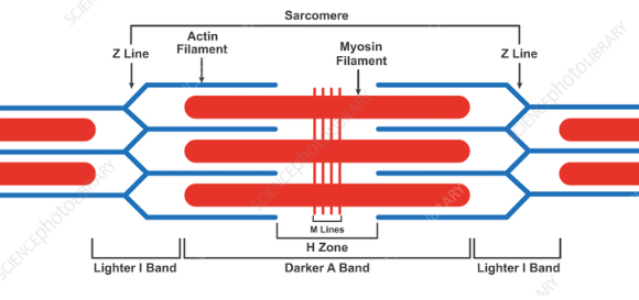
sliding filament theory
neuronmuscular junction
A motor neuron synapse with a muscle fiber to initiate movement
acetylcholine is released and binds to r3ecptors on muscle fiber
the muscle then releases calcium to initiate contraction
Step 1 and 2
myosin heads hydrolyze ATP and become reoriented and energized (in the right position)
myosin heads bind to the actin forming cross bridges (they are connected)
step 3
myosin head rotates toward the center of the sarcomere (power stroke)
this is its natural position
step 4
myosin heads bind ATP, the cross bridges detach from the actin
bond broken and now it’s in its start position
the caclium released in synapse binds to …….
troponin
troponin moves tropomyosin aside exposing the binding sites
these binding sites allow for crossbridges to form
cardiac muscle vs skeltal muscle
cardiac muscle:
short, fewer nuclei, branched, connected by intercalated discs, has sarcomeres but less pronounced
skeletal muscle:
long, many nuclei, has dark pronounce sarcomeres
titin
a massive protein in the sarcomere that recoils after contraction
prevents overstretching
the recoiling action helps antagonistic muscle movements