Lecture 8: Diagnosis and Management of Ligament and Tendon Disorders
 Call Kai
 Learn
Learn Practice Test
 Spaced Repetition
 Match
Match Flashcards
 Knowt Play
1/93

There's no tags or description
Looks like no tags are added yet.
Name | Mastery | Learn | Test | Matching | Spaced | Call with Kai | Chat |
|---|
No analytics yet
Send a link to your students to track their progress
94 Terms
- swelling = localized to a site of injury
- heat = warm to touch due to inflammation
- tenderness = palpation may elicit pain and discomfort
- thickening = fibrosis causes firmer or thicker tissues
- altered posture
- lameness when asked to move or flex affect limb
- joint instability in severe cases
What are the common clinical signs for soft tissue injury and what do they look like?
- they are refusing to bear weight on the affected limb
Why can a horse have altered posture with soft tissue injury?
- strain
- contusion
- laceration
What are ddx for a muscle injury?
- tendonitis or desmitis
What are ddx for a tendon or ligament injury?
- synovitis
- tenosynovitis
- bursitis
What are ddx for an injury to a synovial structure?
- edema
- cellulitis
- seroma
- hematoma
What are ddx for an injury to the skin and fascia?
- 11-46% prevalence of tendon and ligament injuries in performance horses
- most common soft tissue injury in racing TB
- 43-93% risk of reinjury
What is the prevalence of tendon and ligament injuries in horses?
essential in movement, load distribution, and joint stability
tendons and ligaments are crucial for equine locomotion
1. superficial digital flexor tendon (tendonitis)
2. suspensory ligament (desmitis)
what are the first and second most frequently injured soft tissue structures injured in the equine lower limb?
- connects muscle to bone
- transfers muscle force
- induces joint mobility
What is a tendon? What are its functions?
- connects bone to bone
- provides joint stability
What is a ligament? What are its functions?
- composed mostly of collagen fibers
- fibers arranged in parallel bundles, which creates linear pattern seen on ultrasonography
What is the microscopic anatomy of tendons and ligaments?
- collagen (type 1) for tensile strength
- proteoglycans = for viscoelasticity
What are the 2 type of collagen fibers that tendons and ligaments are composed of and this function?
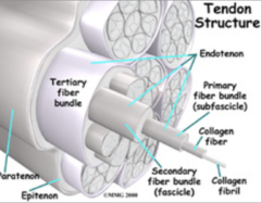
endotenon
- surrounds bundles of collagen fibers
- path for blood vessels, nerves, lymphatics
paratendon
- surrounds entire tendon, synovial cells
- provides movement or gliding
synovial sheath
- present in select areas to reduce friction
What are the connective tissue covering of tendons and ligaments?
- digital flexor tendon sheath
- carpal flexor tendon sheath
- tarsal flexor tendon sheath
Where are synovial sheaths found?
- musculotendinous junction
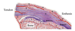
- bone and tendon/lig attachment site (enthesis and enthesophytes from here)
- intratendinous (endotendon)
- tendon sheaths (mesotendon)
vascular supply in ligaments and tendons is not good, where are they found?
- golgi tendon organ
Tendons and ligaments have a rich nerve supply what is responsible for the following?
- proprioception
- detect stretch and tension
- detects changes in joint position
- free nerve ending
Tendons and ligaments have a rich nerve supply what is responsible for the following?
- nociception
- detect noxious stimulus
- transmit forces from muscles to bones to induce joint movement
- resists applied forces to maintain joint alignment and provide joint stability
- stores and releases elastic energy by acting as shock absorbers
What are the biomechanics of tendons?
- ability to store and release elastic energy
Tendons exhibit varying degrees of stiffness. What does this affect?
- stiff = better suited for controlled joint movement = walking, turning
- compliant = store elastic energy, aid in locomotion efficiency = running or jumping
When is it better to have stiff or complaint (stretchy) tendons and ligaments?
- critical shock absorption and elastic energy storage
- undergo high strain during galloping, making them prone to injury
What is the function of the digital flexor tendons (SDFT, DDFT) and when do they undergo high strain?
- prevents hyperextension of the fetlock to help support it
What is the function of the suspensory ligaments?
1. overuse
2. acute overloading
3. conformation
4. genetic disorders
What are the 4 common etiologies of tendon and ligament injuries?
- accumulated microtrauma from chronic repetitive use without rest and repair
How do tendon and ligament injuries occur from overuse?
- sudden onset during high-impact activity or due to lacerations of infections
- tendon strength is ~2x muscle strength so injury location is usually musculotendinous junction
How do tendon and ligament injuries occur from acute overloading?
- bone and joint anomalies may increase stress
- foot and hoof issues from poor hoof balance or improper trimming
How do tendon and ligament injuries occur from conformation?
- collagen defect
- increased extensibility, reduced tissue healing
How do tendon and ligament injuries occur from genetic disorders?
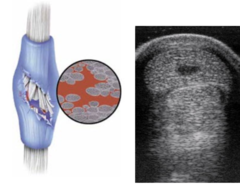
- tends to occur within the center of the tendon (core lesions), because they are sites of increased tissue loading with limited blood supply (hypoxia)
Where do tendon injuries commonly occur? Why?
- slow and inadequate tissue healing
- increased risk of reinjury
What is tendon healing like?
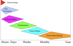
1. acute inflammation --> 1-2 weeks
2. proliferation --> 3-4 weeks
3. remodeling and maturation --> 6 wks - 12 mnths
What are the 3 stages of tendon healing and when do they occur?
- occurs at 1-2 weeks
- inflammatory products, blood, debris, fibrin
What is the acute inflammation stage of tendon healing?
- 3-6 weeks
- collagen synthesis (type 3) = disorganized, weak
What is the proliferation stage of tendon healing?
- early mobilization (>3 weeks); detrimental to collagen orientation = too early
- mobilization at >3 weeks; increases tensile strength, enhances healing = goal
What are the parts of collagen synthesis during the 2nd stage of tendon healing?
- 6 weeks-12 months
- matrix remodels over months to years =collagen type 1
What is the remodeling and maturation stage of tendon healing?
- linear arrangement of collagen fibrils
What does ideal healing of a tendon look like?
- alters local tissue strain -> risk of reinjury
When a tendon is healing how can fibrosis impact it?
- torn ligament -> injured nerves (neuromuscular dysfunction)
- altered proprioception (poor muscle activation and coordination
- pain and ligamentous instability (muscular co-contractions; flexor-extensor imbalance)
- chronic muscle hypertonicity (compensatory gait patterns)
What are common ligamentous injuries in horses?
- superficial digital flexor tendon (SDFT)
- deep digital flexor tendon (DDFT)
Which tendons does contracted tendons commonly occur at lead to limb deformities?
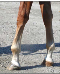
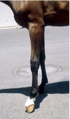
- SDFT contracture
- foot on ground but contraction in fetlock since that is where it attached
What is wrong with this horse? How can you tell?
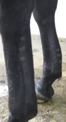
- DDFT contracture
- foot elevates off ground since it attaches to P3
What is wrong with this horse? How can you tell?
- large forces applied and repetitive trauma
- secondary joint instability
- high mechanical loading and inability to unload during healing
- poor vascularity = limited healing
- collagen density = scar tissue formation (fibrosis)
- slow healing process = remodeling
Tendon and ligament injuries are common in athletic horses and can be difficult to treat successfully, why is this?
- observe: swelling, lameness
- palpate for pain, heat, soft tissue swelling, irregular contours like firmness
- gait evaluation: lameness and joint stiffness (variable), positive response to flexion tests
- diagnostic local anesthesia (prn)
How do you diagnose tendon and ligament injuries?
- look at the consistency is it firm or soft
How can you characterize tendon swelling?
- fluid accumulation within tissues (subcutis)
- area will be diffusely swollen
- soft and squishy
What is edema? What does it feel like?
bacterial infection of the dermis or subcutis
What is cellulitis?
- localized to a structure like a joint capsule, bursa, or tendon sheath
What is effusion?
- ultrasonography
What is the preferred method for imaging tendons and ligaments?
- location of structure affected
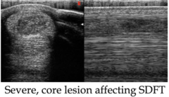
- type of fiber disruption (diffuse, core lesion)
- extent of damage (area and length)
What is U/S used for when diagnosis a tendon or ligament injury?
zones (each about 4cm in length and can be subjective)
- 1A, 1B: SDFT, DDFT, origin of the suspensory ligament, origin of accessory ligament
- 2A, 2B: SDFT, DDFT, accessory ligament, suspensory ligament
- 3A, 3B: branches of suspensory ligament
- 3C: proximal sesamoid bones
location by length distal to the accessory carpal bone (in cm)
how can you localize lesions in tendon and ligaments?
red = SDFT
blue = DDFT
green = distal accessory ligament
yellow = suspensory ligament
This is a transverse ultrasound image, what are these structures?
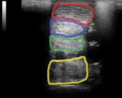
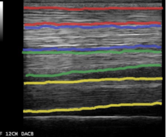
red = SDFT
blue = DDFT
green = distal accessory ligament
yellow = suspensory ligament
This is an longitudinal ultrasound image, what are the different structures?
- echogenicity and consistency
- fiber alignment = linear and uniform
- cross sectional area
- shape = bulging, irregular, or loss of normal profile
- margins = smooth vs irregular, sharp vs poorly defined
- depth = correctly identify lesions relative to bone and adjacent structures
What are the things you assess when looking at the integrity of a tendon on U/S?
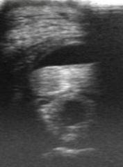
- hypoechoic = fluid accumulation, fiber disruption
- hyperechoic = fibrosis or mineralization
How do you monitor tendon integrity based on echogenicity on U/S?
- Note: the zones, % of area affected, symmetricallyness
- decreased = remodeling
- increased = chronic thickening, edema, fibrosis
How do you assess cross sectional area with U/S for a tendon/ligament injury?
- re-examine every 4-6 weeks
- examine echogenicity
- examine cross-sectional area
- examine fiber pattern realignment
When you are monitor a healing tendon, how do you monitor it?
- gradual return of linear pattern shows maturation of repair
When monitoring the healing of a tendon or ligament how do you assess fiber pattern realignment?
- looking for other injuries
- identify possible bony lesions such as fractures
- enthesopathy (ossification of ligaments or tendon insertion site)
- contrast rads to visualize joints, bursa, and tendon sheath
Why might using radiographs for tendon and ligament injuries be useful?
- detailed view of internal structures
- easier to see subtle or deep injuries
Why might using MRIs for tendon and ligament injuries be useful?
- most frequent soft tissue injury of the lower limb
- associated with high impact injuries from overuse, trauma, and high intensity exercise
- mostly occurred in the forelimbs
- low percentage is bilateral
What is superficial digital flexor tendonitis in horses like?
- relatively easy to diagnose
- lame
- characteristic swelling
- pain on palpation
- ultrasound for definitive diagnosis
How is superficial digital flexor tendonitis diagnosed?
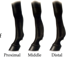
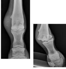
- requires a minimum of 6 months off with periodic ultrasound monitoring
What is wrong with this horse? What should be done when it is healing?
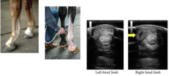
- high speed injury
- acute trauma
- wear and tear
What is the etiology for deep digital flexor tendonitis?
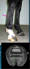
- not as obvious as SDFT lesion
- lameness, less prominent swelling, pain on palpation
- may be associated with navicular syndrome, due to anatomy
- MRI = definitive diagnosis
How do you diagnose deep digital flexor tendonitis?
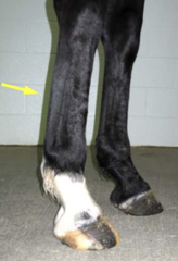
- constriction of the palmar annular ligament (like carpal tunnel-like syndrome)
What is digital flexor tendon sheath tenosynovitis?
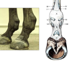
- may not heal readily because synovial fluid impedes tendon healing
- may require surgical intervention by transecting the palmar annular ligament
- prognosis: decreases with chronicity
What should you consider when dealing with digital flexor tendon sheath tenosynovitis?
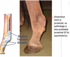
- 2nd most common soft tissue injury of the lower limb
- inflammation and tearing --> chronic lameness and prolonged recovery
- common in show jumpers and dressage
What is suspensory ligament desmitis like in horses?
- due to chronic and repetitive
- high speed activities
- poor conformation
- inadequate warm up
What are the causes of suspensory ligament desmitis in horses?
- common in performance horses due to working on uneven footing or conformation (straight hocks or hyperextended fetlocks)
Who commonly gets proximal suspensory ligament desmitis of the forelimb? Why?
- intermittent and subtle lameness that worsens on soft ground and with limb on outside of circle
- may be associated with a "splint" exostosis of second or fourth metacarpal bones
What are the clinical signs of forelimb proximal suspensory ligament desmitis?
- recurrence is common and horses often do not return to former level of work
What is the prognosis for forelimb proximal suspensory ligament desmitis?
- pain on deep palpation
- U/S: irregular fibers at the attachment side
- rads: bone irregularity
- nuclear scintigraphy or MRI
How do you diagnose forelimb proximal suspensory ligament desmitis?
- insidious chronic lameness with shortened cranial phase of stride
- typically worse on soft ground and with affected limb on outside of circle
- can be positive to upper limb flexion
- perineural anesthesia: deep branch of the lateral plantar nerve
proximal suspensory ligament desmitis of hindlimb is harder to diagnose than in the forelimb, how is it done?
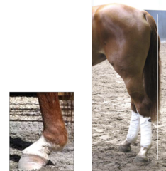
- most common in the forelimb
- similar etiology, dx, tx, and prognosis as digital flexor tendinitis
What is mid body suspensory ligament desmitis?
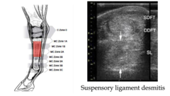
- hyperextension or fetlock injury
- galloping - forelimb
- dressage and jumping - hindlimb
What is suspensory ligament branch desmitis caused by?
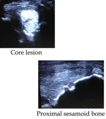
- moderate lameness
- thickening on palpation
- +/- positive to lower limb flexion
- ultrasound shows insertional lesions and core lesions
- rads may show concurrent proximal sesamoid bone irregularity (sesamoiditis)
How is suspensory ligament branch desmitis diagnosed?
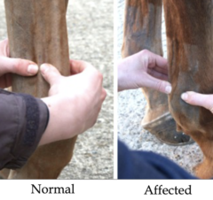
- fair ~40% return to full use but reinjury is possible
What is the prognosis for suspensory ligament branch desmitis?
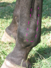
- a congenital defect of the collagen fibers leading to progressive ligament degeneration
What is the cause of degenerative suspensory ligament desmitis?
- aged broodmares (Andalusina, Peruvian paso, Paso fino)
What is the signalment for degenerative suspensory ligament desmitis?
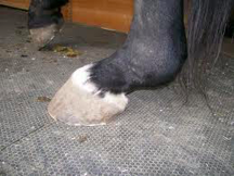
- most commonly bilateral hind limbs
- straight hock conformation
- fetlock hyperextension --> may progress to touching the ground
- stand against wall with their heels elevated
What are the most common clinical signs for degenerative suspensory ligament desmitis?
- corrective shoeing, fetlock arthrodesis
How do you treat degenerative suspensory ligament desmitis?
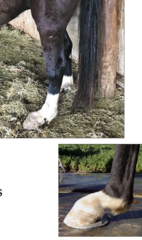
- arthroscopy: intra-articular ligaments
- tenoscopy: flexor tendon sheath
What are tendon and ligament surgical interventions that lead to a diagnosis?
- tendon repair
- ligament repair
- tendon sheath debridement
- tendon splitting of core lesion
- tenotomy for tendon contractures
What are surgical intervention treatment?
- common injury either partial or complete transection
- surgical repair and wound management = #1
- heals well with conservative treatment (stall rest 6-8 wks, may need a splint or cast depending on wound severity)
- good prognosis
Extensor tendon laceration is like what in horses?
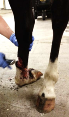
- treat like fracture with external coaptation, 4-6 weeks min
- prognosis good if no underlying joint damage, guarded for some joints because of difficulty in reducing the luxation (ex tarsocrural joint)
How do you treat collateral ligament ruptures and what is the prognosis in horses?
- rest: may require complete stall rest or limited hand walking
- compression wrap: reduces swelling and edema, protect tissues
- cryotherapy: reduce swelling and acute inflammation
- NSAIDs: phenylbutazone to manage pain and reduce inflammation
- surgical intervention: laceration or fracture repair
What are treatments during the acute inflammation stage of tendon/ligament repair (1-2 weeks)?
- supportive bandaging: provides mechanical support during healing
- controlled exercise: gradual reintroduction to exercise, stimulate healing, strengthen tendon, and prevent stiffness remodeling and maturation (1-12 months)
- physiotherapeutic modalities: low-level laser therapy to enhance cellular regeneration and reduce pain; shockwave therapy for chronic pain and tendinitis
- manual therapies: passive ROM, stretching, massage to improve flexibility
- orthobiologics: stem cells, platelet rich plasma (PRP) to deliver growth factors
What are the treatments that can be used during proliferation (3-6 weeks) stage of tendon/ligament repair?
- gradual return to exercise: increase tissue loading for tissue realignment and controlled hand walking followed by trotting
- progressive exercise intensity and duration: increased tissue load for tendon strength
- supportive care: proper hoof trimming and balance to optimize tissue loading
- regularly scheduled monitoring: track the healing progress and adjust rehab protocols
What are the treatments that can be used during the remodeling and maturation (1-12 months) stage of tendon/ligament repair?
- warm up and cool down: optimize musculoskeletal function and prevent tendon overload
- rest and recovery: sufficient period between intense training sessions
- monitoring training load: avoiding sudden increases in workload, intensity, and duration
How do you prevent tendon and ligament injuries?
- severity of injury
- location
- response to treatment
What are the factors that prognosis depends on?
- generally good for return to work with appropriate rest and rehabilitation
What is the prognosis of acute and mild tendonitis?
- can often return to work but require more time for healing and a more controlled rehabilitation program
What is the prognosis for moderate tendonitis?
- severe fiber disruption or tendon rupture may lead to guarded or poor prognosis and horses never returning to full performance
What is the prognosis for severe tendon injuries?
- require long term management and monitoring
What is the prognosis for chronic tendonitis?