Genetic tooth disorders - Dr.Gonzalez
 Call Kai
 Learn
Learn Practice Test
 Spaced Repetition
 Match
Match Flashcards
 Knowt Play
1/110

Name | Mastery | Learn | Test | Matching | Spaced | Call with Kai | Chat |
|---|
No analytics yet
Send a link to your students to track their progress
111 Terms
Tooth development results from:
-Reciprocal induction: recipirocal epithelial-mesenchymal interactions
-cross-talk between cells from oral epithelium and cells from underlying ectomesenchyme
-structural gene expression at different stages of tooth formation
Hypodontia is also called:
Oligodontia
define: Hypodontia
having fewer than regular number of teeth
-missing third molars and upper lateral incisors is common
Mutations that cause Hypodontia
-Pax9
-Msx1
Pax9 and MSX1
Mutations of both PAX9 and MSX1 cause arrested tooth formation and clinical hypodontia
2 gene mutations that can cause supernumerary teeth
Runx2
APC
Runx2 and APC
These mutations cause supernumerary teeth
Define: Anodontia
congenitally missing ALL primary or permanent teeth
_____________ is frequently seen in Ectodermal dysplasia
Anodontia
Define Homeobox genes:
contains 180bp homeodomain that binds DNA and regulates transcriptional cascade of downstream genes early in development
PAx9 gene is expressed in _______________ of tooth bud
Ectomesenchyme
Mutation of human Pax9 gene result in ______________
molar hypodontia
Pax9 acts on ________________ Stage of tooth development to cause hypodontia
Morphogenesis pathway
--> between bud stage and cap stage
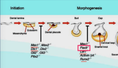
MSX1 haploinsufficiency affects:
Development of ALL teeth
MSX1 Haploinsufficienty affects development of __________ teeth
ALL teeth
Haploinsufficiency - (only half of protein level is present)
MSX1 is a transcriptional (activator/repressor)
repressor
Patients with MSX1 mutation have :
Orofacial cleft - cleft lip/palate
Tooth agenesis - Hypodontia
____________ mutation causes complete absence of MSX1 homeodomain
Ser105Stop mutation
(type of nonsense mutation - premature termination)
Ser105Stop mutation in MSX1 gene affects __________________ in homeodomain
complete absence of MSX1 homeodomain (no functional protein at all)
2 types of tooth agenesis
-Hypodontia
-Anodontia
Ectodermal Dysplasia
-group of inherited disorders characterized by aplasia or dysplasia of tissues of ectodermal origin
- skin, hair, nails, teeth, sweat glands, nerves, constituent parts of ear and eye
2 forms of clinical conditions of Ectodermal dysplasia
-Hidrotic
-Anhidrotic (reduced number of functional sweat glands)
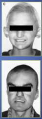
Extra-oral signs of Ectodermal dysplasia
-Sparse fine/coarse curly hair
-abnormally developed nails
-frontal bossing (protruding forehead; enlargement of frontal bone)
-prominent lips
-depressed midface and nasal bridge
-soft, thin, dry skin - prone to eczema
-Hypodontia occurs in 80% of cases

Anhidrotic Ectodermal dysplasia 's pattern of inheritance:
X-linked recessive
-Deletions within EDA gene on chromosome X
_______________ causes reduced number of functional sweat glands
Anhidrotic ED
Manifestations of Anhidrotic ED
-Hypodontia
-Hypohidrosis (reduced sweating)
-Hypotrichosis (sparse hair)
-prominent forehead
-wide eyebrows
-saddle-shaped nose
-thick everted lips
-dry skin
-fine sparse hair
Treatment options for ED Hypodontia :
-Orthognathic surgical procedures
--> jaw positioned correctly
--> Bone grafting procedure or Le Fort I combined with bone grafting
-Implant placement for denture retention (implant denture)

Ectodermal dysplasia causes (hypodontia/anodontia)
BOTH
Supernumerary teeth
-teeth that are additional to normal complement
-primary AND permanent dentitions can be affected
-More common in Males

Sequelae (consequence) of Supernumerary teeth
-normal eruption or FAILURE of eruption can both occur
-displacement or rotation of teeth
-crowding
-abnormal diastema / premature space closure
-dilaceration, delayed or abnormal root development of permanent teeth
-Cystic formation
-Eruption into nasal cavity
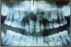
Tooth eruption into nasal cavity is associated with ______________ condition
Supernumerary teeth
Classification of Supernumerary Teeth
-conical
-tuberculate (barrel-shaped)
-supplemental
-Odontoma
-Mesiodens
-Paramolar
-Distomolar
Conical supernumerary tooth
small peg-shaped conical tooth that presents as a mesiodens between maxillary central incisors that rarely erupts labially
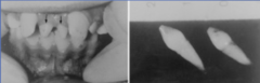
Mesioden supernumerary teeth
supernumerary teeth arising in the midline between two central incisors

paramolar supernumerary teeth
-supernumerary molar, usually small and rudimentary
-situated buccally or lingually to one of the maxilary molars or in the interproximal space buccal to 2nd and 3rd molar
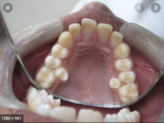
Distomolar supernumerary teeth
-distal to 3rd molar, usually small and rudimentary
-delays or impedes eruption of normal tooth

Tuberculate supernumerary tooth
-larger than conical tooth
-has more than 1 cusp
-barrel-shaped
-stunted root formation
-often paired in two
-on the palatal aspect of maxillary central incisors
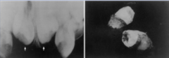
Supplemental Supernumerary tooth
-duplication of teeth in normal series
-found at the end of tooth series
-permanent maxillary lateral incisor is most common (supplemental molars occur as well)

Odontoma supernumerary teeth
-Unorganized mass of tissue ; benign tumor that is composed of normal dental tissue that has grown in an irregular way

Etiology of Syndromic vs. Non-syndromic Supernumerary teeth
Syndromic - genetic mutation of Runx2, APC
Non-syndromic - Hereditary ; localized and independent hyperactivity of dental lamina
Local hyperactivity of dental lamina results in :
Non-syndromic supernumerary teeth
Supplemental supernumerary teeth result from:
Local, independent, conditioned hyperactivity of dental lamina ;
-lingual extension of additional tooth bud leads to supplemental tooth (e.x. Distomolar)
Good example of supplemental supernumerary tooth that develops from lingual extension of additional tooth:
Distomolar
Rudimentary (dysmorphic) supernumerary tooth result from:
proliferation of epithelial remnants of dental lamina
--> This is why it's "dysmorphic" - does not look like a real tooth
Eumorphic vs. Dysmorphic form of supernumerary tooth are called:
Eumorphic - Supplemental
Dysmorphic - Rudimentary
2 forms of supernumerary teeth that result from Local hyperactivity of dental lamina:
*supplemental (eumorphic)
*rudimentary (dysmorphic)
Difference in effect caused by mutations that occur early on vs. Later on such as during mineralization or root formation stage:
Early on - affect Number of teeth
Later on - affect Quality of teeth (but teeth# will be normal)
List 3 syndromic supernumerary teeth :
-Cleft lip/ palate
-Cleidocranial Dysplasia (CCD)
-Gardner's Syndrome (FAP)
______________ syndrome is associated with BOTH hypodontia and supernumerary tooth
Cleft lip/palate
Clinical features of Cleidocranial Dysplasia
-Short stature
-late closure of fontanels & sutures
-Aplasia of clavicles (no clavicles so they're able to touch their shoulders together)
-Hypertelorism (wide eyes)
-Low nasal bridge
-supernumerary teeth
_____________ is caused by mutation of Runx2
Cleidocranial dysplasia
Cleidocranial dysplasia 's pattern of inheritance:
-Autosomal Dominant pattern of inheritance
(only one copy of chromosome mutated will result in CCD)
-Heterozygous mutation of Runx2
Runx2
Transcription factor
-essential for bone and tooth development
Oral features of Cleidocranial Dysplasia:
-Supernumerary teeth
-delayed eruption
-malformed roots
-no cellular cementum
-high palate
-submucosal cleft (palate cleft)
Syndromic vs. Non-syndromic supernumerary teeth
Syndromic - supernumerary teeth syndrome being only one of the conditions of certain disease (ex. Cleidocranial dysplasia); these are always accompanied by other syndromes as well
Non-syndromic - having just supernumerary teeth syndrome and nothing else accompanied.
______________ syndrome result in Familial Adenomatous polyposis accompanied with Oral manifestations
Gardner's syndrome
Familial Adenomatous Polyposis (FAP)
-develop multiple premalignant colorectal adenomas
-100% chance that it will progress to colorectal cancer around age of 39
-Jaw Osteomas (bone tumor in jaw), Odontomas (tooth-derived tumor) and supernumerary teeth
-Oral signs precede GI symptoms --> early diagnosis is life saving
Pattern of Inheritance of Familial Adenomatous Polyposis
-Autosomal dominant
-mutations in APC-gene (Adenomatous polyposis Coli gene) - which is a tumor suppressor gene
Gardner's syndrome
FAP + Oral manifestations (supernumerary teeth)
Mutation in _________________ causes Familial Adenomatous Polyposis (FAP)
Adenomatous Polyposis coli gene (APC)
-tumor suppressor gene
(1/3 cases are due to spontaneous mutations - not through inheritance)
Ways to diagnose Familial Adenomatous polyposis (FAP)
-100 or more colorectal adenomas
-mutation in APC gene
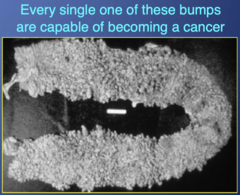
Spontaneous mutations in APC gene can cause __________________
Familial Adenomatous polyposis
Germline mutations in APC gene are located in __________
Exon 15
%_______ of FAP patients will have supernumerary teeth
30
Mutations of _________________ region of APC gene is associated with oral manifestations
3' region
How does site of mutation correlate with clinical phenotype in patients with FAP vs. Gardner's syndrome ?
Mutation on 3' region of APC gene will correlate with oral manifestations
(so 30% of patients with FAP will have Gardner's syndrome, which is associated with Oral manifestations)
Proteins associated with mineralized tissues are all encoded on chromosome #_____
4
2 main ECM proteins in enamel
Ameloblastin
Enamelin
Composition of Enamel ECM
95% Mineral
4% protein (90% Amelogenin; 10% Enamelin and Ameloblastin)
1% water
Dentin ECM proteins expressed in both Bone and Dentin :
SIBLINGS
(small integrin-binding ligand, N-linked Glycoprotein)
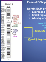
SIBLINGS
Dentin ECM proteins that are expressed in both bone and dentin on chromosome 4
-small regions of similarity between them
-AA sequence highly conserved across species

Amelogenins are encoded by genes on __________________
X& Y chromosomes
-so in males, two types of Amelogenin
-in females, one type of Amelogenin
Function of Amelogenin, Enamelin, Ameloblastin:
Amelogenin - Regulates growth of crystals in length
Enamelin - Regulates crystal elongation
Ameloblastin - cement protein that integrates enamel and dentin at DEJ
Cause of Amelogenesis Imperfecta
-Inheritance
-may be associated with syndrome
Subclasses of Amelogenesis Imperfecta depend on:
-mode of inheritance (ex. Autosomal dominant)
-Enamel appearance
-correlation of genotype with phenotype
3 major categories of Amelogenesis Imperfecta (AI)
1. Hypoplastic - defect in amount of enamel
2. Hypomaturation - defect in removal of proteins from enamel
3. Hypocalcified - defect in calcification
Hypoplastic Amelogenesis Imperfecta
-rough pitted surface
-(+) calcification
-enamel is hard
-very thin enamel
-cannot detect enamel on radiograph

Conserved Chromosomal Synteny
Organization of the Cluster of genes (ex: proteins associated with biomineralization in teeth) are conserved across all species
2 Genetic findings/mutations in Hypoplastic AI
-Enamelin on Chromosome 4
-Amelogenin on Chromosome X&Y
Hypoplastic AI is caused by genetic mutation in Which two Enamel ECM proteins?
-Enamelin
-Amelogenin
Chromosome 4 contains:
-Enamel ECM proteins
- Ameloblastin and Enamelin
-Dentin ECM proteins - SIBLINGS
Amellogenin is NOT here (it's at X&Y)
Hypomaturation AI
-defect in mineralization of ECM
-Enamel is softer than unaffected enamel, but stronger than hypocalcified AI
-Mottled Enamel w/ brown pigment
-Snow-capped on incisal/occlusal surface

Genetic mutations that cause Hypomaturation AI
-Enamelysin (MMP20)
-Kallikrein-4 (Klk4)
2 enzymes that are mutated causing Hypomaturation AI
-MMP20 (Enamelysin)
-Klk4 (kallikrein)
Hypocalcified AI
-Normal thickness of enamel but No calcification at all
-Soft enamel - "Swiss Cheese"
-Brown due to extrinsic stains
-NOT prone to caries
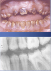
Gene that is associated with Hypocalcified Amelogenesis imperfecta
-Gene FAM83H
(associated with secretory MATRIX vesicles)
List of questions to ask during Diagnosis of Amelogenesis Imperfecta
1. Observe/ describe clinical findings
2. Primary vs. Permanent dentition affected?
3. "Does this run in your family?"
4. Identify which type of AI it is.
Collagen types present in Dentin ECM
Collagen type I, III, V, VI
List all Non-Collagenous proteins in Dentin ECM
DSP
DPP
DGP
BSP
OSP
OCN
OSN
MEPE
(DSP, DPP, DGP encoded by DSPP)

Cause of Dentinogenesis Imperfecta
-Inheritance (Autosomal dominant)
Pattern of Inheritance for Dentinogenesis Imperfecta
-Autosomal dominant
In ___________, teeth appear blue-grey or amber-brown and opalescent
Dentinogenesis Imperfecta
Dentinogenesis Imperfecta
-teeth appear blue-grey or amber-brown and opalescent
-narrow roots with little to no pulp chamber
-bulbous crowns
-Enamel splits readily from dentin when subjected to force
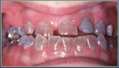
3 Types of Dentinogenesis Imperfecta (DGI)
1. Type I Osteogenesis Imperfecta
--> (+) bone defects
2. Type II Opalescent
--> (-) bone defects
3. Type III Brandywine Isolate
--> (-) bone defects
Type I Osteogenesis Imperfecta
-brittle bones
-blue sclera
-bitemporal bossing
-bowing of the limbs
Mutation in _____________ causes DGI Type I. Osteogenesis Imperfecta
Type I Collagen
Mutation in type I collagen causes:
Dentinogenesis Imperfecta
Clinical oral manifestations of DGI type I
-Enamel splits away from dentin
-amber translucent color is common
-severe attrition
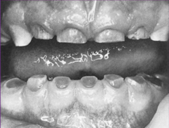
Radiographic findings of DGI Type I
-Obliteration of pulp chamber
- filled with reparative dentin
-small underdeveloped roots
-roots + fractures
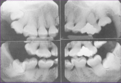
Obliteration of pulp chamber and small underdeveloped roots are manifestations of __________________
Dentinogenesis Imperfecta type I (Osteogenesis Imperfecta)