Cardiovascular system
 Call Kai
 Learn
Learn Practice Test
 Spaced Repetition
 Match
Match Flashcards
 Knowt Play
1/83

Earn XP
Description and Tags
RBC, WBC , platlets , Blood groups
Name | Mastery | Learn | Test | Matching | Spaced | Call with Kai | Chat |
|---|
No analytics yet
Send a link to your students to track their progress
84 Terms
What are the general functions of blood?
-Blood transports dissolved gases, nutrients, hormones and metabolic wastes;
.
-regulates pH and ion composition of interstitial fluids;
.
- restricts fluid loss at injury sites;
.
-defends against toxins and pathogens;
.
-stabilizes body temperature.
constituents of plasma

What are the major plasma proteins?
The major plasma proteins are albumins, globulins and fibrinogen.
-Albumins make up about 55–60%;
.
-globulins about 35–38%; and
.
- fibrinogen about 4–7% of plasma proteins.
What are the functions of albumin?
Albumin maintains plasma osmotic pressure
.
-transports fatty acids thyroid hormones, some steroid hormones and other substances.
,
-most of blood vol
Why does albumin contribute to osmotic pressure?
most abundant
Osmotic pressure is the pulling force on water caused by solutes and hydrostatic pushing force due to presence of more fluid in one region
Indication of diff albumin levels
-If lower albumin level:
may have malnutrition or liver disease, kidney disease, or an inflammatory disease
.
-If higher albumin level:
may be caused by acute infections, burns, and stress
from surgery or a heart attack
.
-Higher than normal
albumin levels:
may be a sign of dehydration, which may be caused by severe diarrhea or other conditions.
What are globulins and categories
-Globulins are plasma proteins that include enzymes [Immunoglobulin m (IgM) , (IgA)antibodies] and fibrous or contractile proteins
.
- and are divided into alpha beta and gamma globulins.
What substances can transport globulins carry and ie?
Transport globulins can bind small ions ,hormones and compounds that may be lost at the kidneys or have very low solubility in water;
.
examples include thyroid-binding proteins transferrin apolipoproteins and steroid-binding proteins.
Globulin test
Used to diagnose kidney,
liver, or immune disorders.
Different levels of globulin indication
-High/Low Levels?
High levels may indicate
autoimmune disease, infections
or cancer.
.
-Low globulin reading may be a
sign of liver or kidney disease.
Low fibrinogen symptoms
-Bleeding gums
.
-Bleeding in your
gastrointestinal tract
.
-Blood in your urine or stool
.
-Coughing up blood
.
-Excessive bruising
.
- Frequent nosebleeds.
- Ruptured spleen
Types of Fibrinogen Deficiency
Afibrinogenemia: no fibrinogen in
your blood; extremely rare (1 in 1mil)
.
Hypofibrinogenemia: low fibrinogen
levels; more common than
afibrinogenemia.
.
Dysfibrinogenemia:
typical fibrinogen levels, but fibrinogen
doesn’t function properly; a large
number of people without
symptoms - but more common than
afibrinogenemia.
High vs low fibrinogen
High
> 700 mg/dL may mean
danger of forming clots
that could harm heart or
brain
.
Low
< 50 mg/dL: may mean
danger of bleeding
after surgery.
How to get rid of excess fibrinogen
-smoking cessation most
effective
.
-weight or stress
reduction or an increase
in regular physical
activity may have less
pronounced effects
-Erythrocytes transport oxygen through hemoglobin and
,
-transport carbon dioxide on a smaller scale; (CO2 is transported by binding to the protein portion of hemoglobin.)
What is the hematocrit?
Hematocrit is the percentage of whole blood volume contributed by formed elements
.
-percentage of whole blood volume consisting of RBC (bk0
packed cell vol
percentage of all formed elements within a sample of whole blood
process formed elements are produced
Hemopoiesis or hematopoiesis
Describe various conditions due to varying hematocrit levels
Dehydration: increase in hemocrit due to decreases plasma volume of stimulation of hormone erythropoietin.
.
-decrease due to internal bleeding or RBC formation problems

Hemopoiesis and site
Formation of blood cells (formed elements)
.
occurs in red bone marrow

Erythropoiesis
Formation of red blood cells in red bone marrow/ myeloid tissue stimulated directly by erythropoietin; indirectly androgen, thyroxine and growth hormones

What gives urine its yellow color
Urobilins
What gives faces its brown color
urobilins
.
stercobilins

structure of hemoglobin
-Quaternary structure: 2 alpha and 2 beta-chains
.
-Each HB chain contains a heme group
.
-Each Heme unit holds Fe2+ so it can interact with O2
Agglutinogen
Specific surface antigens on RBC plasma membranes
.
They are integral membrane glycoproteins or
glycolipids
List the 4 diff blood types

Difference btwn antibodies and antigens
Antigens are present on RBC and in the plasma membrane used to determine blood type
.
-Antibodies detect the presence of foreign RBC and attach to those antigens causing agglutination and lysis of RBC
what is the Rhesus blood typing system
Blood group based on the presence or absence of a Rh surface antigen
.
Rh pos (Rh+) presence of RH factor / Rh neg (Rh-) no Rh factor
.
( term RH usually emitted ie : O+ , A- etc
Rhesus System Antibodies
Only the Anti-Rhesus
Antibody exists in the
Rhesus System
Characteristics of WBC
lack hemoglobin
Migrate out of the blood stream
Capable of Amoeboid movement ( gliding motion due to the flow of cytoplasm )
Attracted to specific chemical stimuli ( positive chemotaxis) guides WBC to invading pathogens , damaged tissues and other WBC
Defends body against pathogens removing toxins , waste and abnormal or damaged cells
Explain the migration of WBC out of the bloodstream and another name
Diapedesis
.
WBC are activated and adhere to the vessel walls in a process called margination
.
WBC then squeeze btwn adjacent endothelial cells and enter the surrounding tissue ( emigration ) leaping motion
What are the two major categories of leukocytes and how they’re identified ?
Granulocytes and agranulocytes.
.
Appearance after Wright’s stain or Giemsa stain

A: Monocyte
B: RBC
C: lymphocyte
D: platelet
E: neutrophil
f: Eosinophil
g: Basophil
What distinguishes granulocytes from agranulocytes?
Granulocytes contain azurophilic primary granules and specific secondary granules while agranulocytes only have primary granules
What are the main structural features of neutrophils?
-Neutrophils are about 12 µm in diameter
.
-2 – 5 lobed nucleus
.
-Granules not stained well by either acidic or basic dyes
.
-Granules contain lysosomal
enzymes and bactericidal agents

What are the main functions of neutrophils?
Neutrophils are usually first at injury sites and attack engulf and digest bacteria
.
they can produce H2O2 and O2 to kill bacteria and release prostaglandins: capillary permeability and leukotrienes : attract phagocytes and coordinate
immune response
What are the main structural features and functions of eosinophils?
-Eosinophils have a bilobed nucleus
.
- large reddish granules;
.
-They engulf antibody-coated bacteria protozoa and cellular debris
.
-releases cytotoxic substances
destroying parasites (flukes, parasitic roundworms)
.
-Increase during allergic reactions
.
-Attracted to injury sites; release enzymes to
reduce inflammation

What are the main structural features and functions of basophils?
-Rare cells with large purple-blue granules takes up basic stain
,
- obscured bilobed nucleus

Function of Basophils
Migrate to injury sites and intensify inflammation
.
-Histamine: dilates blood vessels, increase capillary
permeability resulting in edema/swelling
.
-Heparin: prevents clotting
.
-Chemicals: that attract eosinophils and other
basophils
What are the main structural features of monocytes?
-Spherical
.
may exceed 15 um
.
-Large oval or kidney-bean shaped nucleus
.
-bluish-gray cytoplasm.

Functions of monocytes
they remain in circulation for about 24 hours before entering tissues and becoming macrophages
.
-engulfs debris and pathogens release chemicals that attract and stimulate
neutrophils, monocytes and fibroblasts


What are the main structural features of lymphocytes?
-spherical cells
.
-small lymphocyte : 6-9 μm in dia., thin rim of cytoplasm , round nuclei
(densely stained heterochromatin)
.
-Large : – 9-15 μm in dia., more cytoplasm than sml lymphocytes; nuclei
contain euchromatin and heterochromatin.
.
- thin layer of cytoplasm surrounding the nucleus
.
-slightly larger than RBC
.
-Continuously move between bloodstream and peripheral tissues
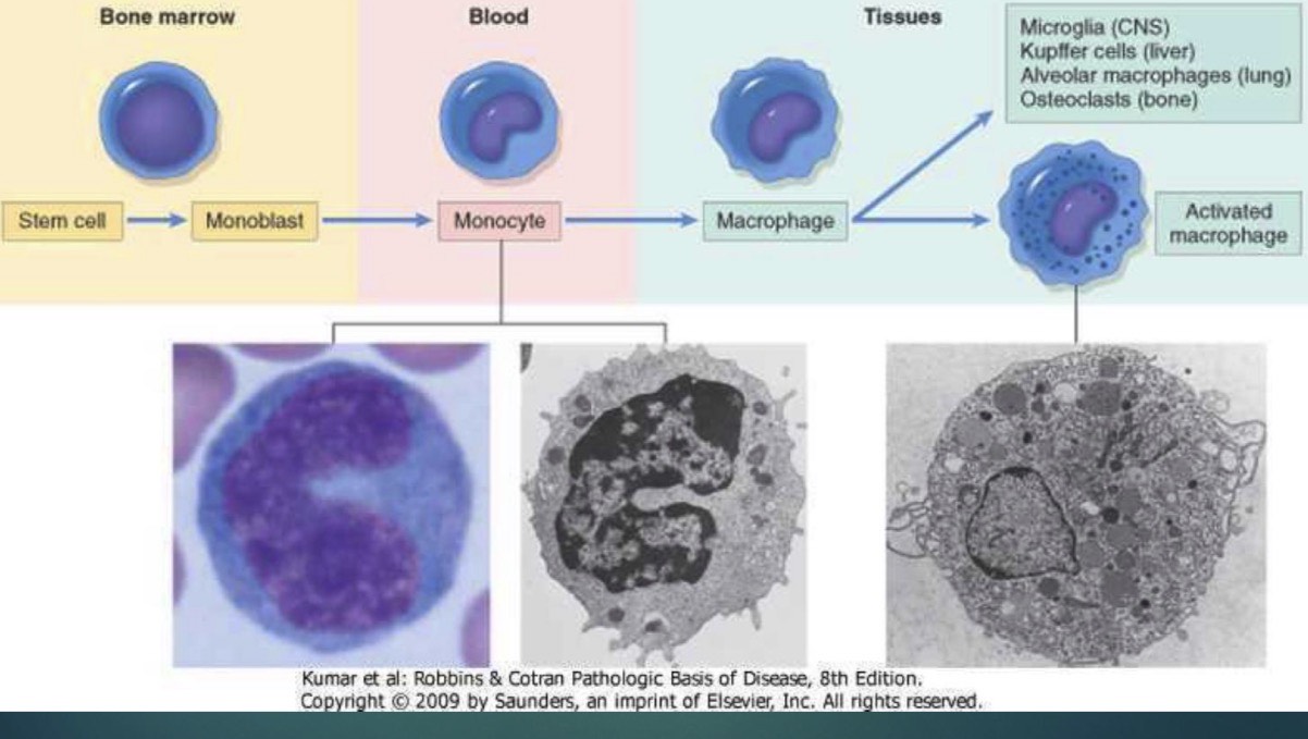
Explain
Origin and Differentiation:
Macrophages originate in the bone marrow from stem cells that differentiate into monoblasts, which then develop into monocytes circulating in the blood.
.
* Tissue Migration:
Monocytes migrate from the bloodstream into various body tissues, where they mature into resident macrophages.
* Specialized Tissues:
Depending on their destination tissue, resident macrophages take on specialized forms and names, including:
* Microglia in the central nervous system (CNS)
* Kupffer cells in the liver
* Alveolar macrophages in the lungs
* Osteoclasts in bone
.
*Activation:
Upon encountering pathogens or inflammatory signals, tissue macrophages can transform into activated macrophages
3 types of Lymphocytes
T Cells (T-lymphocytes):
-cell-mediated immunity specific defense against invading foreign cells and tissues
-attack peripheral tissues invading foreign cells & directly control activity of other lymphocytes
.
B cells (B-lymphocytes):
-humoral immunity (antibody-mediated) involves production of antibodies for defense
-Attack foreign antigens; Activated B-cells Differentiate into Plasma cells which produce antibodies
.
Natural Killer cells:
-Detect and destroy abnormal tissue cells
.
-Helps prevent cancer
How do the functions of neutrophils eosinophils basophils monocytes and lymphocytes differ?
Neutrophils mainly attack bacteria
.
-eosinophils defend against parasites and participate in allergic reactions;
.
-basophils intensify inflammation; monocytes become macrophages that perform phagocytosis
.
- lymphocytes provide specific and immune-surveillance functions.