Pathology of the Scrotum
 Call Kai
 Learn
Learn Practice Test
 Spaced Repetition
 Match
Match Flashcards
 Knowt Play
1/144

Earn XP
Description and Tags
PG 440
Name | Mastery | Learn | Test | Matching | Spaced | Call with Kai | Chat |
|---|
No analytics yet
Send a link to your students to track their progress
145 Terms
Cryptorchidism describes the condition of
having an undescended testis
Cryptorchidism, which is found in — of full-term births
3% to 4%
Cryptorchidism is associated with
infertility and an increase in the risk for malignancy in the involved testis
— is the most common cancer found in an undescended testis
seminoma
undescended testis are most often found in the
inguinal canal
surgical correction of an undescended testis is referred to as
orchiopexy
sonographfically, the undescended testis will appear — to the normal testis
hypoechoic
clinical findings of cryptorchidism
one or both testicles not palpable within the scrotum
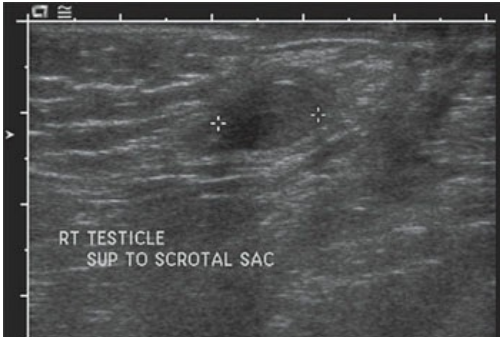
Cryptorchidism. This right testicle (calipers) was noted within the inguinal canal.
sonographic findings of cryptorchidism
testis located outside of the scrotum (most likely the inguinal canal)
the cryptorchid testis will appear hypoechoic to the normal testis
testicular torsion may also be referred to as
spermatic cord torsion
testicular torsion occurs when the —- secondary to the twisting of the testicular axis
arterial blood supply to the testicle is cut off
a — will result in blocked venous drainage and arterial supply
360 degree torsion
a 360 degree torsion will lead to
ischemia within the testis
the amount of ischemic damage is directly related to
the degree of the torsion
testicular torsion occurs more often during the ages of —
12 and 18
with torsion, salvage rates range from 80%-100% if the patient is treated within — of symptom onset
6 hours
with torsion, the testicle is usually not salvageable after
24 hours
testicular torsion can be associated with
trauma, strenuous exercise, and sexual activity
bell-clapper deformity is a
congenital abnormality that describes the situation in which the patient lacks the normal posterior fixation of the testis and epididymis to the scrotal wall
ball-clapper deformity is classically
bilateral
intravaginal torsion is the
most common form of testicular torsion
bell-clapper deformity is part of what is termed
intravaginal torsion
with intravaginal torsion, the tesis, which is not affixed to the scrotal wall, is permitted to —
migrate and twist freely within the scrotum
extravaginal torsion occurs when the
neonatal period or in utero and is related to torsion of the spermatic cord within the inguinal canal
patients suffering from testicular torsion are often awakened in the middle of the night by —
severe scrotal pain
chronic testicular torsion is torsion that has lasted —
longer than 10 days
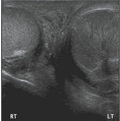
Left testicular torsion. Transverse image of both testicles. The left testicle (LT) had no detectable Doppler signals. Note that it has a heterogeneous echotexture compared to the normal right testicle (RT).
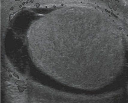
Testicle torsion. Left testicle torsion was diagnosed because no intratesticular flow was detected with color Doppler analysis. (Color image provided online.)
A torsed testis can either — compared to the normal testis
lack flow completely or have decreased flow
clinical findings of acute testicular torsion
acute onset of testicular pain (often during sleep)
possible pain within the lower abdomen and inguinal region
swollen testis/scrotum
nausea and vomitting
higher postioned, painful testis with a horizontal position
sonographic findings of acute testicular torsion
enlargement of the spermatic cord, epididymis, and testis
thickened scrotal wall
hypoechoic or heterogeneous testis
reactive hydrocele
no intravascular flow
decreased intratesticular flow (as compared to the asymptomatic testis)
sonographic findings of chronic testicular torsion
enlargement of the spermatic cord, epididymis, and testis
no intratesticular flow
hyperemic flow around the testis
heterogeneous testis with areas of necrosis
— is the most common cause of acute scrotal pain in prepubertal boys
torsion of the appendix testis
appendages of the testis:
appendix testis, appendix epididymis, and the appendix vas
testicular appendages are structures said to be the embryologic remnants of the
mullerian duct, wolffian duct, and mesonephric duct
the appendix testis is located between the
head of the epididymis and the superior pole of the testis
the appendix epididymis is located at the
head of the epididymis
appendages may be seen in the presence of a
hydrocele or other scrotal fluid collections
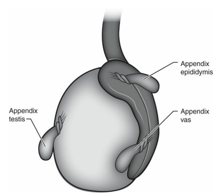
Locations of the testicular appendages.
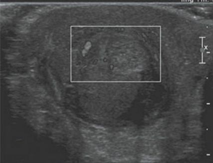
Torsion of the appendix testis. This heterogeneous, avascular mass was seen adjacent to the superior pole of the testis. It was proven to be a torsed appendix testis.
the “blue dot” sign is the observable clinical finding associated with
torsion of the appendix testis
clinical findings of torsion of the testicular appendage
acute testicular pain
pain localized to the superior pole of the testis
“blue dot” sign
sonographic findings of torsion of the testicular appendage
normal intratesticular flow
small, avascular, hypoechoic or hyperechoic mass adjacent to the superior pole of the testis
reactive hydrocele
scrotal wall thickening
a simple fluid collection within the scrotum is referred to as a
hydrocele
hydroceles are found between the two layers of the — and often displace the testicles posteriorly
tunica vaginalis
hydroceles can be idiopathic or described as reactive hydroceles when found in the presence of
scrotal infections, testicular torsion, trauma, or tumors
hydroceles are often accompained by
scrotal wall thickening
clinical findings of a hydrocele
transilluminates light
painless scrotal swelling
may present with pain when found in the presence of scrotal infections, testicular torsion, trauma, or a tumor
sonographic findings of a hydrocele
simple fluid anterior to the testis
scrotal wall thickening
chronic hydroceles may have internal debris and septations
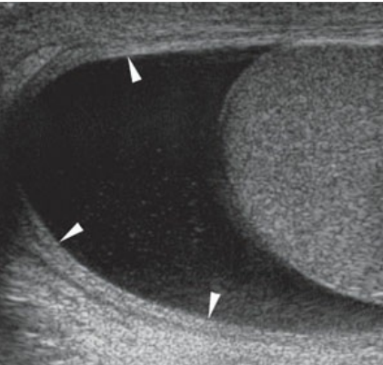
Hydrocele. Anechoic fluid can be noted surrounding the testicle. The scrotal wall (arrowheads) appears to be thickened as well.
a —, which is said to be the most common scrotal mass, is a cyst found most often in the head of the epididymis
spermatocele
spermatocele is composed of
momviable sperm, fat, cellular debris, and lymphocytes
an — appears similar to a spermatocele and can be seen anywhere along the length of the epididymis
epididymal cyst
— are located anywhere along the periphery of the testicle, within the tunica albuginea
tunica albuginea cysts
clinical findings of a spermatocele, epididymal cyst, and tunica albuginea cyst
if large enough, they may be palpable
typically not painful
sonographic findings of a spermatocele, epididymal cyst, and tunica albuginea cyst
round, anechoic mass with acoustic enhancement
may contain some layering debris
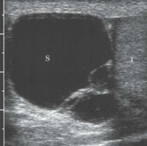
Spermatocele. Large cystic extratesticular mass (S) is noted superior to the testis (T) and is characteristic of a spermatocele.
a dilated group of veins found within the scrotum is termed
a varicocele
varicoceles are caused by
incompetent or abnormal valves within the pampiniform plexus
because of the increased heat released by excess blood within the scrotum, overheating of the — can occur
fragile spermatoza
varicoceles have been cited as the
most common cause of correctable male infertility
the two types of varicocele
primary and secondary
primary varcioceles are most often found in the — and are palpable during physical examination
left
primary varcioceles high incidence on the left is thought to be caused by the
elevated vascular pressure on the left side or possibly as a result of the exteneded length of the left testicular vein, and the sharp angle at which it enters the left renal vein
if a varicocele is found on the right side it is termed
secondary varicocele
secondary varicoceles may be associated with
a hepatic mass, marked hydronephrosis, hepatomegaly, or retroperitoneal neoplasm like a right sided renal mass
nutcreacker syndrome is an
anomaly where left renal vein entrapment occurs between the SMA and the abdominal aorta
one predisposing facor for a varicocele is
nutcraker syndrome
varicoceles will have a dilated vein measuring greater than —
2 mm
clinical findings of a varicocele
typically painless (large varicoceles can cause discomfort)
palpable extratesticular mass
possible infertility
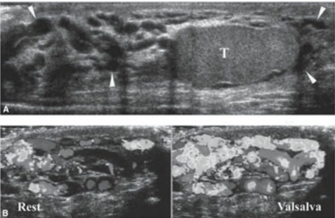
Varicocele. A. Multiple tubular structures (arrowheads) are seen surrounding the testicle (T). B. Color Doppler confirms flow within these structures during rest. With the Valsalva maneuver, the veins enlarged and there is a dramatic increase in flow. (Color image provided online.)
sonographic findings of a varicocele
a group of anechoic, tubular structures located outside of the testis
distended veins that fill with color flow when the valsalva maneuver is performed
dilated veins that measure greater than 2 mm
possibly associated with hydronephrosis, hepatomegaly, or a retroperitoneal neoplasm if found on the right side
a scrotal pearl is an
extratesticular calculus
scrotal pearls are thought to be
remnants of a formerly torsed and displaced testicular appendage
sonographic findings of a scrotal pearl
extremely echogenic, mobile extratesticular structure that produces acoustic shadowing
inflammation of the epididymis is referred to as
epididymitis
epididymitis is the most common cause of
acute testicular pain in adults
inflammation of the testis is referred to as
orchitis
the combination of an infection within the epididymis and testis is termed
epididymo-orchitis
frequent cause of epididymitis is the
spread of bacteria from the prostate or urinary tract
common causes of epididymitis in younger men are the
sexually transmitted diseases chlamydia and gonorrhea
chronic epidiymitis may appear as an
enlarged, hyperechoic epididymis with calcifications
clinical findings of epididymitis and epididymo-orchitis
acute testicular pain
leukocytosis
fever
dysuria
urethral discharge
scrotal wall edema
sonographic findings of epididymitis and epididymo-orchitis
enlargement of the entire epididymis (diffuse)
enlargement of only part of the epididymis (focal)
hypoechoic echotexture of the affected section(s) of the epididymis
hypoechoic testis (with orchitis)
hyperemia within the epi and/or testis
thickened scrotal wall
reactive hydrocele
an abscess that occurs within the testicle is typically the result of
untreated epididymo-orchitis
a — is a complex hydrocele that contains pus
pyocele
a pyocele is often seen in the presence of a
persistent scrotal infection or ruptured testicular abscess
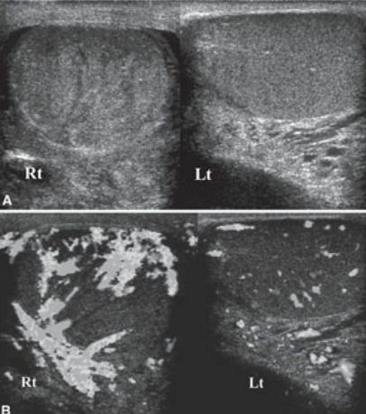
Acute epididymo-orchitis. A. The grayscale sonographic appearance of the testis reveals a right (Rt) testicle that appears heterogeneous compared to the left (Lt) testicle. B. Color Doppler reveals hyperemia in the right (Rt) testicle compared to the left (Lt) testicle, which is consistent with diffuse right-sided epididymo-orchitis. (Color image provided online.)
although intratesticular abscesses lack detected flow within them, they are often surrounded by
hyperemic flow
clinical findings of a testicular abscess
painful, swollen scrotum
fever
leukocytosis
sonographic findings of a testicular abcess
complex intratesticualr mass
mass that has no flow centrally but increased flow around its margins
may have a coexisting pyocele
sonographic findings of a pyocele
complex fluid collection within the scrotum
scrotal wall thickening
may be seen in conjuction with rupture of a testicular abscess
multiple small intratesticular cysts are often seen
along the mediastinum testis
small cysts seen in the mediastinum are clinically insignificant and may be the result of
tubular ectasta of the rete testes
multiple small cysts in the mediastinum are thought ot represent
cystic dilation of the rete testis
epidermoid cysts often appear to have a
whorled or onion skin appearance
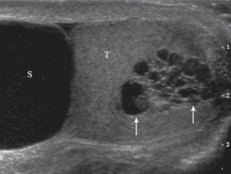
Tubular ectasia of the rete testis. Multiple cysts (arrows) are seen in the area of the mediastinum testes in this testicle (T). A large spermatocele (S) is also noted.
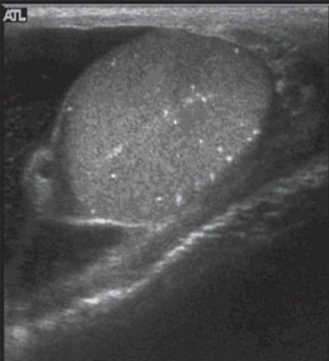
Intratesticular microlithiasis. Multiple echogenic foci with no acoustic shadowing are seen within this testis.