OSPS CVS
 Call Kai
 Learn
Learn Practice Test
 Spaced Repetition
 Match
Match Flashcards
 Knowt Play
1/129

There's no tags or description
Looks like no tags are added yet.
Name | Mastery | Learn | Test | Matching | Spaced | Call with Kai | Chat |
|---|
No analytics yet
Send a link to your students to track their progress
130 Terms

Hyaline Arteriolosclerosis (benign nephrosclerosis)
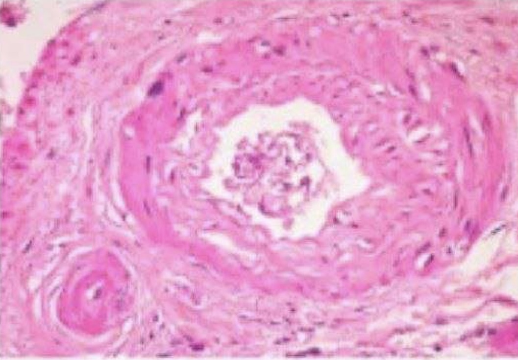
Elastosis (fibroelastic hyperplasia)

Hyperplastic Arteriolosclerosis (Malignant nephrosclerosi)

Hyperplastic Arteriolosclerosis (Malignant nephrosclerosi)


Hyaline arteriolosclerosis (benign nephrosclerosis)

Hyperplastic Arteriolosclerosis (Malignant nephrosclerosi)

Hypertensive heart disease

Hypertensive heart disease

Benign Nephrosclerosis

Malignant Nephrosclerosis

Benign Nephrosclerosis

Benign Nephrosclerosis

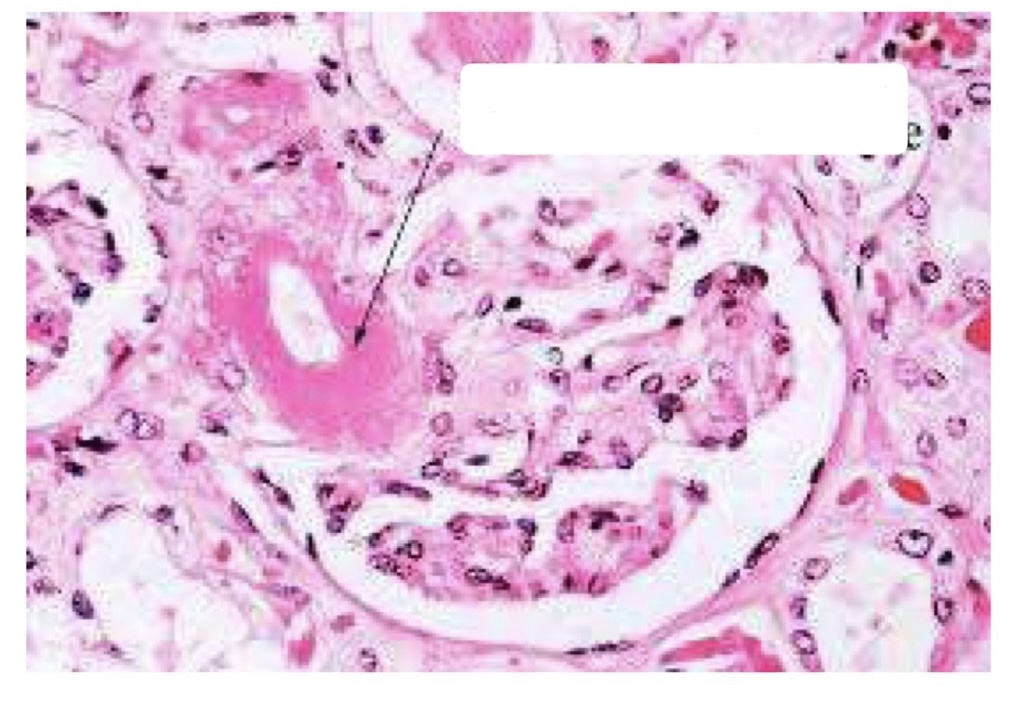
Malignant nephrosclerosis (fibrinoid necrosis)
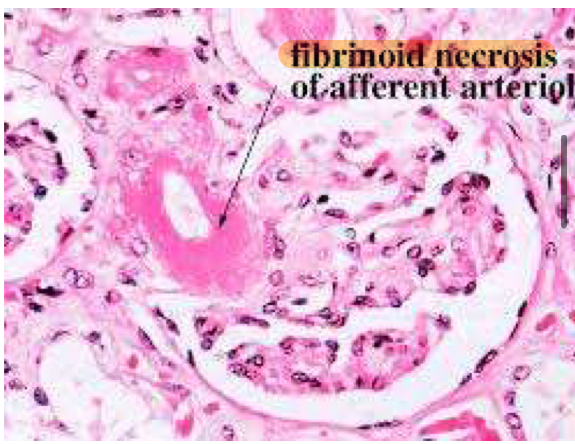

Malignant nephrosclerosis (hyperplastic arteriolosclerosis)
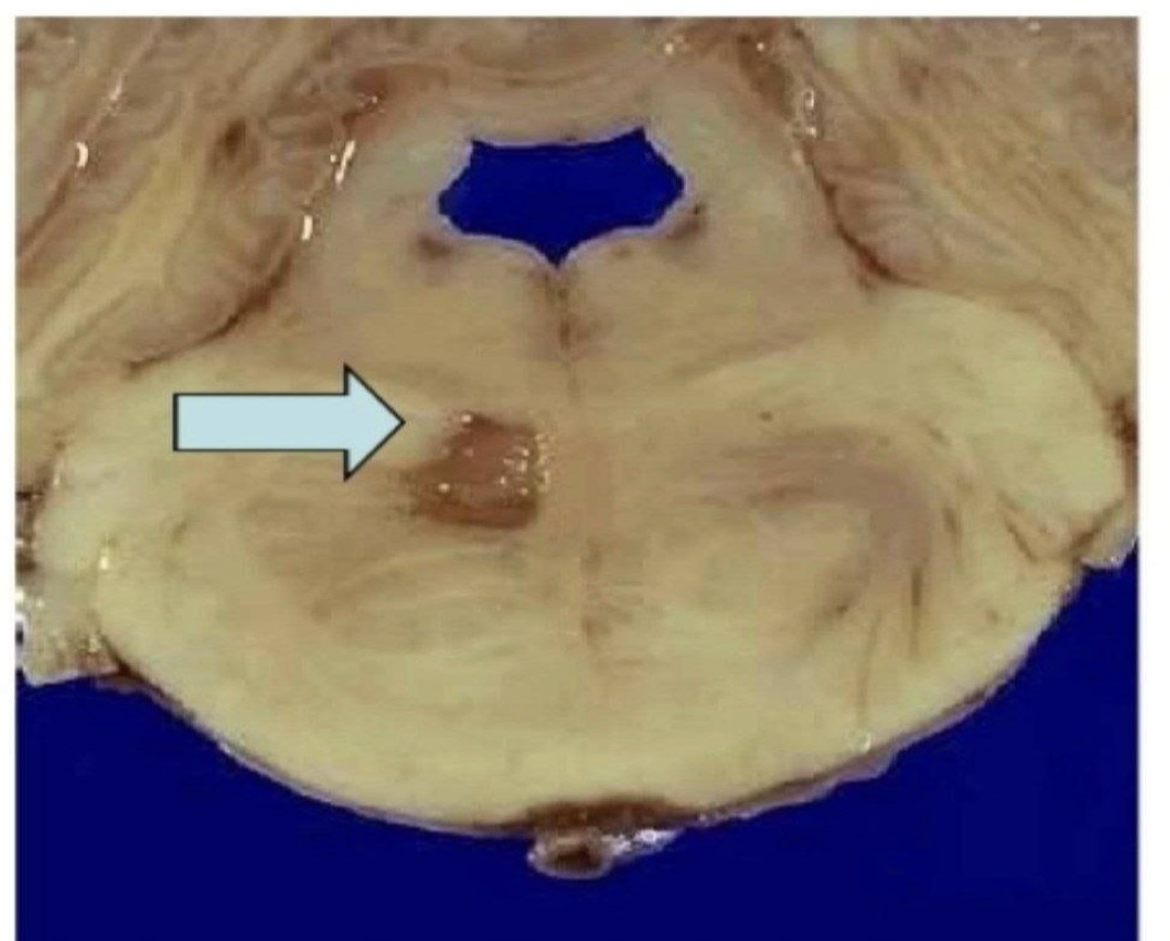
Hypertensive cerebral diseases (ischemic stroke

Hypertensive cerebral diseases


Hypertensive cerebral diseases (Subarachnoid hemorrhage) (hemorrhagic stroke)

Hypertensive cerebral diseases (Cerebral hemorrhage) (hemorrhagic stroke)

Hypertensive cerebral diseases


Hypertensive retinopathy


Regional subendocardial infarction

Regional transmural infarction

MI (0-0.5h)


MI (12-24h)


MI (7-10d)


MI (2-8w)


MI (12-24 hours: Dark mottling)

MI (3-10 days: yellow tan soft center with hemorrhagic borders)

MI (3-10 days: yellow tan soft center with hemorrhagic borders)

MI (2-8 weeks: Grayish-white scar)

TTC (Triphenyl Tetrazolium Chloride) Stain Interpretation


MI (1-4 hours: Wavy myocyte fibers)

MI(4-24 hours: Loss of nucleus, Contraction bands,
Coagulative necrosis)

MI (4-7 days: Macrophages clear necrotic debris with early granulation tissue)

MI (1-4 days: Intense neutrophilic infiltrate)

MI (1-2 weeks: Well organized granulation tissue, capillaries, collagen deposition)

MI (3-6 weeks: Dense fibrous scar)
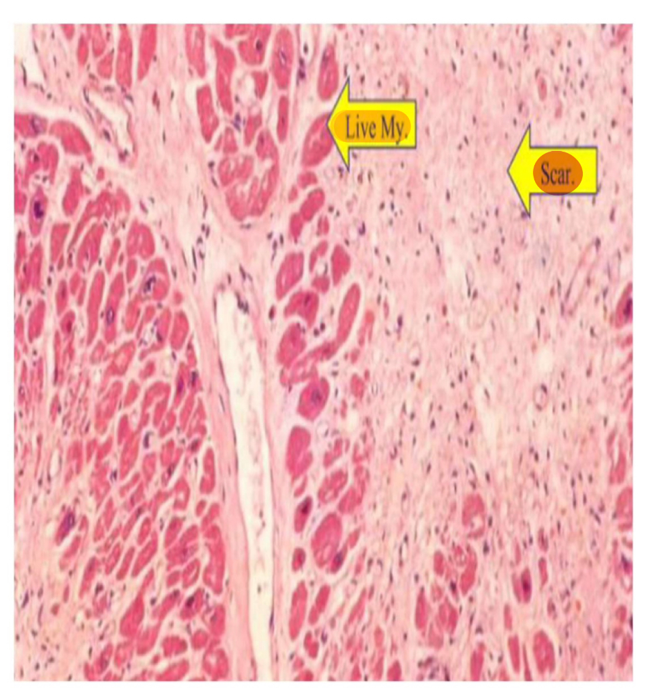

Type of necrosis?
Coagulative necrosis

Type of necrosis?
Caseous necrosis

Type of necrosis?
Fibrinoid necrosis

Type of necrosis?
Liquefactive necrosis

Type of necrosis?
Fat necrosis

Type of necrosis?
Gangrenous Necrosis

Type of cardiomyopathy?
Dilated Cardiomyopathy

Type of cardiomyopathy?
Dilated Cardiomyopathy

Type of cardiomyopathy?
Dilated Cardiomyopathy

Type of cardiomyopathy?
Hypertrophic Cardiomyopathy
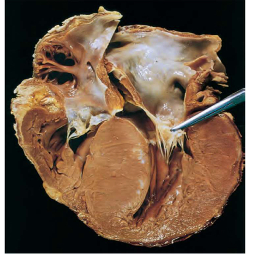
Type of cardiomyopathy?
Hypertrophic Cardiomyopathy

Type of cardiomyopathy?
Hypertrophic Cardiomyopathy

Type of cardiomyopathy?
Restrictive Cardiomyopathy

Type of cardiomyopathy?
Restrictive Cardiomyopathy

Type of cardiomyopathy?
Restrictive Cardiomyopathy

Type of cardiomyopathy?
Restrictive Cardiomyopathy

Concentric Hypertrophy (Pressure Overload)

Eccentric Hypertrophy (Volume Overload)

Concentric Hypertrophy (Pressure Overload)

Eccentric Hypertrophy (Volume Overload)

Myocyte hypertrophy (LV remodeling)

Eccentric Hypertrophy (Volume Overload)

Concentric Hypertrophy (Pressure Overload)

Pulmonary venous congestion (left sided HF)

The arrow points to?
This image represents?
heart failure cells
Pulmonary venous congestion (left sided HF)

Congestive Hepatomegaly ("Nutmeg Liver")
Systemic venous congestion in right sided HF

Peripheral pitting edema
Systemic venous congestion in right sided HF

Ascites (fluid collection in the peritoneum)
Systemic venous congestion in right sided HF

• Congestive Splenomegaly
Systemic venous congestion in right sided HF

• Congestive Splenomegaly
Systemic venous congestion in right sided HF

• Congestive Splenomegaly
Systemic venous congestion in right sided HF

Congestive Hepatomegaly ("Nutmeg Liver")
Systemic venous congestion in right sided HF

Type of myocarditis?
Lymphocytic myocarditis

Type of myocarditis?
Hypersensitive myocarditis

Type of myocarditis?
Giant cell myocarditis

Type of myocarditis?
Chaga’s disease

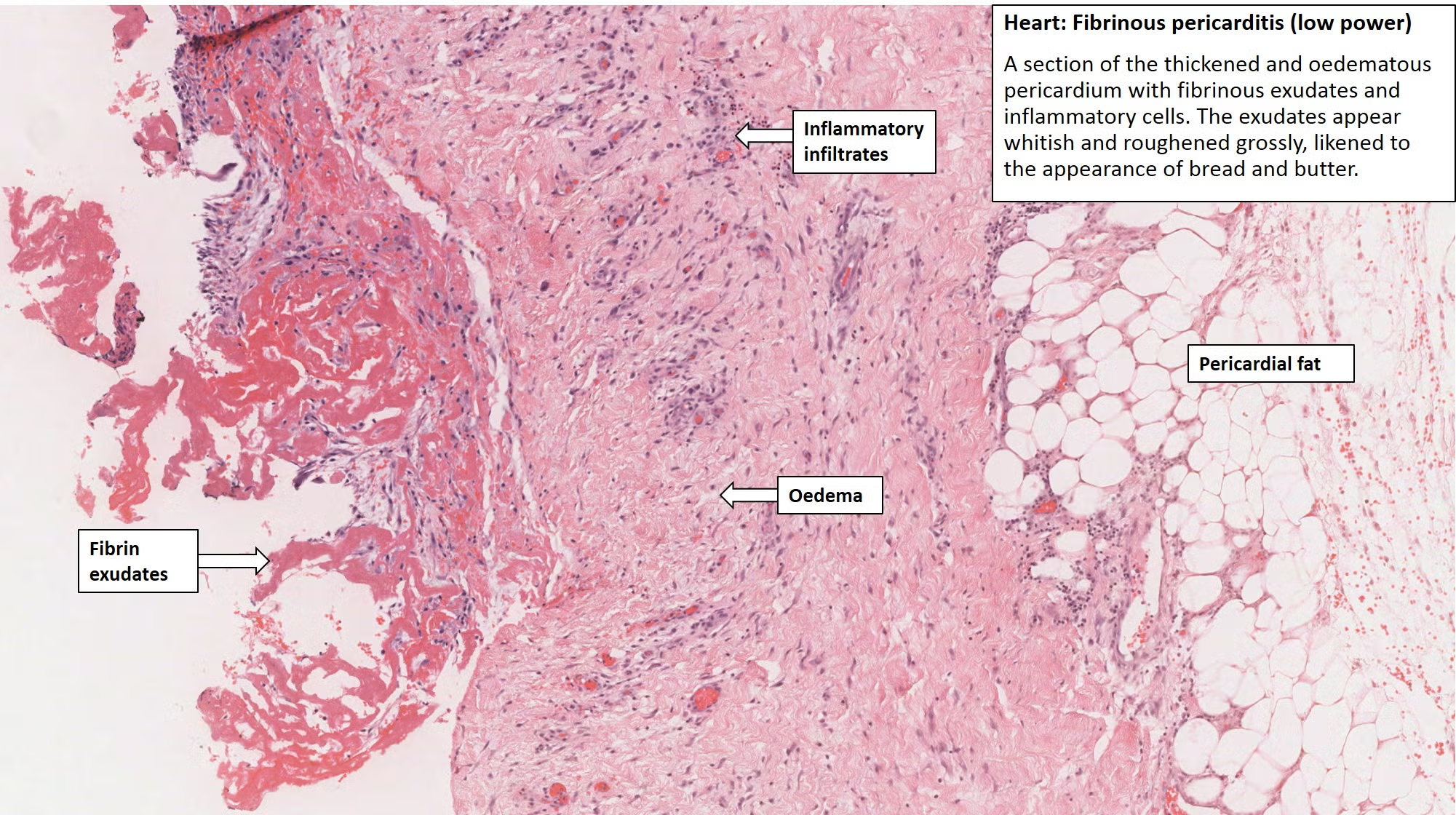
Type of pericarditis?
Fibrinous/ Serofibrinous Pericarditis

Type of pericarditis?
Fibrinous/ Serofibrinous Pericarditis

Hemopericardium

Type of pericarditis ?
Constrictive Pericarditis

Type of pericarditis?
Tuberculous Pericarditis

Type of pericarditis?
Tuberculous Pericarditis

Type of pericarditis?
Hemorrhagic Pericarditis

Type of pericarditis?
Type of pericarditis?
Hemorrhagic Pericarditis

Type of pericarditis?
Suppurative Pericarditis

Type of pericarditis?
Suppurative Pericarditis

Type of pericarditis?
Suppurative Pericarditis

ASCHOFF NODULES

(M/E) – ASCHOFF BODIES
1- anitshkow cells
2- aschoff giant cell


This structure in rheumatic fever represents?
Aschoff giant cells

Which valance is affected?
mitral valve- infective endocarditis

Q: What’s in the image below?
complication of rheumatic valvular endocarditis (mitral stenosis)

Which valve is affected?
The cusps of mitral valve are diffusely involved by tan-yellow, solitary nodular calcification.

severely stenotic aortic valve (there are prominent lipocalcific changes on aortic side of valve cusps (arrow), with sparing of commissures)

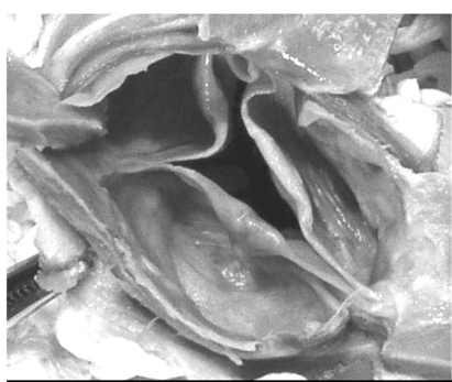
minimally diseased aortic valve

This finding in rheumatic myocarditis represents?
Aschoff bodies

Type of pericarditis (in rheumatic fever)?
Sero-fibrinous pericarditis

Type of pericarditis (in rheumatic fever)?
Sero-fibrinous pericarditis

Type of pericarditis (in rheumatic fever)?
Fibrinous pericarditis (bread & butter) appearance

vegetation found in?
RHD (small, firm, multiple, adherent)

Vegetation found in?
IE (large, polypoid, irregular, yellow, friable)

Vegetation found in?
NBTE (bland, single/multiple)

Type of endocarditis?
Non infective thrombotic endocarditis