Histology- bone, cartilage connective tissue
1/32
There's no tags or description
Looks like no tags are added yet.
Name | Mastery | Learn | Test | Matching | Spaced | Call with Kai |
|---|
No analytics yet
Send a link to your students to track their progress
33 Terms
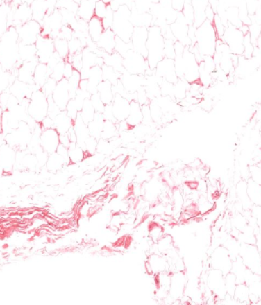
adipocytes
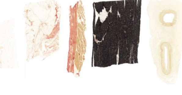
type of connective tissue, notice small amounts of pink/red wispy fibers
collagen (one)
type of connective tissue. Note the different types of fibers and cells
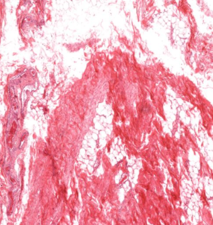
dense connective tissue
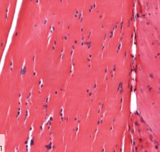
type of connective tissue
notice the packed, parallel arrangement of fibers
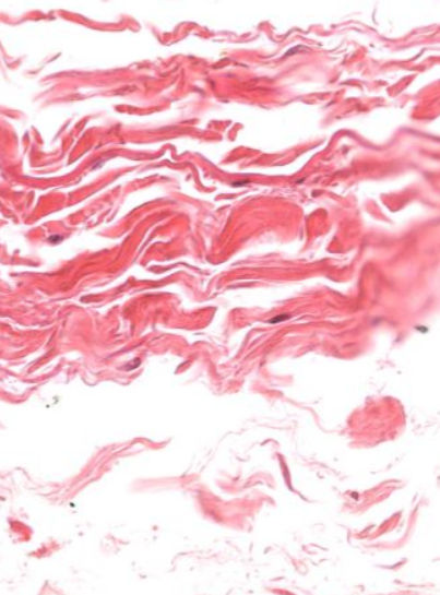
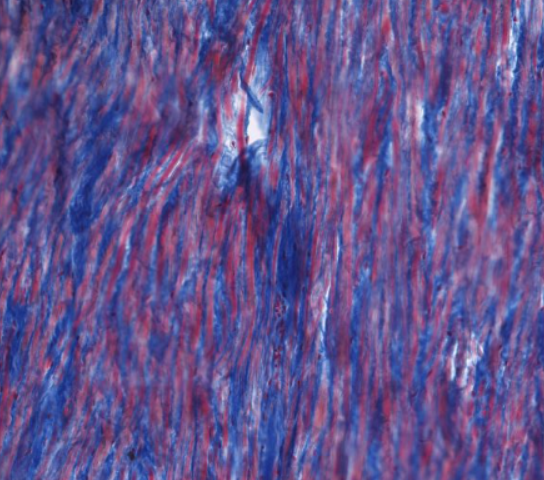
elastic connective tissue
(Collagen stains blue and elastin stains red)
(Azan stain)
embryonic connective tissue
note pink stained fibers
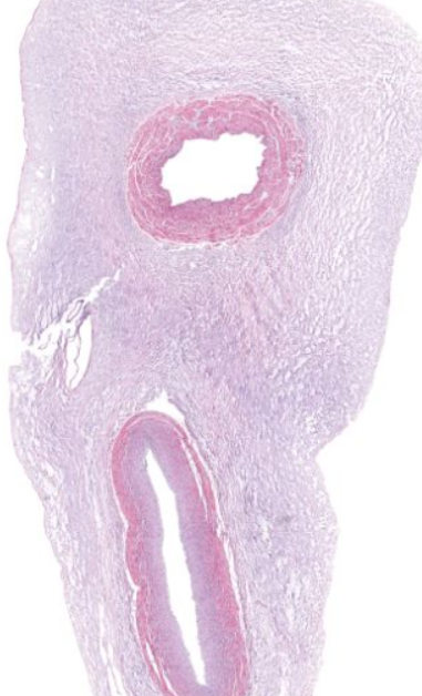
Azan stain
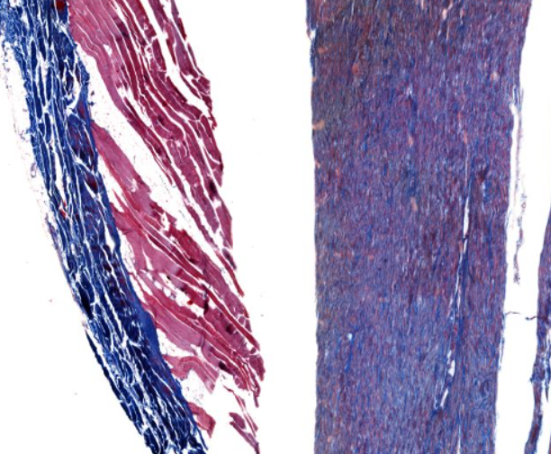
Collagen stains blue and elastin stains red
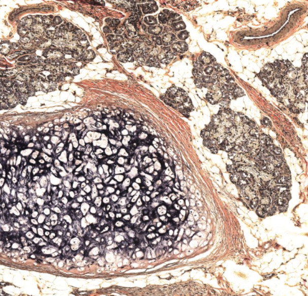
Verhoeff stain
It intensely stains elastic fibers black and lightly stains collagen red.
dense irregular connective tissue
(perichondrium around the trachea)
(pink) type of connective tissue
chondrocytes
the cells within the “holes”
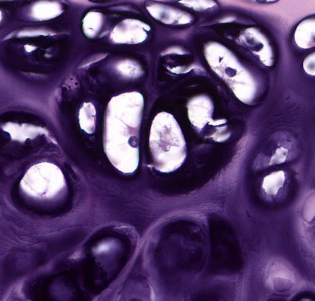
lacunae
the “holes” that contain a type of cell

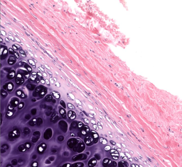
articular/hyaline cartilage
the lighter purple- on the “edge” of a bone
notice the lack of a perichondrium
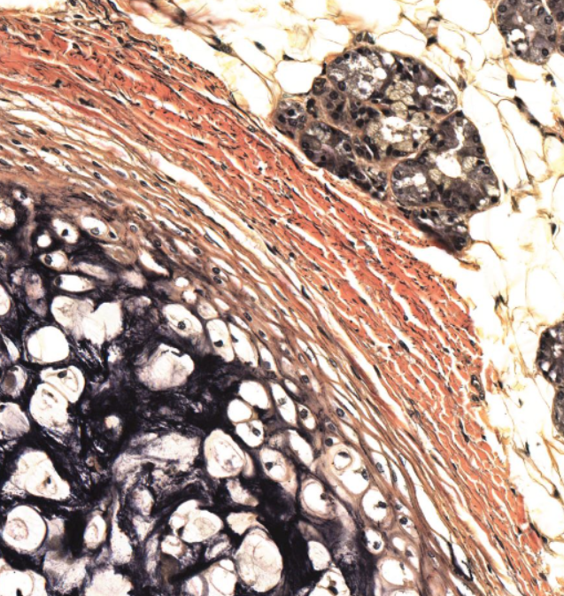
perichondrium
the “reddish” middle layer
notice how it has two layers
outer fibrous layer of the perichondrium
(Dense irregular connective tissue/collagen I)
the more orangey, crackly looking tissue

inner chondrogenic layer of the perichondrium
the “inner”, more stringy looking tissue.
fibrocartilage
notice the mix between dense regular CT and hyaline cartilage
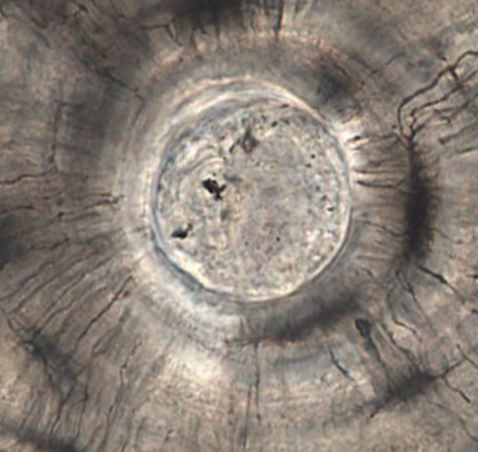
osteon
circular column of bone “building block”
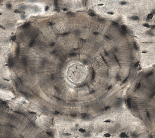
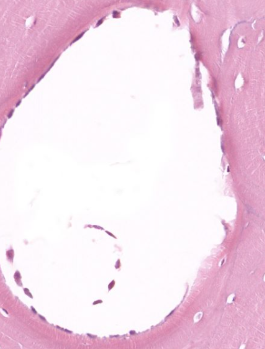
central canal
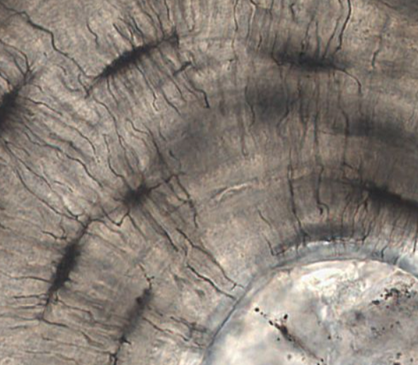
lamellae
layers of intracellular bone matrix
in this photo, they are arranged in rings
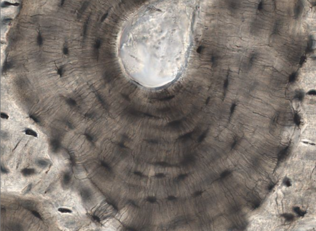
lacunae
darker stained “spaces” that contain a cell

canaliculi
darkly stained small channels
haversian vessels
vasculature in the center of the canal
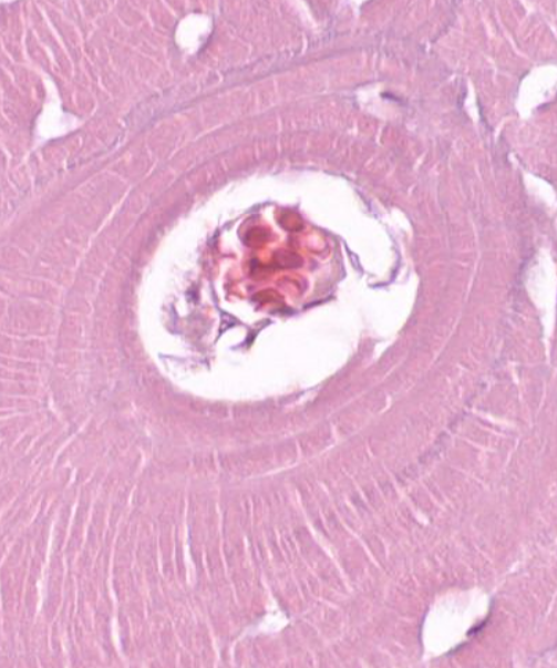
osteoblasts
cells found on the inner surface of some canals
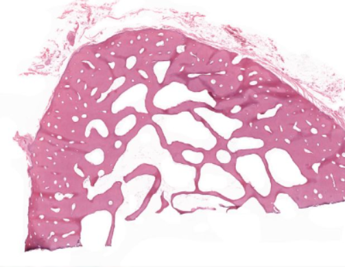
compact bone
the two outer edges
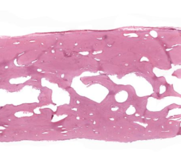
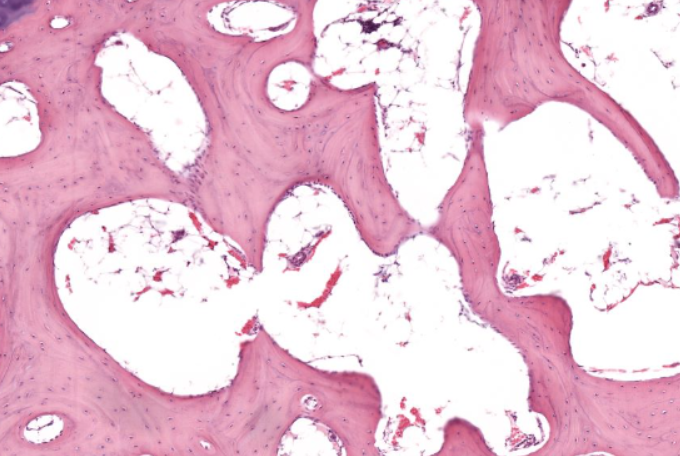
spongy/cancellous/trabecular bone
the “inside” part of the hemisphere
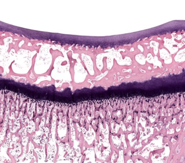
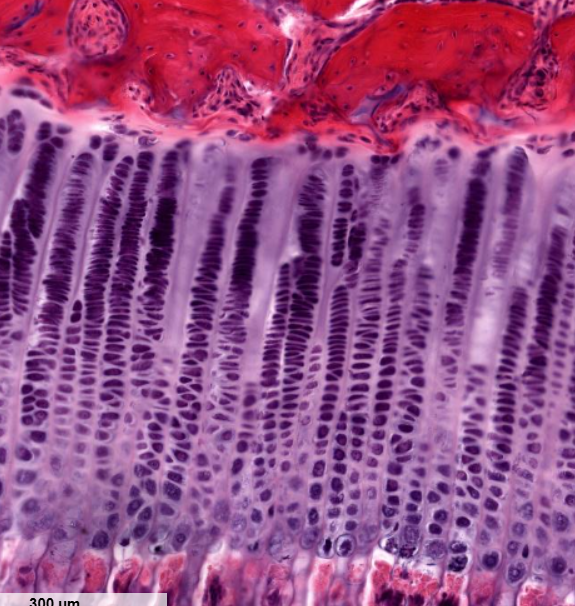
epiphyseal plate
the two purple solid layers
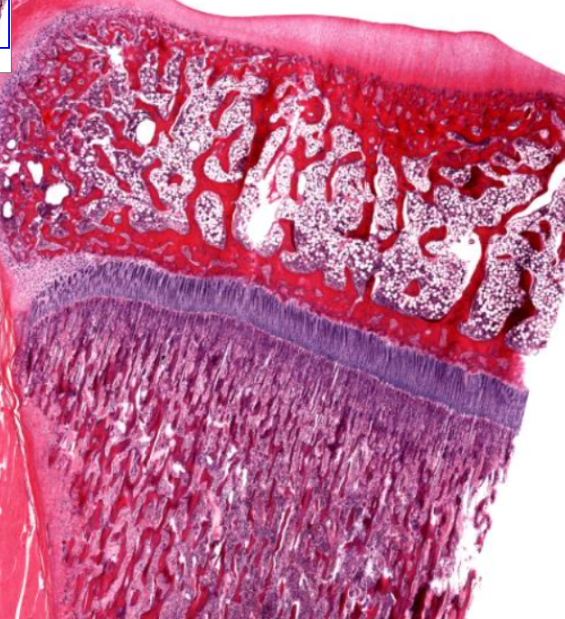
resting zone of the epiphyseal plate
the lighter purple section
part of endochondral ossification
non-dividing chondrocytes
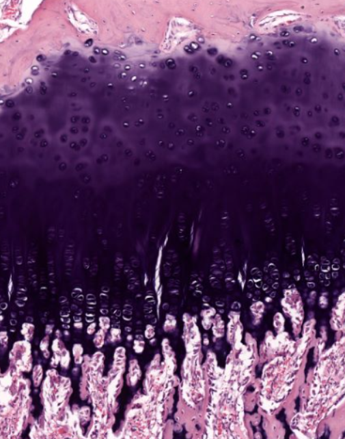
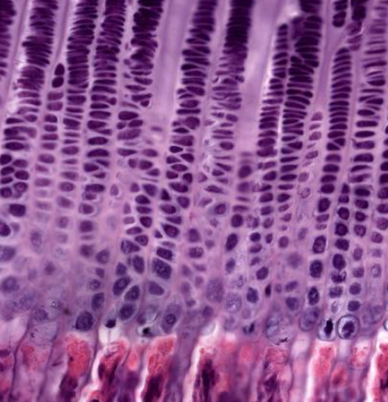
proliferative zone of the epiphyseal plate
area where cells are rapidly dividing
notice the “stacks of coins”
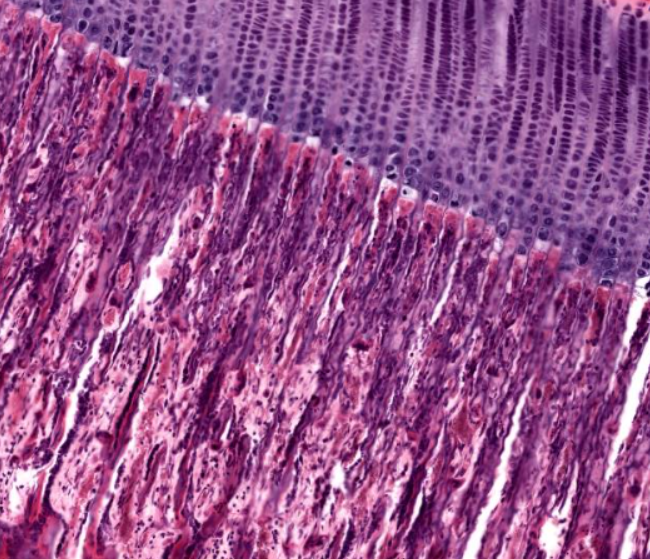
zone of hypertrophy of the epiphyseal plate
note the cells growing larger
zone of calcification/ossification of the epiphyseal plate
the reddish-purple layer
note the change in color→ what substance is this becoming?
osteoclast
type of cell
note the large cell size and the many nuclei
some of these cells have “fuzzy” or “spiky” edges
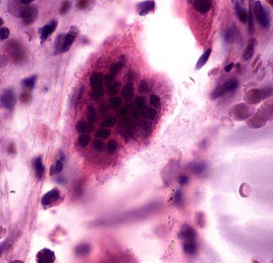
trabeculae
spiky projections that are found in developing bone