12. So you've aquired cardiac disease...
 Call Kai
 Learn
Learn Practice Test
 Spaced Repetition
 Match
Match Flashcards
 Knowt Play
1/72

There's no tags or description
Looks like no tags are added yet.
Name | Mastery | Learn | Test | Matching | Spaced | Call with Kai | Chat |
|---|
No analytics yet
Send a link to your students to track their progress
73 Terms
mitral regurgitation
________________ is the most common of the chronic degenerative valvular diseases
small breed dogs
Who is mitral valve endocarditis normally found in?
left atrial enlargement, volume overload
The earliest radiographic sign of mitral regurgitation is ______________ caused by _____________
hypertrophy, dilation
Radiographically in cases of mitral regurgitation, you will see left ventricular enlargement and rounding of the left ventricular border from ________ in the early stages of the disease, and _______ in later stages
echocardiogram
Because all you can see is the outside of the heart, you can tell it's enlarged but not why... What do you have to do to know why the left ventricle would be enlarged?
dorsal deviation of trachea, left ventricular enlargement, compression/elevation of left mainstem bronchi, left atrial enlargement, distended pulmonary veins
What are the radiographic signs of mitral regurgitation?
cardiac waist, double opacity
Because mitral regurge is a left sided disease, early radiographs will show a straightening of the ________ on the lateral images and a subtle ________ on the VD view
when the mainstem bronchi becomes compressed
When does a mitral regurge patient start coughing?
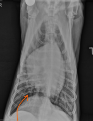
yes (L caudal mainstem bronchus compressed), compensated, not in failure
Is this patient coughing? Is this patient compensated or uncompensated disease? Is there heart failure here?
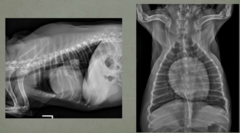
pulmonary venous enlargement,
The first step of pulmonary edema in left sided heart failure is _________
interstitial, hilar, multifocal alveolar,
cardiogenic pulmonary edema starts initially as a _________________ pattern in the _______ region. And then transitions rapidly to ___________________ obscuring the pulmonary vessels
pulmonary venous congestion
What are we seeing here?

diffuse interstitial lung pattern, pulmonary edema, decompensated (cant see the cranial lobar vessels in the lateral view and can see the vesssels on the VD view but cant trace them down the image)
What are we looking at here?

systemic hypertension
___________________ in dogs is typically secondary to underlying clinical disease or conditions like renal disease, cushings, diabetes or an adrenal tumor
hyperthyroidism, renal disease
In cats, ________ and ________ are the most common causes of systemic hypertension
true
true/false: aortic dilation/aneurysm, left ventricular hypertrophy, arrhythmias, epistaxis, and left sided heart failure are all heart/vascular signs of systemic hypertension
aorta, enlarged, undulated
Radiographic changes that indicate systemic hypertension are typically that the ________ looks _________ and _________
enlarged and undulating aorta
What is the abnormality here

cool
Here's that last "enlarged and undulating aorta" image with an outline
Cool?
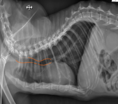
aortic knob, not mass
Cat presented for detatched retinas due to systemic hypertension and a large ascending aorta with undulation/uncoiling was noted. In this case, whats important to remember about the area at the arrow?

enlarged aorta
Whats going on here?

no, yes, enlarged aorta
Does this dog have a grossly enlarged heart? Is there still evidence of hypertension? Why?

dexmedetomidine (in kitty magic)
What drug will make the heart enlarged on radiographs? (specifically in sedation protocols)
yes, yes (interstitial pattern indicated pulmonary edema)
Does this cat have heart disease? Is this cat in heart failure?

dorsally deviated trachea, fluid, carina dorsally deviated
what signs of heart failure are here?

enlarged caudal lobar vessels
What sign of heart failure is here?

fluid
What do we see here?

hyperthyroidism, systemic hypertension
If a cat has hypertrophic phenotype (wall thickening) of the heart what must be ruled out before diagnosing cardiomyopathy?
echocardiogram
In order to tell what type of heart disease a cat has though... you need what?
enlarged caudal lobar arteries, bronchial pattern
In dogs you typically see an enlarged heart with heartworms... what will cats get radiographically?
hypertrophic cardiomyopathy
most common heart disease in cats?
maine coon, ragdoll
hypertrophic cardiomyopathy is heritable in which breeds of cats?
diastolic dysfunction, systolic myocardial failure
Early cardiomyopathy in cats will present as a dysfunction where?? And end-stage will manifest as what type of failure?
left ventricular and atrial enlargement, pulmonary venous congestion, interstitial to alveolar pattern, pulmonary edema
What are radiographic signs of cardiomyopathy?
enlarged left ventricle (apex shifted over), no
What heart disease sign do we see here? Are we in heart failure?

less
Feline restrictive cardiomyopathy is [more/less] common in cats than HCM
systolic, taurine
feline dilated cardiomyopathy is a [systolic/diastolic] dysfunction that was previously related to ______________
left ventricle and left atrial enlargement, no
What abnormalities do we see here? Are we in failure?

right ventricular and atrial enlargement, enlarged CVC, ascites, maybe pleural effusion, but NO LARGE R ATRIUM
Feline-arrhythmogenic right ventricular cardiomyopathy (ARVC) is rare, but what signs would it cause on radiographs?
right heart disease, yes
Which side is affected? Are we in failure?

normal, venous congestion, left heart failure
Canine dilated cardiomyopathy radiographs will appear relatively ________. But because this is a disorder of dilation, it can ultimately cause _______________________ and ___________________
heavy interstitial, patchy alveolar
What lung patterns do we see with canine dilated cardiomyopathy?
grain-free diets
What is canine dilated cardiomyopathy induced by?
straightening of cardiac waist, enlarged left atrium
What is pretty much the only sign you see with canine dilated cardiomyopathy?

VD/DV view
Which view is the "easiest" to document and monitor the clearing of pulmonary edema in large breed dogs?
left caudal mainstem bronchus dorsal and compressed, pleural effusion
What abnormalities do we see here?
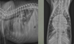
yes
Are we in failure?

true
true/false: grain free diets can create DCM and biventricular failure; legume-rich associated, taurine deficiency is documented in golden retrievers with these diets. (like this one)
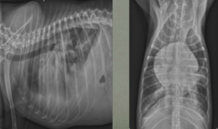
arrythmogenic right ventricular cardiomyopathy
What boxer specific cardiomyopathy can look normal on radiographs but causes arrythmias and syncope
DCM
If boxer specific cardiomyopathy DOES show radiographic signs, what other disease do they mimic?
true
Dogs with ARCM (that boxer thing) that have the DCM form, rarely live long enough to show signs of right heart failure with abdominal and pleural effusion
rounded, neoplasia, right atrium/auricle
Pericardial effusion, will cause the cardiac silhouette to appear ________ and is most commonly related to __________ specifically associated with the ____________
ascites, cardiac tamponade, filling of right atrium/ventricle
Pericardial effusion cases typically present with _______ and signs of right heart failure due to ________ which prevents ______________
enlarged CVC, hepatomegaly, ascites, occasional pleural effusion
What signs ultimately do you see with pericardial effusion?
pericardial effusion/globoid heart
Okay, so whats up here?

no
Are we in failure?
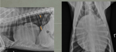
yes
are we in failure now?

stomach axis shifted dorsally
Im telling you this dog has hepatomegally... how can we tell that on this radiograph?

right auricular mass/bulge
Heyyyy buddy. Whats going on there?
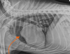
no, ultrasound or other radiographs
Realistically, can we tell exactly whats going on with this herniated right atrium? So what needs to happen?

heart base mass, displaced trachea on BOTH VD and lateral
Whats going on here? How can we tell?

enlarged tortuous pulmonary arteries, right atrial/ventricular enlargement (reverse D), dorsal deviation of trachea, main pulmonary arterial enlargment, pulmonary parynchemal changes, hepatomegally
What radiographic changes do we see with canine heartworms? (the first one listed is what she said is the main thing she looks for)
turbulent flow, pulmonary hypertension
what causes the main pulmonary arterial enlargement in heartworm positive dogs?
pulmonary hypertension, loss of laminar flow
What causes the enlarged or tortuous pulmonary arteries in heartworm positive dogs?
enlarged tortuous pulmonary arteries
What sign is seen here?

no
Is this dog in failure?

reverse D, large tortuous pulmonary arteries, stomach axis shifted back = hepatomegally
There are several signs of heartworms here, what are they?
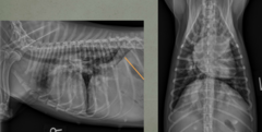
no
Are we in heart failure here?

cool
Here is an example of that tortuous vessel outlined.
Cool?

true
true/false: pulmonary hypertension can sometimes enlarge the vessel only in the cranial portion over the heart, so you have to look at the whole vessel

chronic bacterial endocarditis
________________________ can present with a fever of unknown origin and presents radiographically with left side heart disease, and/or failure, and a showering of the lungs with thromboembolisms
patchy interstitial to alveolar lung pattern, showering of lung with thromboembolism, swollen joints, shifting leg lameness
What are the clinical signs of canine endocarditis?
showing of thromboembolisms
Whats going on here?