histology lab quiz 0
 Call Kai
 Learn
Learn Practice Test
 Spaced Repetition
 Match
Match Flashcards
 Knowt Play
1/11

There's no tags or description
Looks like no tags are added yet.
Name | Mastery | Learn | Test | Matching | Spaced | Call with Kai | Chat |
|---|
No analytics yet
Send a link to your students to track their progress
12 Terms
Histology
Study of the anatomy of cells and tissues of body by using microscopy
Distance maxes for eye, light microscope, electron microscope
Eye: 0.1 mm
Light: 0.1 um
Electron: 0.1 nm

Simple microscope features
Image, object, lens
virtual image
Compound microscope
Light source, condenser lens, specimen, objective lens, ocular lens
inverted image
Need staining, image is not the actual color
2D
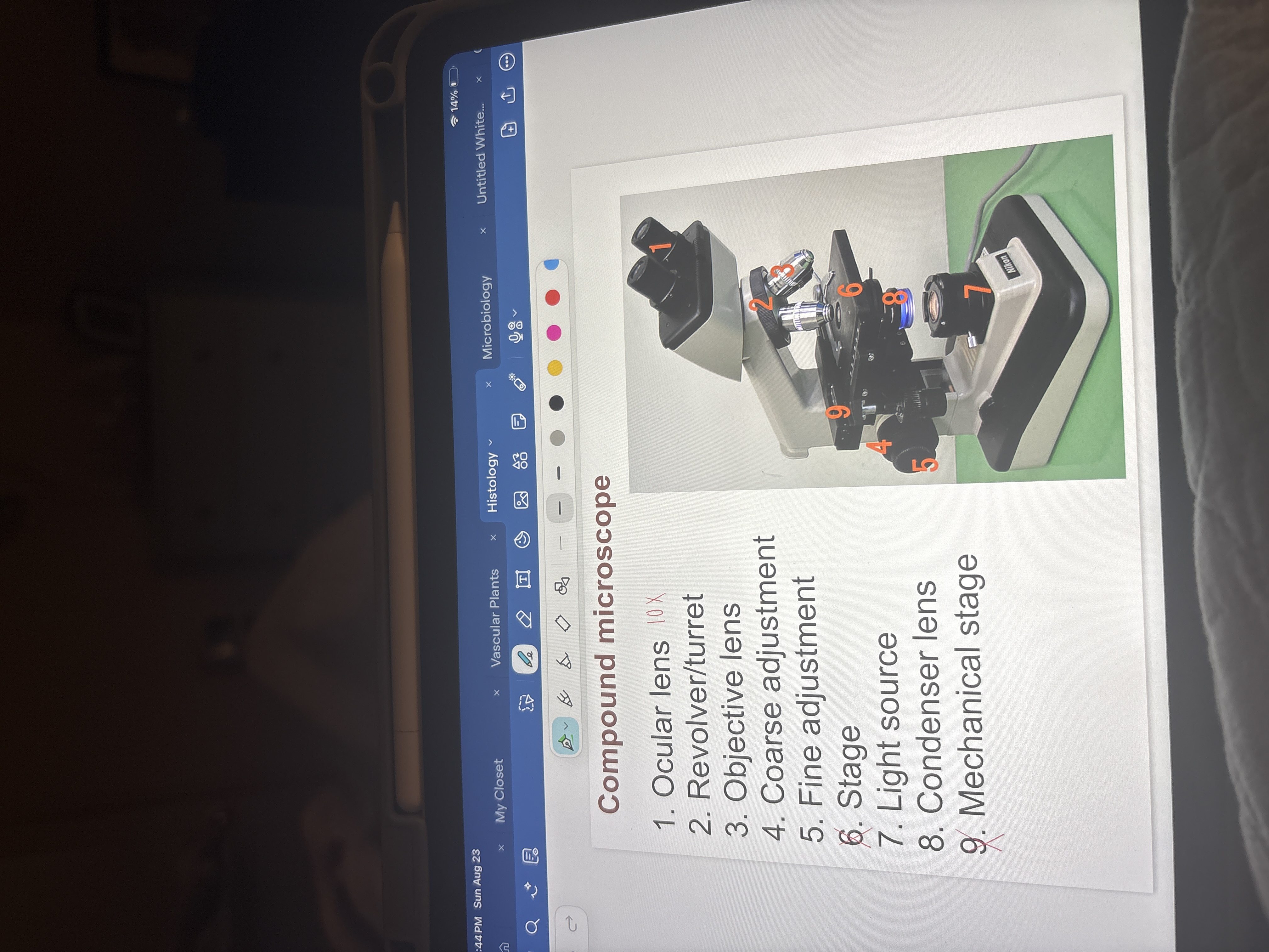
Magnification levels
Ocular: 10x
Low: 4X, 40X total mag.
medium: 10X, 100X total
High: 40x, 400X total
Oil: 100X, 1000X total
Stereo microscope
3D, reflected image, light source on top
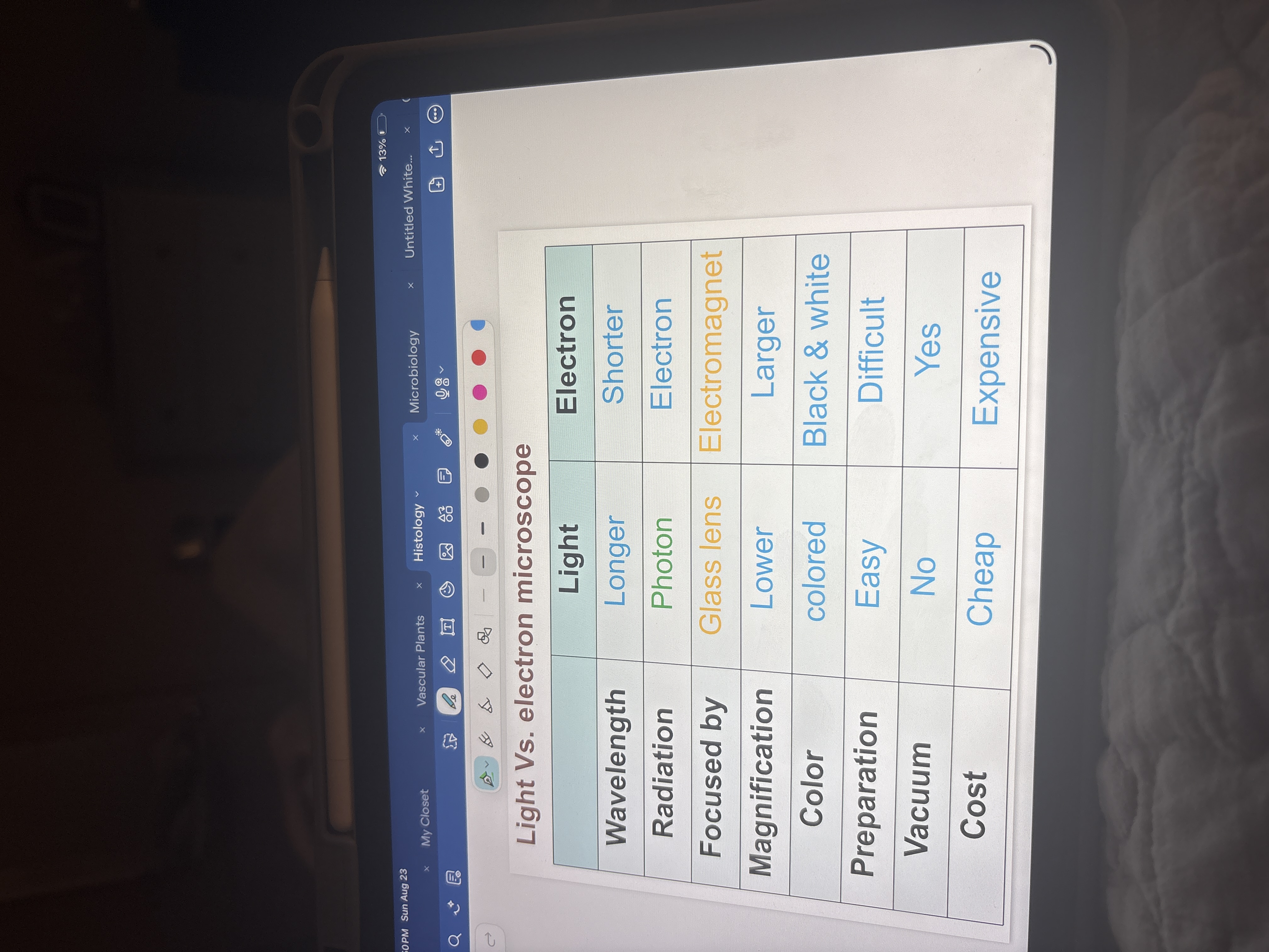
Electron microscope vs light microscope
Light uses photons but electron uses electrons to show (darker means high electron density)
electron has shorter wavelengths, larger magnification, difficult to prepare, has a vacuum, and is expensive
Transmission electron microscope
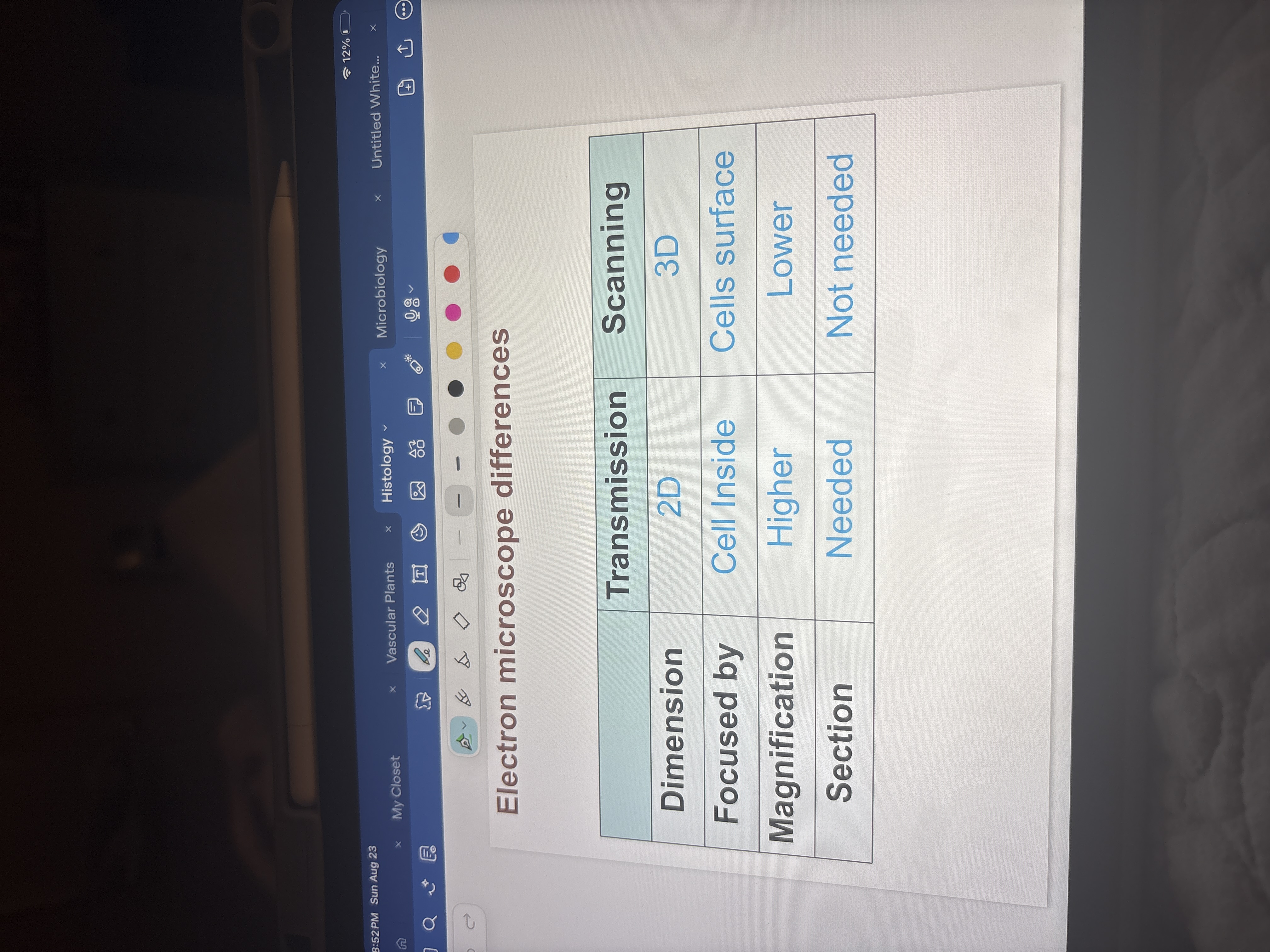
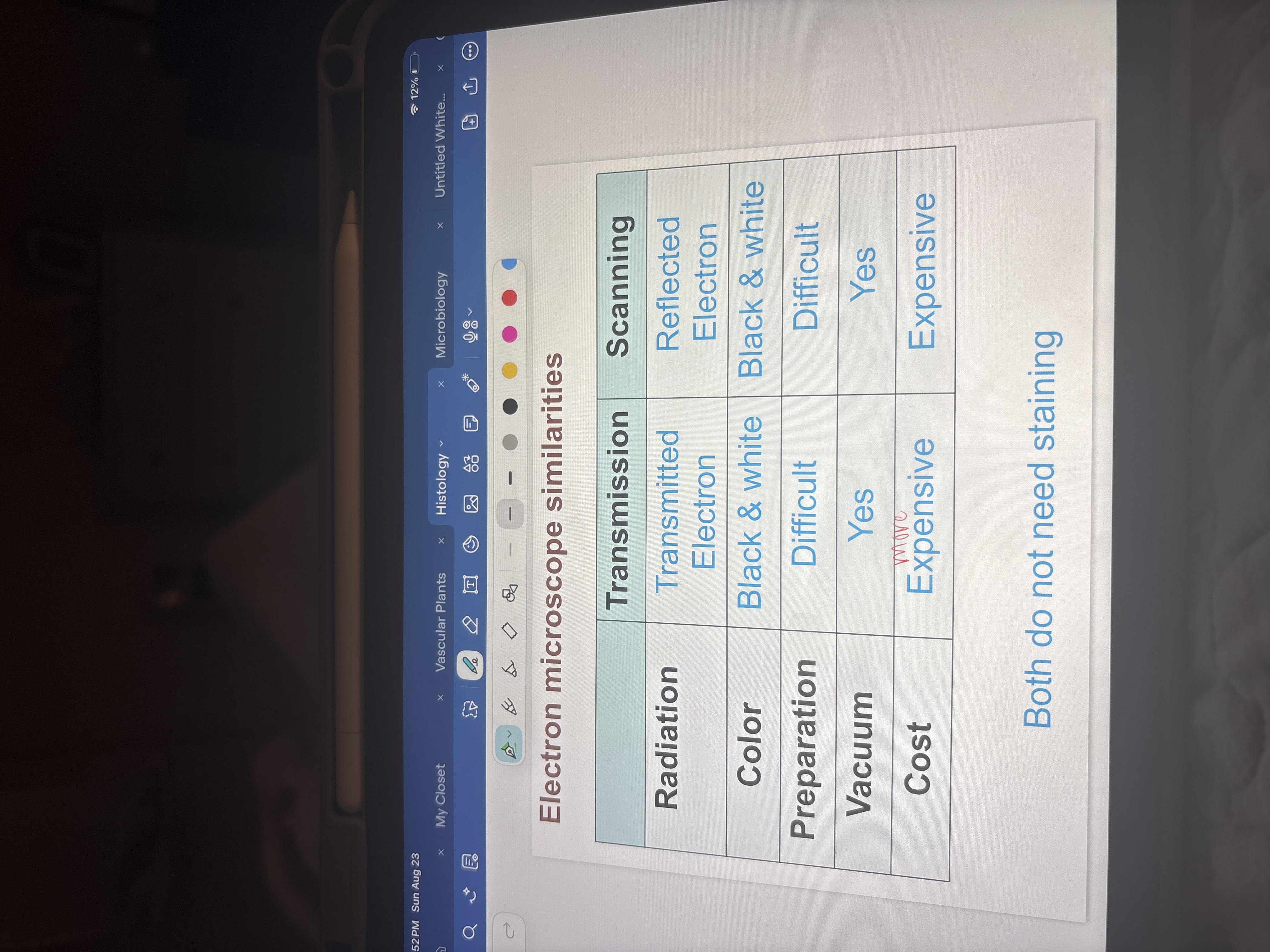
2D, transmitted electrons, black and white
Compound microscope is comparative
Scanning electron microscope
3D, section not necessary, electrons scan over surface of sample
Staining
Only needed for compound microscopes
general and special

H&E staining (general)
Basic dye: hematoxylin, blue, positive, basic
affinity for nuclei (DNA is negative) and RER (basophilic structures)
Acidic dye: eosin, red, negative, acidic
affinity for positive proteins such as mitochondria (acidophilic structures)
Special staining (4)
PAS: stains glycogen (liver)
Trichrome: massons trichrome stains collagen fibers
H&E: mix of blue and red (blue parts are basophilia and red are eosinophilia)
silver stains: stains neurons and neuroglia, stains reticular fibers