Case 4: Have you met Wilhelm Roentgen?
 Call Kai
 Learn
Learn Practice Test
 Spaced Repetition
 Match
Match Flashcards
 Knowt Play
1/49

There's no tags or description
Looks like no tags are added yet.
Name | Mastery | Learn | Test | Matching | Spaced | Call with Kai | Chat |
|---|
No analytics yet
Send a link to your students to track their progress
50 Terms
False
note: in a right lateral, the right side is down, meaning the lungs become atelectic and you actually see the left lung lobes which are "up" and still full of air
In a right lateral projection, the lungs you are evaluating are the right cranial, right middle and the right caudal lung lobes.
TRUE OR FALSE
True
End on blood vessels are often misinterpreted as nodules when looking for metastatic disease in the thorax.
TRUE OR FALSE
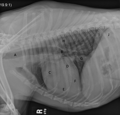
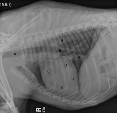
Trachea
What is A?
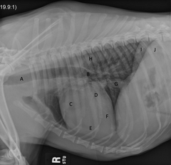
Carina
note: where the trachea comes down and divides into main pulmonary bronchi
What is B?
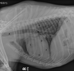
Right atrium
What is C?
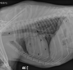
Left atrium
What is D?
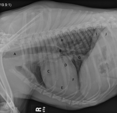
Right ventricle
What is E?
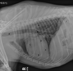
Left ventricle
What is F?
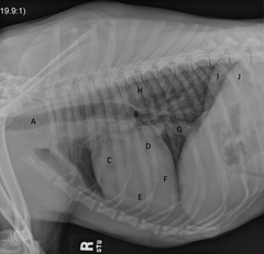
Caudal vena cava
What is G?
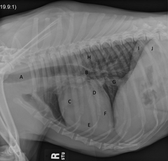
Aorta
What is H?
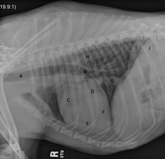
Right Crus of Diaphragm
What is I?
Left Crus of Diaphragm
What is J?
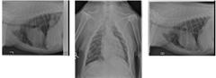
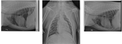
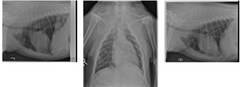
Canine Right lateral, left lateral and VD thoracic projections are available for review.
Describe the study you are seeing here
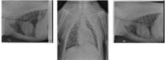
Patient positioning is adequate for interpretation.
Study technique is adequate for interpretation.
Describe patient positioning and technique
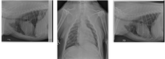
Multiple, variably sized soft tissue nodules are present throughout the pulmonary parenchyma. The cardiac silhouette, other mediastinal structures, pulmonary vasculature, pleural space and extra-thoracic structures are all within normal limits.
Describe your findings (roentgen signs)
Given the presence of a mass within the abdomen and the age of the patient, these findings are most consistent with metastatic disease.
Describe Conclusions: Give your radiographic diagnosis and list possible differential diagnoses for all your findings
If not already performed, abdominal ultrasound could be considered to further investigate the palpated abdominal mass.
Describe Next Steps: Are there any additional imaging suggestions that would help clarify your findings? Are there critical next steps to take from a stabilization/management/treatment plan standpoint?
1. extrathoracic structures (musculoskeletal, trachea, superficial soft tissue structures)
2. pleural space
3. mediastinum (cranial, middle, and caudal)
4. pulmonary parenchyma
What 4 main places do you evaluate with thoracic radiographs?
Pleural space
What structure is outline here?
Cardiac silhouette
What is the most prominent thing that lives in the mediastinum?
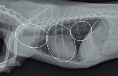
1. lymph nodes (sternal, cranial mediastinal)
2. thymus
3. trachea
4. esophagus
What lives in the cranial mediastinum?
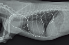
1. esophagus
2. tracheal termination (carina)
3. tracheobronchial lymph nodes
4. cardiac silhouette
What lives in the middle mediastinum?
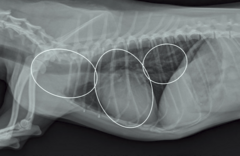
1. esophagus
2. aorta
3. caudal vena cava
What lives in the caudal mediastinum?
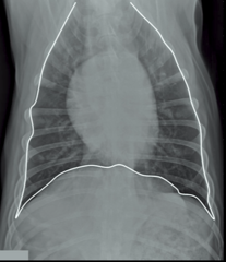
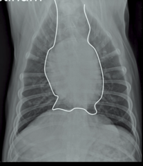
Mediastinum
What is being outline here?
It is a HUMANOID projection
means the scapula are rotated out of the thoracic cavity to remove superimposition of the lungs
note: normally, the legs are moved UP towards the patient's head, but in the humanoid projection, they are moved down towards the side of the body
What is interesting about the VD projection here?
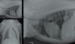
Pleural space is good, lungs extend all the way out to the thoracic body wall
note: it is the pulmonary PARENCHYMA that has the issue
Describe the pleural space here
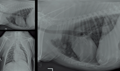
Cranial, middle, and caudal are all within normal limits
Describe the mediastinum
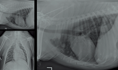
Soft tissue nodules within the pulmonary parenchyma. Diffuse structured interstitial pattern
Describe the pulmonary parenchyma (brief)
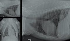
1. that they are soft tissue
2. that they are round
What two thing does the word "nodule" imply?
Metastatic disease
Multiple soft tissue nodules in the lungs are most consistent with what pathology?
Right side of the patient is on the table --> right lung becomes non-aerated (becomes soft tissue opacity) = left lung evaluated
What is a "right lateral"? What lungs are you looking at?
Stomach
Which structure is associated with the left crus?
Caudal vena cava
Which structure is associated with the right crus?
Right lateral
If there are nodules in the left lung, which lateral image will you best see them in?
Normal
Is this a normal or abnormal thorax?
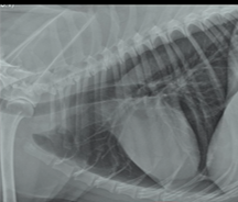
Structured interstitial pattern
Discrete masses within the pulmonary parenchyma best describes WHICH pattern?
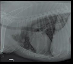
Unstructured interstitial pattern
Diffuse increase in opacity throughout the pulmonary parenchyma, typically from congestive heart failure, best describes WHICH pattern?
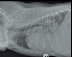
INCREASED
note: there are VERY FEW things that can cause decreased opacity (more lucency): hypovolemia, small vessels in the lungs, overinflation like a cat with asthma
Is it more common for the lungs to be abnormally increased or decreased in opacity?
Expiration (less air in the lung)
On inspiration or expiration, the lung looks more opaque
Bronchial tree
What is being outlined here?
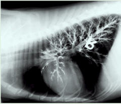
Alveolar pattern
Fluid or cells within the alveoli like pneumonia that causes increase in opacity is WHAT pattern?
Connective tissue, blood vessels, lymphatics
note: alveoli can leak into interstitum to cause a secondary interstitial pattern as well
What lives in the interstitium?
Leaking into interstitium --> may secondarily leak into alveoli
Where does CHF start?
Thickening of the bronchial walls from things like asthma or bronchitis
What causes "train tracks" of the bronchial pattern (fluid or cells associated with the airway)?
Cardiogenic or noncardiogenic pulmonary edema
What kinds of pathologic processes are associated with unstructured interstitial pattern?
False
It is relatively easy to identify pulmonary nodules that are superimposed over the cardiac silhouette.
TRUE OR FALSE
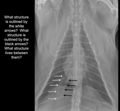
WHITE = caudal lobar pulmonary artery
BLACK = caudal lobar pulmonary vein
BETWEEN = caudal lobar bronchus
What are the white arrows outlining? How about black? What lies between?
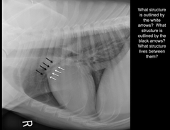
WHITE = cranial lobar pulmonary vein
BLACK = cranial lobar pulmonary artery
BETWEEN = cranial lobar bronchus
What are the white arrows outlining? How about black? What lies between?
True
You can usually determine which lateral you have in a thoracic radiograph using just the anatomy. You do not need to have a marker.
TRUE OR FALSE
False
note: we see nodules because they are soft tissue opacity adjacent to air filled lung
You can only see soft tissue nodules in the lungs that are close to the plate. It is their proximity to the plate that allows us to see them.
TRUE OR FALSE