T1 - Introductory concepts and patterns of disease
 Call Kai
 Learn
Learn Practice Test
 Spaced Repetition
 Match
Match Flashcards
 Knowt Play
1/57

There's no tags or description
Looks like no tags are added yet.
Name | Mastery | Learn | Test | Matching | Spaced | Call with Kai | Chat |
|---|
No analytics yet
Send a link to your students to track their progress
58 Terms
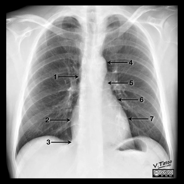
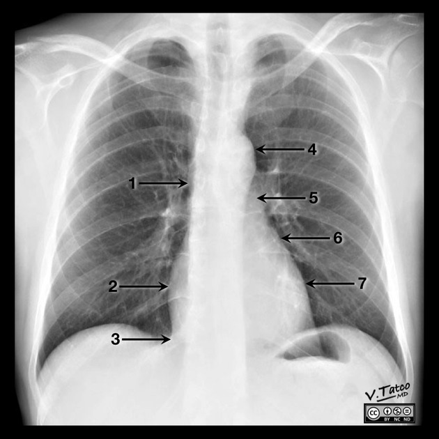
Name structures
1: SVC
2: Right atrium
3: IVC
4: Aortic arch
5: Main pulmonary artery
6: Left atrial appendage
7: Left ventricle
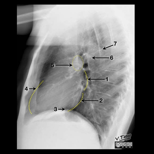
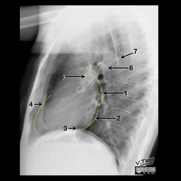
Name structures
1: Left atrium
2: Left ventricle
3: IVC
4: Right ventricle
5: Right pulmonary artery
6: Left pulmonary artery
7: Aorta
Describe intralobular fissures
Minor fissure separates RUL from RML, seen as fine horizontal line on frontal and lateral CXR
Major/oblique fissures seen only on lateral CXR.
On right side: seperates RUL and RML from RLL.
On left side: seperates LUL from LLL.
List most common accessory fissures
Azygous fissure (1%) in presence of azygous lobe
Superior accessory fissure (5%) separates superior and basal segments of RLL.
Inferior accessory fissure (12%, R>L lung) separates medial basal segment from the other basal
segments.
Left minor fissure (8%), separates lingula from LUL
Atelectasis, definition and mechanisms
Loss of lung volume due to decreased aeriation. Synonymous with collapse. May be caused by bronchial obstruction, mucus plugging, external compression (ie. effusion/ptx or small lung volumes)
Direct signs atelecatis
From lobar volume loss include:
- Displacement of fissures
- Plate-like / triangular opacity from collapsed lung
- Vascular overcrowding
Indirect signs atelectasis
Due to effect of volume loss on adjacent structures include:
- Elevation of diaphragm
- Rib crowding on side of colume loss
- Mediastinal shift to side with volume less
- Overinflation of adjacent/contralateral lobes
- Hilar displacement
May there be air bronchograms in atelectasis?
NOT is the cause is central bronchial obstruction.
MAY BE SEEN if the cause is external compression
Describe mechanism og obstructive atelectasis
Alveoar gas absorbed by blood circulating through alveolar capillaries, but is not replaced by inspired air due to bronchial obstruction.
May cause lobal atelectasis.
Epidemiology of obstructive atelectasis
Obstructive atelectasis occurs more quickly when pt is breathing supplemental oxygen since oxygen is absorbed from alveoli more rapidly than nitrogen.
In children, most often due to aspiration of foreign object. In contrast to adults, the affected side becomes hyperexpanded in children due to ball-valve effect.
Subsegmental atelectasis is a subtype of obstructive atelectasis commonly seen after surgery or general illness, due to mucus obstruction of small airway.
Describe mechanism of relaxation atelectasis
Caused by relaxation of lung adjacent to intrathoracic lesion causing mass effect (pleural effusion, ptx, pulmonary mass).
Describe mechanism of adhesive/passive atelectasis
Surfactant deficiency. Most comonly seen in NRDS, may also be seen in ARDS in adults.
Describe mechanism og cicatrical atelectasis
Volume loss from architectural distortion of lung parenchyma by fibrosis.
Pathomechanism in lobar atelectasis
Usually by central bronchial obstruction (obstructive atelectasis) which may be secondary to mucus plugging or obstructive mass.
Lobar atelectasis etiology
If acute - mucus plugging most likely cause.
Mucus plugging most common in lower lobes, least common i LUL
If lobar atelectasis seen in outpatient, an obstructing central tumor must be ruled out.
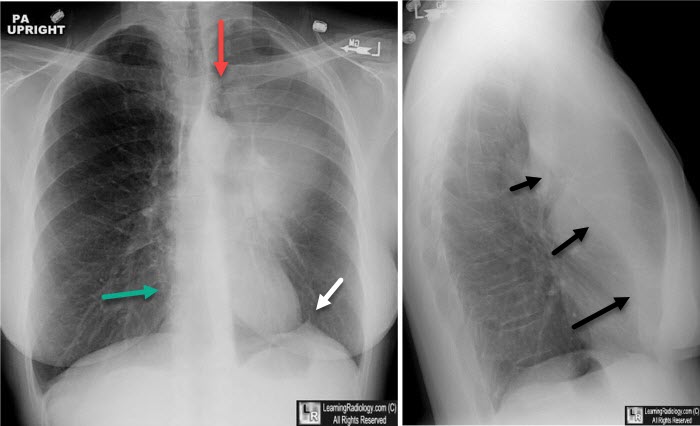
LUL atelectasis CXR
Frontal view: Luftschiel sign, not always present.
Lateral view: anterior displacement of major fissure and anterior collapsed lung
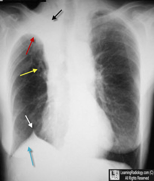
RUL atelectasis CXR
Frontal view: reverse S sign of Golden when underlying obstructive mass. Juxtaphrenic peak sign is a peridiaphragmatic trangular opacity caused by diaphragmatic traction from an inferior accessory fissure og an inferior pulmonary ligament, this is in RUL volume loss from any cause.
Lateral view: Wedge-shaped collapse of RUL.
LLL atelectasis CXR
Frontal view: Triangular retrocardic opacity main feature.
Lateral view: Flat waist sign = flattening of left heart border due to posterior shift of hilar structures and resultant cardiac rotation.
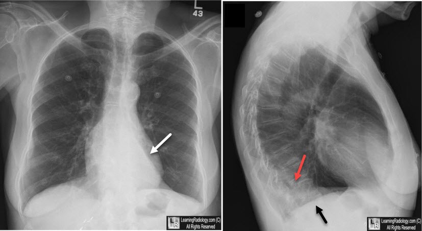
RLL atelectasis CXR
Frontal view: Not well seen as it collapses medially. Right heart border is preserved.
Lateral view: Triangular retrocardiac opacity.
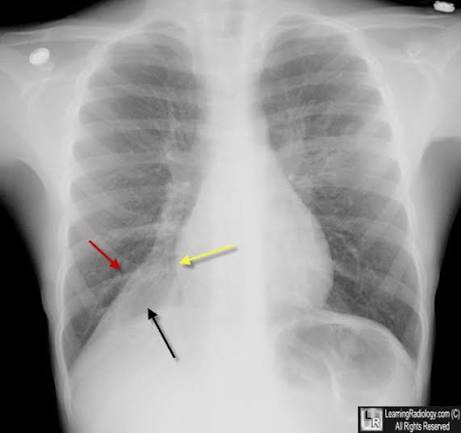
RML atelectasis CXR
Frontal view: Subtle. Silhouetting of right heart border by collapsed medial segment of middle lobe may be only clue.
Lateral view: Triangular opacity anteriorly.
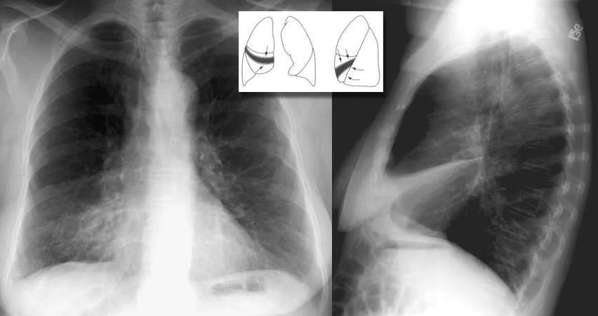
Obstruction of bronchus intermedius leads to atelectasis of which lobes and which findings on CXR
RML and RLL atelectasis, witch obscuration of both right cardiac border and right hemidraphragm, with a linear superior margin directed towards the hilum.
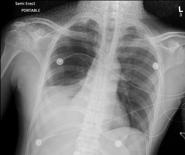
Round atelectasis definition
Focal atelectasis with round morphology almost always associated with an adjacent pleural abnormality (effusion, thickening, plaque).
Most common in posterior lower lobes.
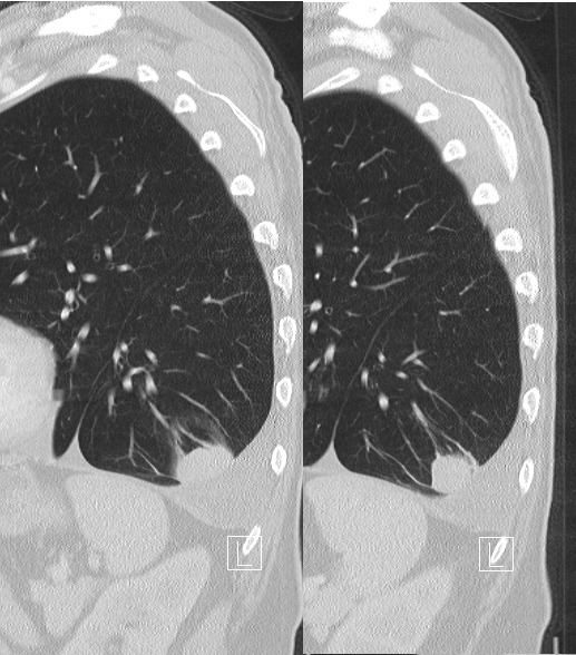
Which 5 imaging features MUST be present to diagnose round atelectasis
1) Adjacent pleura must be abnormal
2) Opacity must be peripheral and in contact with pleura
3) Opacity must be round og elliptical
4) Volume loss must be present in affected lobe
5) Pulmonary vessels and bronchi leading into opacity must be curved, leading to focus of atelectasis (comet tail sign).
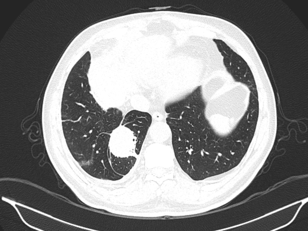
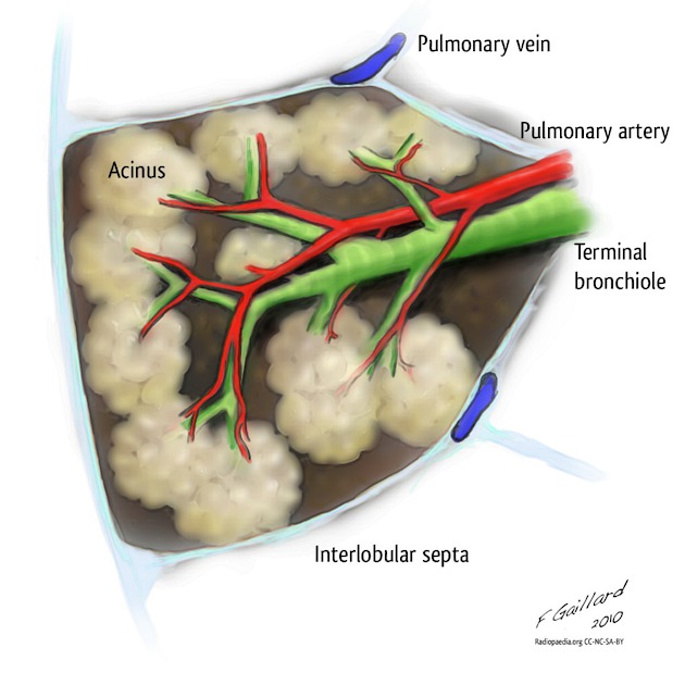
Define/describe the secondary pulmonary lobule (SPL)
The elemental unit of lung function.
Contains central artery and central bronchus, each branching multiple times to ultimately produce acinar arteries and respiratory bronchioles
Pulmonary veins and lymphatics collect in the periphery
Connective tissue, called interlobular septa, encases each SPL.
Each SPL is between 1-2.5 cm in diameter
Generally 12 or fewer acini per SPL
Consolidation and ground glass represent which basic pathological processes (2)
1) Filling of aleoli with fluid (blood, pus, water or cells)
2) Incomplete aeration
Most common differential in acute consolidation
1) Pneumonia (most common cause)
2) Aspiration (may be heterogenous from mucus plugging)
3) Pulmonary hemorrhage (primary pulmonary hemorrhage or aspiration of hemorrhage)
4) ARDS (noncardiogenic pulmonary edema in critically ill pt thought to be caused by increased
capillary permeability)
5) Pulmonary edema (consolidation if severe)
Differential in chronic consolidation
1) Adenocarcinoma
2) Lymphoma
3) Organizing pneumonia (nonspecific response to injury characterized by granulation polyps which
fill the distal airways, producing peripheral rounded and nodular consolidation)
4) Chronic eosinophilic pneumonia (inflammatory process characterized by eosinophils causing
alveolar filling in UPPER-LOBE distribution)
Ground glass opacification definition and what processes it represents(3)
Haziness/opacification where underlying vessels are still visible.
1) Partial filling of alveoli (blood, pus, water or cells)
2) Alveolar wall thickening
3) Reduced aeration
Differentials in acute grond glass
1) Pulmonary edema (central or dependent)
2) Pneumonia (atypical pneumonia, e.g. viral og PJP)
3) Pulmonary hemorrhage (pure GGO in acute phase, subacute phase shows peripheral sparing and
crazy paving)
4) ARDS
Differential in chronic ground glass
1) Lung adenocarcinoma (focal or multifocal)
2) Organizing pneumonia (rounded, peripheral opacities)
3) Chronic eosinophilic pneumonia (upper-lobe predominance)
4) ILD (incl DIP, NSIP, HP)
5) Hypersensitivity pneumonitis (HP) (type III hypersensitivity reaction to inhaled organic antigens. In subacute phase, GGO centrilobular nodules and mosaic attenuation).
Differential in peripheral consolidation or ground glass opacification
1) Organizing pneumonia
2) Chronic eosinophilic pneumonia (upper-lobe predominance)
3) Pulmonary infarction
Interlobular septal thickening mechanism
1) Dilation of pulmonary veins (smooth)
2) Disease involving interstitium/lymphatics (may be more nodular)
Differential in smooth interlobular thickening
1) Pulmonary edema
2) Lymphangitis carcinomatosis
Differential in nodular/irregular/asymmetric septal thickening
1) Lymphangitic carcinomatosis (tumor spread through lymphatics)
2) Sarcoidosis (rarely causes septal thickening)
Definition of crazy paving pattern
INTERlobular septal thickening with superimposed groung glass opacification. It is nonspecific.
First described for alveolar proteinosis, where GGO is caused by filling of alveoli by proteinaceous material and interlobular septal thickening is caused by lymphatics taking up the same material.
Differential diagnosis in crazy paving
1) Pulmonary edema (most common cause)
2) Pulmonary hemorrhage
3) ARDS
4) Pulmonary alveolar proteinosis (idiopathic disease characterized by alveolar filling by
proteinaceous substance. PAP almost always seen with crazy paving.
5) PJP pneumonia
6) Adenocarcinoma (uncommon cause)
7) Lipoid pneumonia (inflammatory pneumonia caused by reaction to aspirated lipids, uncommon cause)
Nodular patterns, how are they divided?
1) Centrilobular (incl. tree-in-bud)
2) Peribronchial nodules
3) Perlymphatic nodules (peribronchial + subpleural nodules)
4) Random nodules
Definition and imaging centrilobular nodules
Opacification of and around centrilobular bronchioles or, less commonly, centrilobular artery.
On CT: small nodules in centers of SPL, NEVER EXTEND TO PLEURAL SURFACE.
May be solid or GGO.
Differential in centrilobular nodules (without tree-in-bud)
1) Viral pneumonia
2) Hypersensitivity pneumonitis (most common)
3) Aspiration
4) Pulmonary capillary hemangiomatosis
5) Metastatic calcification
6) Occasionally, pulmonary edema
7) Exposure-related lung disease (most common)
Definition and imaging of centrilobular tree-in-bud nodules
Multiple small nodules connected to linear branching structures. The linear branching structures represent mucus-impacted bronchioles (usually invisible on CT) and nodules represent impacted terminal bronchioles.
They are due to mucus, pus or fluid impacting bronchioles and terminal bronchioles.
Almost ALWAYS associated with small airway infection or inflammation, e.g. endobronchial spread of Tb.
Differential in tree-in-bud nodules
1) Infectious bronchiolitis (Tb, atypical mycobacteria, viral, bacterial)
2) Aspiration
3) Rarely, lymphangitic carcinomatosis and vascular abnormalities (endovascular metastasis and pulmonary arterial aneurysms)
Perilymphatic nodules definition and imaging
Nodules follow anatomic locations of pulmonary lymphatics: subpleural, peribronchovascular, septal.
Differential perilymphatic nodules
1) Sarcoidosis (typically upper-lobe) - may become confluent, creating galaxy sign which multiple tiny nodules surround central lesion
2) Pneumoconiosis (silicosis and coal workers’ pneumoconiosis - reactions to inorganic dust inhalation. Imaging may look identical to sarcoidosis with perilymphatic nodules, but usually a history of exposure (e.g coal worker, sandblaster who develops silicosis)
3) Lymphangitic carcinomatosis
Random nodules definition and imaging
Multiple nodules with random distribution, usually through hematogenous spread. Includes miliary pattern, which are innumerable tiny random nodules the size of millet seeds.
Differential random nodules
1) Hematogenous metastases
2) Disseminated mycobacteria
3) Disseminated fungal infections
Cavitary lesion definition
Development of air within pre-existing lesion (nodule, mass, consolidation).
A solitary cavitary lesion is most likely cancer or infection
Imaging findings cavitary lesion
Typically has thick, irregular wall, often with solid mural component. Findings of benign and malignant cavitary lesions overlap, but a maximum wall thickness up to 4 mm is usually benign and >15 mm is usually malignant. Spiculated margins also suggest malignancy.
Differential in solitary cavitary lesion
1) Primary bronchogenic carcinoma (SCC and adenocaracinoma, but SCC cavitates more frequently. SCC is never known to cavitate!)
2) Tb (upper-lobe)
3) Fungal pneumonia
4) Cavitary bacterial pneumonia
Differential in multiple cavitary nodules
1) Septic emboli (typically peripheral)
2) Vasculitis (incl. granulomatosis with polyangiitis (GPA)
3) Metastases (classically SCC, but any mets can cavitate)
Pulmonary cyst definition and how to differentiate from emphysema
Air-containing space with thin wall and doesn’t contain any other structures. In contrast with emphysema, which usually has no walls and may have ventral vessels.
Differential in multiple lung cysts
1) Lymphangioleiomyomatosis (LAM)
2) Pulmonary Langerhans cell histiocytosis
3) Lymphoid interstitial pneumonia (LIP)
4) Amyloid
5) Birt-Hogg-Dube syndrome
6) PJP
Lymphangioleiomyomatosis (LAM) is characterised by which findings
Diffuse cystic lung disease due to smooth muscle proliferation of distal airways. LAM causes UNIFORMLY distributed, thin-walled cysts in diffuse distribution. May be associated with chylous effusion.
Pulmonary Langerhans cell histiocytosis are characterized by which features
Irregular cysts and nodules, predominantly distributed in upper lungs
Lymphoid interstitial pneumonia (LIP) is characterised by which findings
Rare disease usually associated with Sjøgrens synrome and characterized by lymphocytic infiltrate and multiple cysts.
Amyloid
Appears similar to LIP: lymphocytic infiltrate and multiple cysts.
Birt-Hogg-Dube syndrome is characterised by which findings
AD-genetic disorder characterized by renal tumors (chromophobe renal carcinoma and renal oncocytoma), renal cysts and pulmonary cysts. Spontaneous ptx may occur as a sequela of pulmonary cysts.
Usually predominantly lower zone and bilateral cysts. Predilection for subpleural lung, incl. paramediastinal and perifissural location. Cysts are usually adjacent to interlobular septa, arteries and veins. Cysts have variable size, may be round or elongated, and may sometimes be multilobulated or multiseptate.
Differential single cyst
1) Bulla (emphysematous space >1 cm. Giant bulla occupies >30% of thoracic volume)
2) Bleb (air-filled cystic structure contiguous with pleura, >1 cm. Rupture of bleb is the most common cause of ptx)
3) Pneumatocele (air-filled space caused by prior lung trauma or infection)
Pulmonary fibrotic changes have different differentials depending on distribution. Name them (upper and lower)
Lower zone
- Usual interstitial pneumonia (UIP) pattern (IPF, RA, asbestosis) - honeycombing.
- Non-specific interstitial pneumonia (NSIP) pattern (commonly associated with vascular disease and drug reaction) - peribronchial reticulation and traction bronchiectasis ± GGO
Upper zone
- End-stage sarcoidosis
- Chronic hypersensitivity pneumonitis
- End-stage silicosis