Cell structure BIO P1
 Call Kai
 Learn
Learn Practice Test
 Spaced Repetition
 Match
Match Flashcards
 Knowt Play
1/50

There's no tags or description
Looks like no tags are added yet.
Name | Mastery | Learn | Test | Matching | Spaced | Call with Kai | Chat |
|---|
No analytics yet
Send a link to your students to track their progress
51 Terms

draw an prokaryotic cell
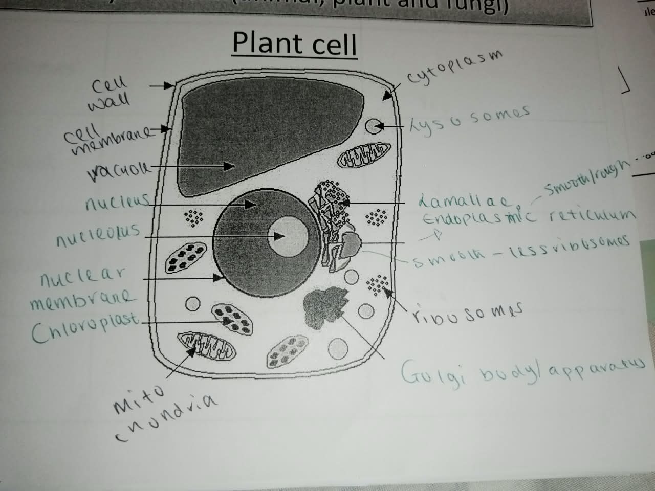
draw eukaryotic cell
what is the size of prokaryotes and eukaryotes
prokaryotes = 1-5 micrometer
eukaryotes = 10 -100 micrometer
what are the components in a nucleus
nucleolus
chromatin
nuclear envelope
describe function of nucleus
stores genetic material of cell
describe the function of the nucleolus
Nucleolus: produces ribosomal RNA (rRNA) and assembles ribosomes, which are needed for protein
synthesis.
site of ribosome production
Q: What is the structure of the nucleolus?
A dense region in the nucleus containing DNA, RNA and proteins.
outline structure and function of chromatin
Structure: consists of DNA and proteins
Function: stores genetic information and controls cell activities by providing instructions for protein synthesis.
describe the structure of the Nuclear envelope
Made of two membranes: an inner and outer membrane.
Contains nuclear pores.
Surrounds the nucleus and separates it from the cytoplasm.
The nuclear envelope surrounds the nucleoplasm.
Nucleoplasm = fluid inside the nucleus
describe the function of the nuclear envelope
Nuclear pores allow relatively large molecules to enter and leave the nucleus.

draw and label componenets of nucleus
structure + function of golgi body
Structure:
Stack of flattened membrane-bound sacs called cisternae
Function:
Modifies and packages proteins into vesicles for transport/secretion.
Forms lysosomes.
structure + function of ribosomes
Structure:
Non-membrane-bound.
Made of rRNA and proteins.
Eukaryotic ribosomes are 80S, made of 60S + 40S subunits.
Prokaryotic ribosomes are 70S, made of 50S + 30S subunits.
Function:
Site of protein synthesis.
structure + function of mitochondria
Structure:
Double membrane.
Inner membrane folded into cristae.
Inner region is the matrix.
Function:
Site of aerobic respiration.
Produces ATP.
structure + function for Lysosomes
Structure:
Spherical vesicles surrounded by a single membrane.
Contain digestive enzymes.
Function:
• Break down/digest materials inside the cell.
structure + function for Chloroplasts?
Structure:
Double membrane.
Contains thylakoid membranes arranged into stacks called grana.
Contains stroma.
Function:
Site of photosynthesis.
Grana contain chlorophyll and are involved in the light-dependent reactions.
Stroma is the site of the light-independent reactions.
structure + function for plasma membrane
Structure:
Phospholipid bilayer containing proteins, cholesterol, glycoproteins and glycolipids.
Function:
Controls movement of substances into and out of the cell.
Proteins act as channels/carriers.
Glycoproteins/glycolipids are involved in cell recognition and signalling.
structure + function for centrioles?
Structure:
Found in pairs.
Made of microtubules.
Function:
Involved in the formation of the spindle fibres during cell division.
structure + function for cell wall
Structure:
Rigid layer outside the plasma membrane.
In plants, made mainly of cellulose.
Function:
Provides support and strength.
Helps maintain the shape of the cell.
Prevents the cell from bursting when water enters by osmosis.
structure + function for cillia
Structure:
Short, numerous projections from the cell surface.
Contain microtubules.
Function:
Beat to move substances across the cell surface.
structure + function for flagella
Structure:
Long, whip-like projections.
In eukaryotic cells, contain microtubules.
Function:
Move the cell through liquid.
Help the cell change direction / propel itself through liquid.
structure + function for Rough Endoplasmic Reticulum (RER)
Structure:
Network of membrane-bound sacs/tubules with ribosomes attached.
Function:
Synthesises proteins using its attached ribosomes.
Transports proteins within the cell.
Transports proteins to the Golgi apparatus in vesicles for further modification and packaging.
structure + function for Smooth Endoplasmic Reticulum (SER)
Structure:
• Network of membrane-bound tubules with no ribosomes attached.
Function:
Synthesises lipids.
list the Prokaryotic cell components
Cell wall
Plasma membrane
Ribosomes
Flagella
list the Eukaryotic cell components
Nucleus
Nucleolus
Nuclear envelope
RER
SER
Golgi apparatus
Ribosomes
Mitochondria
Lysosomes
Chloroplasts
Plasma membrane
Centrioles
Cell wall (plant cells)
Cilia
Flagella (some eukaryotic cells)
What is the interrelationship between organelles involved in the production and secretion of proteins?
1. Nucleus the gene coding for the protein is transcribed into mRNA.
2. mRNA leaves the nucleus through nuclear pores and moves to ribosomes on the RER.
3. Ribosomes translate the mRNA instructions and assemble the protein.
4. The protein enters the cisternae of the RER and moves along the RER.
5. Vesicles containing the protein bud off from the RER and travel to the Golgi apparatus via microtubules
and motor proteins.
6. Vesicles fuse with the Golgi, where the protein may be modified.
7. Vesicles bud off from the Golgi containing the protein and travel to the plasma membrane.
8. Mitochondria provide ATP for processes involved in vesicle movement.
The vesicles fuse with the plasma membrane and the protein is released outside the cell by exocytosis.
describe 3 components of the cytoskeleton
Microfilaments - involved in changing cell shape and cell movement.
Microtubules move chromosomes during mitosis and act as tracks for organelles such as vesicles.
Intermediate filaments provide mechanical strength/support to cells.
What is the importance of the cytoskeleton?
The cytoskeleton provides mechanical strength and support to the cell.
It helps maintain and change cell shape.
It allows organelles to move within the cell, e.g. vesicles.
It is involved in the movement of chromosomes during mitosis.
It allows cilia and flagella to move.
ATP provides energy for cytoskeleton movement.
Q: What is the ultrastructure of a prokaryotic cell?
Capsule may be present; provides protection.
Cell wall → made of peptidoglycan (murein); provides strength and support.
Plasma membrane controls movement of substances into and out of the cell.
Cytoplasm = site of chemical reactions.
70S ribosomes → site of protein synthesis.
Nucleoid contains the main circular DNA; not surrounded by a membrane.
Plasmids → small circular pieces of DNA that may carry additional genes and can be transferred
between bacteria.
Flagellum may be present; allows the cell to move.
What are the similarities and differences between prokaryotic and eukaryotic cells?
Both have a plasma membrane.
Both have cytoplasm.
Both contain ribosomes for protein synthesis.
Both contain DNA.
What are the differences between prokaryotic and eukaryotic cells?
Prokaryotes have no nucleus; their circular DNA is found in the nucleoid.
Eukaryotes have a nucleus containing linear DNA.
Prokaryotes have no membrane-bound organelles; eukaryotes have membrane-bound organelles.
Prokaryotic ribosomes are 70S; eukaryotic cytoplasmic ribosomes are 80S.
Prokaryotic cell walls are made of peptidoglycan (murein); plant cell walls are made of cellulose.
Prokaryotes may contain plasmids; eukaryotic cells generally do not.
Prokaryotes are generally smaller than eukaryotic cells.
what does the light microscope do?
Used to observe whole cells and tissues.
Shows less detailed structure than electron microscopes.
Transmission electron microscope (TEM)?
Shows internal ultrastructure and organelle detail.
Electrons pass through the specimen.
Scanning electron microscope (SEM)?
Shows surface detail of specimens.
Electrons interact with the surface.
Recognising microscope images?
Light microscope - whole cells/tissues
TEM → internal structures/organelles
SEM - surface structures
what are the 3 types of microscopes
Light microscope
Transmission electron microscope (TEM)
Scanning electron microscope (SEM)
How does a light microscope work?
A light microscope uses light to illuminate the specimen.
Light passes from the light source underneath, through the specimen, through the objective lens and into the eye.
Start on the lowest magnification.
Start with the stage closest to the objective lens.
Use the coarse focus to focus the specimen at the lowest magnification.
Once in focus, increase the objective lens magnification.
Use the fine focus to refocus at the higher magnification.
Facts about light microscopе?
The specimen needs to be thin enough for light to pass through.
It is used to look at whole cells and tissues.
Light microscopes have the lowest magnification and resolution of the three.
Facts about transmission electron microscope
Uses electrons rather than light.
The electron beam passes through the specimen.
The specimen must be thin enough for electrons to pass through.
Electron microscopes normally use a vacuum, so specimens are usually dead.
TEM is used to see internal detail of the specimen.
Can show detailed organelle structure.
TEM has the highest magnification and resolution.
Why might TEM images of the same organelle look different?
specimens may have been cut along different planes.
An artefact is a feature or distortion caused by specimen preparation, making the specimen appear different from its natural state.
Facts about Scanning electron microscope?
Uses electrons.
The electron beam does not pass through the specimen.
Electrons bounce off/interact with the surface.
Used to see surface detail.
SEM has lower magnification and resolution than TEM, but higher than a light microscope.
Order of magnification and resolution
TEM → SEM → light microscope
Highest → lowest magnification and resolution.
How to Prepare a light microscope slide?
The sample should be thin enough for light to pass through.
Place the sample towards one side of the slide.
Place the coverslip down at an angle.
Facts about Eyepiece graticule, stage micrometer and Calibration ?
An eyepiece graticule is used to measure the specimen.
A stage micrometer is used to calibrate the eyepiece graticule.
Calibration needs to be carried out at the same magnification used to measure the specimen.
How to do a Graticule calculation?
1. Determine the value of one eyepiece graticule division.
2. Measure the specimen in graticule divisions.
3. Calculate the actual size.
Actual size = number of divisions x value of one division
The eyepiece graticule must be calibrated using a stage micrometer before measuring the specimen.
Facts about staining ?
Staining is used when preparing a microscope slide.
The stain makes the specimen more visible.
A key purpose is to improve contrast.
Staining can allow particular organelles/cellular components to be seen.
Different examples of stains ?
Acetic orcein (stain) → DNA (what it stains) → Dark red (colour)
Eosin (stain) → Cytoplasm (what it stains) → Pink (colour)
Sudan black (stain) → Membranes and other lipids (what it stains) → Black (colour)
Different units and conversions ?
1 mm = 1000 µm
1 µm = 1000 nm
1 mm = 1,000,000 nm
difference between Magnification and resolution ?
Magnification is the number of times larger the image is than the actual object.
Resolution is the ability to distinguish between very small structures that are close together; in detail.
Calculations for magnification ?
Magnification = image size / actual size
Image size = magnification x actual size
Actual size = image size / magnification
Graticules vs magnification
For a graticule:
Actual size = number of divisions x value of one division
For a magnification calculation:
Magnification = image size / actual size