Chapters 1-5 EOC Questions some VT
1/226
There's no tags or description
Looks like no tags are added yet.
Name | Mastery | Learn | Test | Matching | Spaced | Call with Kai |
|---|
No analytics yet
Send a link to your students to track their progress
227 Terms
Knowledge and Comprehension
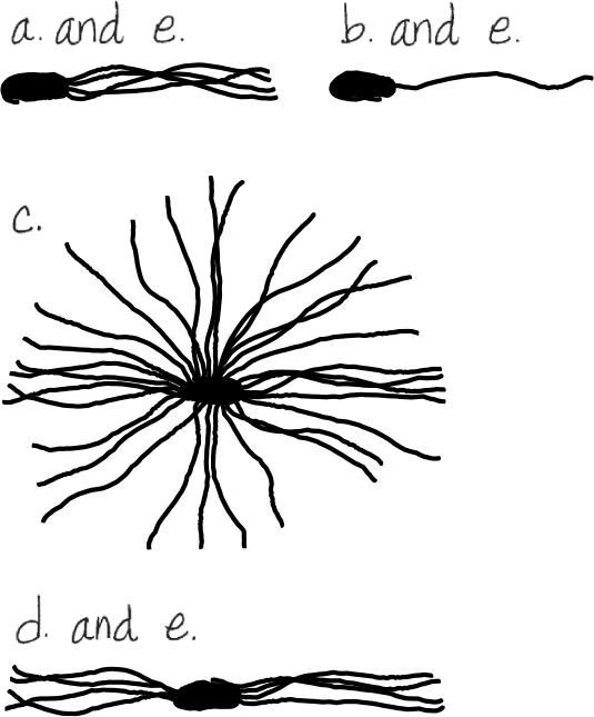
DRAW IT Diagram each of the following flagellar arrangements:
lophotrichous
Atrichous bacteria
Bacteria that lack flagella
Arrangements of bacterial flagella
peritrichous
distributed over the entire cell
polar
at one or both poles or ends of the cell
monotrichous
a single flagellum at one pole
lophotrichous
a tuft of flagella coming from one pole
amphitrichous
flagella at both poles of the cell

DRAW IT Diagram each of the following flagellar arrangements:
monotrichous
Atrichous bacteria
Bacteria that lack flagella
Arrangements of bacterial flagella
peritrichous
distributed over the entire cell
polar
at one or both poles or ends of the cell
monotrichous
a single flagellum at one pole
lophotrichous
a tuft of flagella coming from one pole
amphitrichous
flagella at both poles of the cell

DRAW IT Diagram each of the following flagellar arrangements:
peritrichous
Atrichous bacteria
Bacteria that lack flagella
Arrangements of bacterial flagella
peritrichous
distributed over the entire cell
polar
at one or both poles or ends of the cell
monotrichous
a single flagellum at one pole
lophotrichous
a tuft of flagella coming from one pole
amphitrichous
flagella at both poles of the cell
DRAW IT Diagram each of the following flagellar arrangements:
amphitrichous
Atrichous bacteria
Bacteria that lack flagella
Arrangements of bacterial flagella
peritrichous
distributed over the entire cell
polar
at one or both poles or ends of the cell
monotrichous
a single flagellum at one pole
lophotrichous
a tuft of flagella coming from one pole
amphitrichous
flagella at both poles of the cell

DRAW IT Diagram each of the following flagellar arrangements:
polar
Atrichous bacteria
Bacteria that lack flagella
Arrangements of bacterial flagella
peritrichous
distributed over the entire cell
polar
at one or both poles or ends of the cell
monotrichous
a single flagellum at one pole
lophotrichous
a tuft of flagella coming from one pole
amphitrichous
flagella at both poles of the cell

Endospore formation is called (a) ________. It is initiated by (b) ________. Formation of a new cell from an endospore is called (c) ________. This process is triggered by (d) ________.
A
sporogenesis
Endospore formation is called (a) ________. It is initiated by (b) ________. Formation of a new cell from an endospore is called (c) ________. This process is triggered by (d) ________.
B
certain adverse environmental conditions
Endospore formation is called (a) ________. It is initiated by (b) ________. Formation of a new cell from an endospore is called (c) ________. This process is triggered by (d) ________.
C
germination
Endospore formation is called (a) ________. It is initiated by (b) ________. Formation of a new cell from an endospore is called (c) ________. This process is triggered by (d) ________.
D
favorable growth conditions
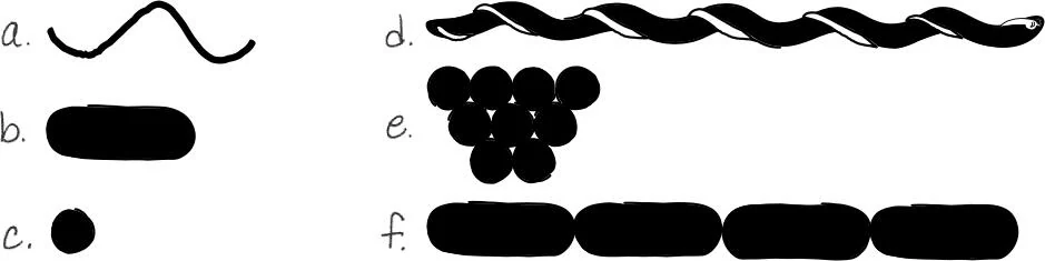
DRAW IT Draw the bacterial shapes listed in (a), (b), and (c). Then draw the shapes in (d), (e), and (f), showing how they are special conditions of a, b, and c, respectively.
spiral
bacillus
coccus
spirochetes
staphylococci
streptobacilli

A
a. 3
B
b. 5

C
c. 1

D
d. 2

E
e. 1,4

F
f. 2,8

G
g. 7

H
h. 6
Why is an endospore called a resting structure?
Allows cell to “rest” or survive as opposed to grow and reproduce.
Of what advantage is an endospore to a bacterial cell?
Protective endospore wall allows bacterium to withstand adverse environmental conditions
Compare and contrast the following:
simple diffusion and facilitated diffusion
Both allow materials to cross the plasma membrane from a high concentration to a low concentration without expending energy. Facilitated diffusion requires carrier proteins.
Compare and contrast the following:
active transport and facilitated diffusion
Both require transport proteins to move materials across the plasma membrane. In active transport, energy is expended.
Compare and contrast the following:
active transport and group translocation
Both move materials across the plasma membrane with an expenditure of energy. In group translocation, the substrate is changed after it crosses the membrane.
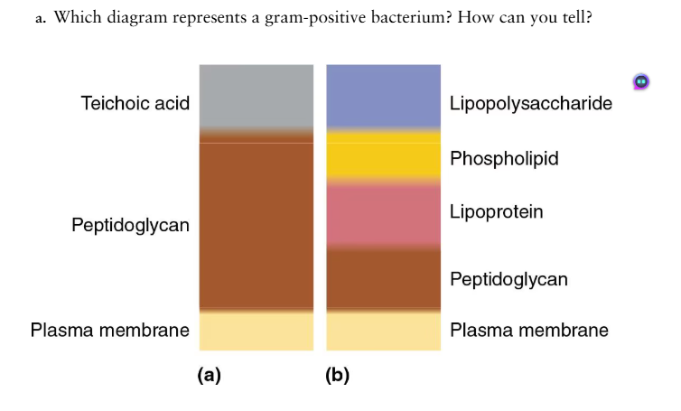
Answer the following questions using the diagrams provided, which represent cross sections of bacterial cell walls.
a. Which diagram represents a gram positive bacterium? How can you tell?
Diagram (a) refers to a gram-positive bacterium because the lipopolysaccharide–phospholipid–lipoprotein layer is absent.

Answer the following questions using the diagrams provided, which represent cross sections of bacterial cell walls.
b. Explain how the Gram stain works to distinguish these two types of cell walls
The gram-negative bacterium initially retains the violet stain, but it is released when the outer membrane is dissolved by the decolorizing agent. After the dye–iodine complex enters, it becomes trapped by the peptidoglycan of gram-positive cells.

Answer the following questions using the diagrams provided, which represent cross sections of bacterial cell walls.
c. Why does penicillin have no effect on most gram-negative cells?
The outer layer of the gram-negative cells prevents penicillin from entering the cells.

Answer the following questions using the diagrams provided, which represent cross sections of bacterial cell walls.
d. How do essential molecules enter cells through each wall?
Gram +: Essential molecules diffuse through cell wall
Gram -: Porins + specific channel proteins allow passage of small water-soluble molecules.

Answer the following questions using the diagrams provided, which represent cross sections of bacterial cell walls.
e. Which cell wall is toxic to humans?
Gram-negative.
Starch is readily metabolized by many cells, but a starch molecule is too large to cross the plasma membrane.
How does a cell obtain the glucose molecules from a starch polymer?
An extracellular enzyme (amylase) hydrolyzes starch into disaccharides (maltose) and monosaccharides (glucose).
Starch is readily metabolized by many cells, but a starch molecule is too large to cross the plasma membrane.
How does the cell transport these glucose molecules across the plasma membrane?
Maltose: A carrier enzyme (maltase) hydrolyzes maltose and moves one glucose into the cell.
Glucose: can be transported by group translocation as glucose-6-phosphate.
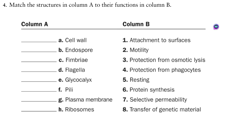
Match the characteristics of eukaryotic cells in column A with their functions in column B.
a. Pericentriolar matrix |
|
b. Chloroplasts |
|
c. Golgi complex |
|
d. Lysosomes |
|
e. Mitochondria |
|
f. Peroxisomes |
|
g. Rough ER |
|
a. 3
b. 4
c. 7
d. 1
e. 6
f. 2
g. 5
DRAW IT What group of microbes is characterized by cells that form filaments, reproduce by spores, and have peptidoglycan in their cell walls?
Actinomycetes
Multiple Choice
Which of the following is not a distinguishing characteristic of prokaryotic cells?
They usually have a single, circular chromosome.
They have 70S ribosomes.
They have cell walls containing peptidoglycan.
Their DNA is not associated with histones.
They lack a plasma membrane.
5
Which statement best describes what happens when a gram-positive bacterium is placed in distilled water and penicillin?
No change will result; the solution is isotonic.
Water will move into the cell.
Water will move out of the cell.
The cell will undergo osmotic lysis.
Sucrose will move into the cell from an area of higher concentration to one of lower concentration.
4
Which statement best describes what happens when a gram-negative bacterium is placed in distilled water and penicillin?
No change will result; the solution is isotonic.
Water will move into the cell.
Water will move out of the cell.
The cell will undergo osmotic lysis.
Sucrose will move into the cell from an area of higher concentration to one of lower concentration.
2
Which statement best describes what happens when a gram-positive bacterium is placed in an aqueous solution of lysozyme and 10% sucrose?
No change will result; the solution is isotonic.
Water will move into the cell.
Water will move out of the cell.
The cell will undergo osmotic lysis.
Sucrose will move into the cell from an area of higher concentration to one of lower concentration.
1
Which of the following statements best describes what happens to a cell exposed to polymyxins that destroy phospholipids?
In an isotonic solution, nothing will happen.
In a hypotonic solution, the cell will lyse.
Water will move into the cell.
Intracellular contents will leak from the cell.
Any of the above might happen.
4
Which of the following is false about fimbriae?
They are composed of protein.
They may be used for attachment.
They are found on gram-negative cells.
They are composed of pilin.
They may be used for motility.
5
Which of the following pairs is mismatched?
glycocalyx—adherence
pili—reproduction
cell wall—toxin
cell wall—protection
plasma membrane—transport
2
Which of the following pairs is mismatched?
metachromatic granules—stored phosphates
polysaccharide granules—stored starch
lipid inclusions—poly-β-hydroxybutyric acid
sulfur granules—energy reserve
ribosomes—protein storage
5
You have isolated a motile, gram-positive cell with no visible nucleus. You can assume this cell has
ribosomes.
mitochondria.
an endoplasmic reticulum.
a Golgi complex.
all of the above
1
The antibiotic amphotericin B disrupts plasma membranes by combining with sterols; it will affect all of the following cells except
animal cells.
gram-negative bacterial cells.
fungal cells.
Mycoplasma cells.
plant cells.
2
Analysis
How can prokaryotic cells be smaller than eukaryotic cells and still carry on all the functions of life?
Prokaryotic cells have a good surface area to volume ratio. They contain 1 circular chromosome and no membrane bound organelles. All of their chemical activity occurs in the cytoplasm and cell membrane. They have all they need at this minimal functional level
The smallest eukaryotic cell is the motile alga Ostreococcus. What is the minimum number of organelles this alga must have?
Nucleus, mitochondrion, chloroplast, flagellum
more likely to be nucleus, mitochondrion, golgi apparatus, chloroplast

Two types of prokaryotic cells have been distinguished: bacteria and archaea. How do these cells differ from each other? How are they similar?
archaea - no cell wall or cell wall without peptidoglycan
Archaea and bacteria are alike in that they are both prokaryotes, meaning they are single-celled organisms without a nucleus. They both reproduce asexually through binary fission and have similar sizes and shapes.
In 1985, a 0.5 mm cell was discovered in surgeonfish and named Epulopiscium fishelsoni (see Figure 11.20). It was presumed to be a protozoan. In 1993, researchers determined that Epulopiscium is actually a gram-positive bacterium.
Why do you suppose this organism was initially identified as a protozoan?
What evidence would change the classification to bacterium?
Probably because it
1. Is gigantic for a bacterial cell. 80um (almost a milimeter)
2. Has strange morphological features, like invaginations, vesicles, and tubules.
3. Reproduces strangely.
but they performed rRNA analysis by Pace, et al in 1993 confirmed that it was a member of the bacteria.
When E. coli cells are exposed to a hypertonic solution, the bacteria produce a transporter protein that can move K+ (potassium ions) into the cell. Of what value is the active transport of K+, which requires ATP?
increases intracelluar solute concentration bringing it closer to that of the hyperosmotic extracellular environment & thus reducing the severity of plasmolysis.
Clinical Applications and Evaluations
Clostridium botulinum is a strict anaerobe; that is, it is killed by the molecular oxygen O2 present in air. Humans can die of botulism from eating foods in which C. botulinum is growing.
How does this bacterium survive on plants picked for human consumption?
Why are home-canned foods most often the source of botulism?
Typically humans do not die from infection by the bacteria. In fact, by the time the food is consumed the bacteria is usually dead. During bacterial growth, a toxin is produced. This toxin is among the most toxic substances known.
The bacteria grow in the soil and sporulates upon unfavorable conditions. The spores are transferred in the soil deposited on the plants. If the canning temperatures are sufficient then the bacteria will die. If not, the bacteria will begin to grow under the anaerobic conditions inside the can/jar. The nutrients are eventually exhausted and the bacteria die, leaving the toxin. Home-canned foods are most often a source because the canning temperatures are less rigorously monitored.
A South San Francisco child enjoyed bath time at home because of the colorful orange and red water. The water did not have this rusty color at its source, and the water department could not culture the Acidithiobacillus bacteria responsible for the rusty color from the source.
How were the bacteria getting into the household water?
What bacterial structures make this possible?
bacteria can oxidizes iron and inorganic sulfur compounds. The oxidation process can be harmful, as it produces sulfuric acid, which is a major pollutant.
The bacteria are usually found in rivers, canals, mine drainage effluents and mining areas. So there may be some source of these bacilli near their house.
They require inorganic sources to grow and contain an enzyme called ferrous oxidase that allows them to metabolize iron. They eat iron pipes leading into households and produce a thick slime layer, biofilm.
Live cultures of Bacillus thuringiensis (Dipel®) and B. subtilis (Kodiak®) are sold as pesticides.
What bacterial structures make it possible to package and sell these bacteria?
For what purpose is each product used?
(Hint: Refer to Chapter 11.)
These bacillus thuringensis species of bacteria secrete protein crystals that are toxic to most insects hence used in agriculture to kill pests that attack the crops. This is also the reason the genes responsible for this protein crystal is incorporated in crops (transgenic crops) to make them resistant to pests.
B. subtilis is important in the nutrient cycle and availing nutrient to plants hence promote plant growth
Both species are able to survive harsh conditions and remain dormant for long periods of time as spores hence their suitability in application in agriculture, their endospores allow them to be packed + survive packing
VoiceThread Questions
Where is the mechanism of action for/what is affected by penicillin on peptidoglycan?
interferes with final linking of peptidoglycan rows by peptide cross-bridges
Penicillin inhibit the synthesis of peptidoglycan in bacterial cell walls, thus weakening them, and increasing the likelihood of the cell bursting (lysis). Penicillin inhibits peptide bridges and thus does not allow for synthesis. penicillin inhibits bacterial cell wall synthesis. Penicillin binds to and inhibits the activity of enzymes called transpeptidases that are responsible for cross-linking the peptide chains. Without proper cross-linking of the peptidoglycan layer, the bacterial cell wall becomes weak and loses its structural integrity leading to cell lysis. Penicillin inhibits peptide bridging which in turn does not allow for synthesis of peptidoglycan, weakening the cell wall of the bacteria and making the cell more susceptible to burst
What is NOT a structure found in all bacteria?
Structures that are NOT found in ALL bacterial cells can include: cell wall, capsule, plasmid, fimbriae, flagella, cilia. As a reverse answer common types of inclusions include: storage granules, carboxysomes, gas vesicles and magnetosomes
Which type of bacteria moves by axial filaments?
Spirochetes/spiral cells
Method that bacteria use to get to an attractant is called
chemotaxis
In a hypertonic solution, what is likely to happen to a protoplast?
in a hypertonic solution, there is more solute outside of the cell than inside, causing the net movement of water to be out of the cell. Because a protoplast is a gram-positive cell that already lacks a cell wall, it would be even easier for water to move out of it, leading it to shrink.
Which is NOT a type of inclusion?
list of inclusions:
metachromatic granules
polysaccharide granules
lipid granules
sulfur granules
carboxysomes
gas vacuoles
magnetosomes
Which is a feature that is exclusively eukaryotic?
membrane bound nucleus
DNA associated with histones
membrane bound organelles ((Mitochondria (site of ATP production via aerobic respiration), Endoplasmic Reticulum (ER) (protein and lipid synthesis), Golgi Apparatus (protein modification and sorting), Lysosomes (intracellular digestion), Chloroplasts (in photosynthetic eukaryotes))
How do fimbriae and pili differ?
Fimbriae and Pili differ in function, as Fimbriae allows for attachment and is present in gram-negative cells, while pili is involved in motility and DNA transfer
In bacteria, photosynthetic pigments are found in...
chromatophores
Knowledge and Comprehension

Fill in the following blanks.
A
a. 10^-6 m

Fill in the following blanks.
B
b. 1 nm
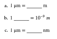
Fill in the following blanks.
C
c. 10³ nm
Which type of microscope would be best to use to observe each of the following?
a stained bacterial smear
Compound light microscope
Which type of microscope would be best to use to observe each of the following?
unstained bacterial cells: the cells are small, and no detail is needed
Darkfield microscope
Which type of microscope would be best to use to observe each of the following?
unstained live tissue when it is desirable to see some intracellular detail
Phase-contrast microscope
Which type of microscope would be best to use to observe each of the following?
a sample that emits light when illuminated with ultraviolet light
flourescence microscope
Which type of microscope would be best to use to observe each of the following?
intracellular detail of a cell that is 1 micrometers long
electron microscope
Which type of microscope would be best to use to observe each of the following?
unstained live cells in which intracellular structures are shown in color
Differential interference contrast microscope

DRAW IT Label the parts of the compound light microscope in the following figure, and then draw the path of light from the illuminator to your eye.
A

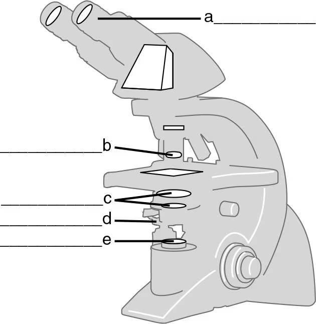
DRAW IT Label the parts of the compound light microscope in the following figure, and then draw the path of light from the illuminator to your eye.
B
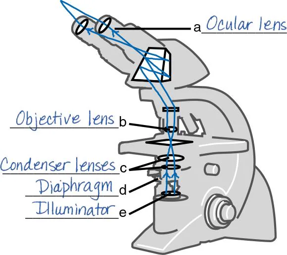

DRAW IT Label the parts of the compound light microscope in the following figure, and then draw the path of light from the illuminator to your eye.
C


DRAW IT Label the parts of the compound light microscope in the following figure, and then draw the path of light from the illuminator to your eye.
D


DRAW IT Label the parts of the compound light microscope in the following figure, and then draw the path of light from the illuminator to your eye.
E

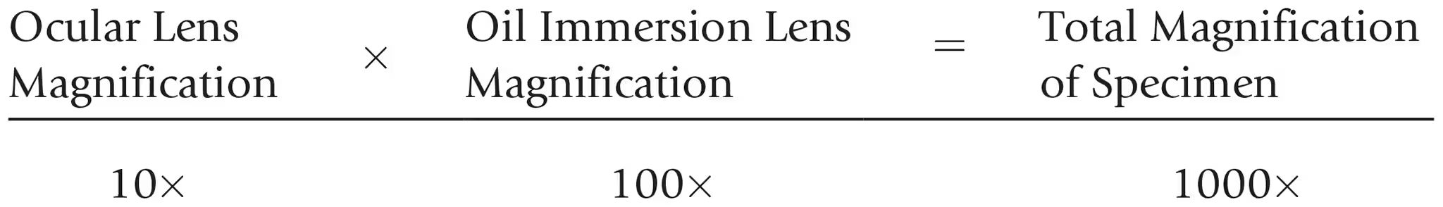
Calculate the total magnification of the nucleus of a cell being observed through a compound light microscope with a 10x ocular lens and an oil immersion lens.
The maximum magnification of a compound microscope is (a) ________; that of an electron microscope, (b) ________. The maximum resolution of a compound microscope is (c) ________; that of an electron microscope, (d) ________. One advantage of a scanning electron microscope over a transmission electron microscope is (e) ________.
A
1,500x
The maximum magnification of a compound microscope is (a) ________; that of an electron microscope, (b) ________. The maximum resolution of a compound microscope is (c) ________; that of an electron microscope, (d) ________. One advantage of a scanning electron microscope over a transmission electron microscope is (e) ________.
B
10,000,000x
The maximum magnification of a compound microscope is (a) ________; that of an electron microscope, (b) ________. The maximum resolution of a compound microscope is (c) ________; that of an electron microscope, (d) ________. One advantage of a scanning electron microscope over a transmission electron microscope is (e) ________.
C
0.2 micrometers
The maximum magnification of a compound microscope is (a) ________; that of an electron microscope, (b) ________. The maximum resolution of a compound microscope is (c) ________; that of an electron microscope, (d) ________. One advantage of a scanning electron microscope over a transmission electron microscope is (e) ________.
D
10 picometers
The maximum magnification of a compound microscope is (a) ________; that of an electron microscope, (b) ________. The maximum resolution of a compound microscope is (c) ________; that of an electron microscope, (d) ________. One advantage of a scanning electron microscope over a transmission electron microscope is (e) ________.
E
Seeing 3D detail
Why is a mordant used in the Gram stain?
Mordant combines with the basic dye to form a complex that will not wash out of gram-positive cells.
Why is a mordant used in the flagella stain?
Mordant accumulates on the flagella so that they can be seen with a light microscope.
What is the purpose of a counterstain in the acid-fast stain?
A counterstain stains the colorless non–acid-fast cells so that they are easily seen through a microscope.
What is the purpose of a decolorizer in the Gram stain?
Decolorizer removes the color from gram-negative cells.
What is the purpose of a decolorizer in the acid-fast stain?
Decolorizer removes the color from non–acid-fast cells.

Fill in the following table regarding the Gram stain:
A + E
purple, purple

Fill in the following table regarding the Gram stain:
B + F
purple, purple
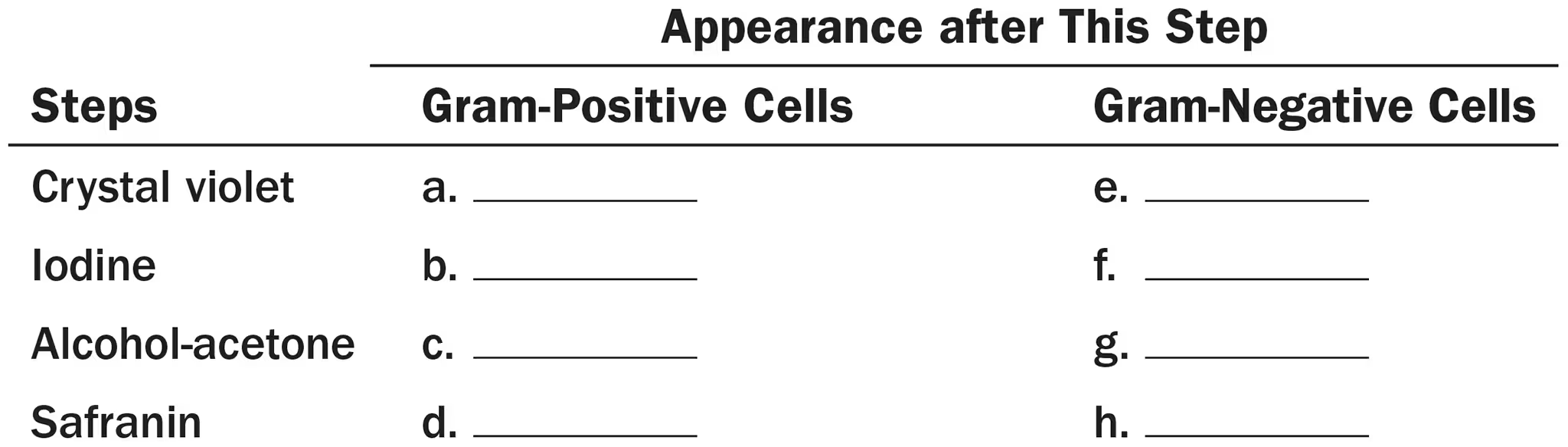
Fill in the following table regarding the Gram stain:
C + G
purple, colorless

Fill in the following table regarding the Gram stain:
D + H
purple, red
DRAW IT A sputum sample from Calle, a 30 year old Asian elephant, was smeared onto a slide and air dried. The smear was fixed, covered with carbolfuchsin, and heated for . After washing with water, acid-alcohol was placed on the smear for 30 seconds. Finally, the smear was stained with methylene blue for 30 seconds, washed with water, and dried. On examination at 1000x, the zoo veterinarian saw red rods on the slide. What microbe do these results suggest?
An acid-fast bacterium (Mycobacterium)
Multiple Choice
Assume you stain Bacillus by applying malachite green with heat and then counterstain with safranin. Through the microscope, the green structures are
cell walls
capsules.
endospores.
flagella.
impossible to identify.
3
Three-dimensional images of live cells can be produced with
darkfield microscopy.
fluorescence microscopy.
transmission electron microscopy.
confocal microscopy.
phase-contrast microscopy.
4
Carbolfuchsin can be used as a simple stain and a negative stain. As a simple stain, the pH is
2
higher than the negative stain.
lower than the negative stain.
the same as the negative stain.
2
Looking at the cell of a photosynthetic microorganism, you observe the chloroplasts are green in brightfield microscopy and red in fluorescence microscopy. You conclude:
chlorophyll is fluorescent.
the magnification has distorted the image.
you’re not looking at the same structure in both microscopes.
the stain masked the green color.
none of the above
1