Exam 2 Biochem
 Call Kai
 Learn
Learn Practice Test
 Spaced Repetition
 Match
Match Flashcards
 Knowt Play
1/250

There's no tags or description
Looks like no tags are added yet.
Name | Mastery | Learn | Test | Matching | Spaced | Call with Kai | Chat |
|---|
No analytics yet
Send a link to your students to track their progress
251 Terms
Carbohydrates are polyalcohols containing either a ____ or an _____ group.
keto; aldehyde
Monosaccharides
the building blocks of all carbohydrates
Basic structure of a monosaccharide
a chain of carbons with a hydroxyl group at each carbon except one, which forms a carbonyl group
Aldoses
contain an aldehyde group
Ex: glucose and glyceraldehyde
Ketoses
contain a keto group
Ex: fructose and dihydroxyacetone
Triose
3 carbons
Ex: D-glyceraldehyde and dihydroxyacetone
Tetrose
4 carbons
Pentose
5 carbons
Ex: Ribose
Hexose
6 carbons
Ex: Glucose
Heptose
7 carbons
The most important monosaccharide because it serves as the primary metabolic fuel for cellular respiration
D-glucose

Epimers
monosaccharides that differ in the orientation of substituents around one of their asymmetric carbons
D-Mannose
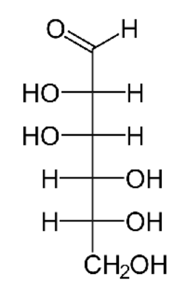
D-Glucose

D-Galactose

Fischer projection structure

Haworth projection

When rings form, carbon 1 of glucose becomes
asymmetric
In glucose, carbon 1 (the aldehyde carbon) is this and in ketoses, the keto carbon (usually carbon 2) is this
anomeric carbon
D and L convention
refers to the orientation of the substituents at the asymmetric carbon farthest from the carbonyl group.
The most prevalent orientation
D-isomers
Isomer diversity
D-glucose, an aldohexose, has 4 asymmetric carbons (C2, C3, C4, C5), allowing for 16 possible optical isomers
D-Mannose is a ____ epimer of glucose
C2
D-Galactose is a ___ epimer of glucose
C4
Hemiacetal
formed from an aldehyde and an alcohol
Hemiketal
formed from a ketone and an alcohol
Pyranose
A 6-membered ring (favored by D-glucose)
Furanose
A 5-membered ring (favored by D-fructose)
Mutarotation
when anomers interconvert spontaneously
Mutarotation is greatly accelerated in the presence of
acids or bases
Upon ring closure, the carbonyl carbon (C1 in aldoses & C2 in ketoses) becomes a new asymmetric center called
anomeric carbon
Monosaccharides combine into larger molecules by forming
glycosidic bonds
Once the glycosidic bond is formed, _____ is no longer possible
mutarotation
Disaccharides
two monosaccharides
Ex: maltose, lactose, sucrose
Oligosaccharides
a few (3-6) monosaccharides
Polysaccharides
many (7+) monosaccharides
Ex: amylose, glycogen, cellulose
Glycoproteins
contain carbohydrate covalently bound to amino acid side chains
Glycolipids
carbohydrate is covalently bound to a lipid
O-glycosidic
if the sugar binds its partner through an oxygen atom
N-glycosidic
if the sugar binds its partner through a nitrogen atom
Carbohydrate behavior is determined by
size and functional groups
____ are highly water-soluble because they can form multiple hydrogen bonds with water
monosaccharides, disaccharides, and oligosaccharides
Many polysaccharides are insoluble because
their large size increases opportunities for intermolecular interactions
The ___ group of a monosaccharide has reducing properties (it can donate electrons)
free carbonyl
A sugar is a reducing sugar only if it has a ___ not involved in a glycosidic bond.
free anomeric carbon
____ is a notable non-reducing sugar because both of its anomeric carbons are used in the glycosidic bond
sucrose
Which of the following is considered the simplest aldose?
D-Glyceraldehyde
How many asymmetric carbons are found in the open-chain form of D-glucose?
4
What is the primary difference between D-glucose and D-galactose?
They are C-4 epimers
The process by which 𝛼-D-glucose and 𝝱-D-glucose interconvert in water is called:
Mutarotation
Why is sucrose classified as a non-reducing sugar?
Both anomeric carbons are involved in the glycosidic bond
Which polysaccharide serves as the most abundant biomolecule on earth and contains 𝝱(1-4) glycosidic bonds?
Cellulose
serves as the primary transported carbohydrate in the human body
Glucose
Glucose uptake from the blood and extracellular fluids occur via
facilitated diffusion
Facilitated diffusion is mediated by
the GLUT family of carriers
Energy requirement for facilitated diffusion
no energy required
Facilitated diffusion driving force
high concentrations of glucose in the blood and extracellular fluids naturally drive glucose into the cells through transporter proteins
For the generation of a metabolic energy, all major nutrients are degraded to
acetyl-CoA
Where is GLUT 1 expressed?
most tissues
Where is GLUT 2 expressed?
liver, intestine, pancreatic B cells
Where is GLUT 3 expressed?
brain
Where is GLUT 4 expressed?
muscle, adipose tissue, heart
Where is GLUT 5 expressed?
intestine
What is the function of GLUT 1?
Basal glucose uptake
What is the function of GLUT 2?
High-capacity glucose uptake
What is the function of GLUT 3?
Neuronal glucose uptake
What is the function of GLUT 4?
Insulin-dependent glucose uptake
What is the function of GLUT 5?
fructose transport
____ is the only insulin-dependent transporter
GLUT 4
Post-Prandial Response
after a carbohydrate-rich meal, insulin levels rise. insulin binds to receptors on the surface of muscle and adipose tissue
Signaling cascade
insulin binding: insulin attaches the the insulin receptor
autophosphorylation: receptor undergoes tyrosine-phosphorylation
Akt signaling: a signaling cascade (involving Akt) is triggered
vesicle fusion: carrier-containing vesicles fuse with the plasma membrane, increasing the number of active GLT4 transporters
Metabolic shift
increases the Vmax of glucose transport. During fasting, when insulin is low, these tissues minimize glucose uptake to ensure the limited supply is redirected to glucose-dependent tissues like the brain and erythrocytes.
Which glucose transporter is specifically responsible for high-capacity uptake in the liver and pancreatic 𝝱-cells?
GLUT 2
Why is the transport of glucose into most cells considered “cost-free” in terms of energy?
There is a high concentration of glucose (100 mg/dL) maintained in the blood
In the absence of insulin, where are the majority of GLUT 4 transporters located in a muscle cell?
Within the membranes of intracellular vesicles
Which of the following is the first step in the insulin signaling cascade that leads to increased glucose uptake?
Insulin receptor autophosphorylation
During fasting, which tissues reduce their glucose uptake to preserve the supple for the brain?
Muscle and adipose tissue
Some individuals are born with a partial deficiency of pyruvate dehydrogenase in all tissues. What tissue suffers most from this abnormality?
Brain
Anaplerotic reaction
a reaction that “fills up” or replenishes metabolic intermediates, such as those in the TCA cycle
Glycogen
the stored form of carbohydrate in the human body
Heteroplasmy
the presence of more than one type of organellar genome (mitochondrial DNA) within a single cell or individual.
Insulin
a hormone produced by the pancreas that regulates the amount of glucose in the blood by promoting its uptake into skeletal muscle and adipose tissue.
Vmax
the maximum velocity of an enzymatic or transport reaction when the system is saturated with substrate.
____ is the cytoplasmic catabolic pathway that converts one molecule of glucose (6 carbons) into two molecules of pyruvate (3 carbons). All cells in the human body are capable of performing this process
glycolysis
Glycolysis Step 1:
Intermediate: Glucose → Glucose-6-Phosphate
Enzyme: Hexokinase
Type: Irreversible (ATP-dependent)
Glycolysis Step 2:
Intermediate: Glucose-6-Phosphate → Fructose-6-Phosphate
Enzyme: Phosphoglucose Isomerase
Type: Reversible
Glycolysis Step 3:
Intermediate: Fructose-6-Phosphate → Fructose-1,6-Bisphosphate
Enzyme: Phosphofructokinase-1
Type: Irreversible (committed step)
Glycolysis Step 4:
Intermediate: Fructose-1,6-Bisphosphate → DHAP + G3P
Enzyme: Aldolase
Type: Reversible (Cleavage)
Glycolysis Step 5:
Intermediate: DHAP ⇌ Glyceraldehyde-3-Phosphate (G3P)
Enzyme: Triose Phosphate Isomerase
Type: Reversible
Glycolysis Step 6:
Intermediate: G3P → 1,3-Bisphosphoglycerate
Enzyme: G3P Dehydrogenase
Type: Reversible (Yields NADH)
Glycolysis Step 7:
Intermediate: 1,3-Bisphosphoglycerate → 3-Phosphoglycerate
Enzyme: Phosphoglycerate Kinase
Type: Reversible (Yields ATP)
Glycolysis Step 8:
Intermediate: 3-Phosphoglycerate → 2-Phosphoglycerate
Enzyme: Phosphoglycerate Mutase
Type: Reversible
Glycolysis Step 9:
Intermediate: 2-Phosphoglycerate → Phosphoenolpyruvate (PEP)
Enzyme: Enolase
Type: Reversible
Glycolysis Step 10:
Intermediate: PEP → Pyruvate
Enzyme: Pyruvate Kinase
Type: Irreversible (Yields ATP)
Hexokinase
the first step, which traps glucose in the cell as glucose-6-phosphate. It is inhibited by its product (glucose-6-phosphate) in most tissues
PFK-1
the "committed step." Once fructose-1,6-bisphosphate is formed, the molecule is destined for glycolysis. It is the most important regulated enzyme, inhibited by high energy (ATP), citrate, and low pH, while being stimulated by AMP, ADP, and insulin
Pyruvate Kinase
the final step, inhibited by ATP in the liver
Glycolysis is divided into two distinct functional phases:
the preparatory (investment) phase and the payoff (generation) phase
Energy Investment (Preparatory) Phase
involves the first three steps of the pathway, where the cell "spends" energy to prime the glucose molecule for cleavage.
ATP Consumption: Two molecules of ATP are used (one by Hexokinase and one by PFK-1).
Purpose: Phosphorylation prevents glucose from leaving the cell and creates a symmetric molecule (fructose-1,6-bisphosphate) that can be cleaved into two three-carbon sugars.
Energy Generation (Payoff) Phase
occurs after the six-carbon fructose is split into two three-carbon molecules (G3P). Because there are now two molecules moving through the pathway, all products are doubled.
ATP Production: Four molecules of ATP are produced via substrate-level phosphorylation (two by Phosphoglycerate Kinase and two by Pyruvate Kinase).
NADH Production: Two molecules of NADH are produced by G3P Dehydrogenase.
Net Yield:
2 ATP (4 generated minus 2 invested).
2 NADH (In aerobic conditions, these proceed to the respiratory chain).
2 Pyruvate.