Stage 2 Bio - Cells as the Basis of Life
1/77
There's no tags or description
Looks like no tags are added yet.
Name | Mastery | Learn | Test | Matching | Spaced |
|---|
No study sessions yet.
78 Terms
what is an endosymbiotic event?
An endosymbiotic event refers to a relationship, where one organism lives inside another, with mutual benefits for both parties.
what is the endosymbiosis theory?
The endosymbiosis theory suggests that the key organelles such as the mitochondria and chloroplast found in eukaryotic cells, originated as independent prokaryotic cells, which were later engulfed by a early eukaryotic cell through endocytosis.
what are the evidence supporting endosymbiosis?
multiple evidence support endosymbiosis. One of this evidence being the organelles double cell membrane, where the inner membrane is prokaryotic in nature, while the outer membrane is eukaryotic in nature. this structural feature suggests that the organelles must of had their own cell membrane, but later gained the host’s membrane when they got engulfed through endocytosis. another strong piece of evidence is the presence of ribosomes and DNA separate to that of the host cell inside the mitochondria and chloroplast. this evidence supports the endosymbiosis theory, as the DNA and ribosome would entail that organelles can make their own protein, a feature necessary for many independent cells to sustain life. Another key evidence, is the organelles ability to divide in a manner similarly to that of binary fission (a prokaryotic form of reproduction), independently to that of the host cell.
describe the structural differences between prokaryotic cell
The structural differences between eukaryotic and prokaryotic cells, lie in the presence of membrane bound organelles, membrane bound nucleus, complexities, size, and shape of DNA. Prokaryotic cells range between 0.5-5 micrometers, while eukaryotic cells ranges between 5-100 micrometers. eukaryotic cells have membrane bound organelles such as the mitochondria, while prokaryotic cells do not. prokaryotic cells also have circular unbounded DNA, found in the cytosol (prokaryotes don’t have a nucleus), while eukaryotic cells have linear DNA bound to proteins called histones, found in the nucleus. unlike eukaryotic cells, prokaryotic cells have a plasmid, which is a small region of seperate bacterial DNA.
describe binary fission
binary fission is a form of aesexual reproduction, commonly found in single celled organisms. In this process, the cells make a copy of its DNA, and then split into two identical daughter cells. 1) The process begins with DNA getting replicated, creating two identical copies. 2) the cells begin elongate, and the two copies of DNA travel to opposite ends of the cell. 3) the cell membrane of the cell begins to pinch, forming a cross wall. 4) then, the cells separate into two genetically identical daughter cells
describe mitosis
mitosis is a form of aesxual reproduction, commonly found in somatic cells. In this process, somatic cells are split into two genetically identical daughter cells, each with the same number of chromosomes as the original. mitosis helps an organism, grow, repair damaged tissue, or replace cells. There are four stages in mitosis (PMAT)
Explain why the amount of DNA in a cell
doubles before division.
The DNA is duplicated so each daughter cell can receive identical DNA, and the complete set of genetic information to that of the host cell.
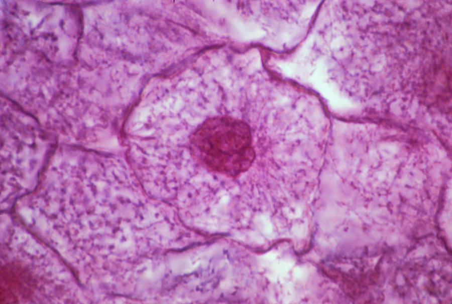
what is occurring in this image?
This image showcases, the S stage in the cell cycle, where DNA is being replicated, into two identical copies of each other. In this stage, the nucleolus is visible, the DNA is uncoiled in a chromatin network, and the nuclear envelope is present.
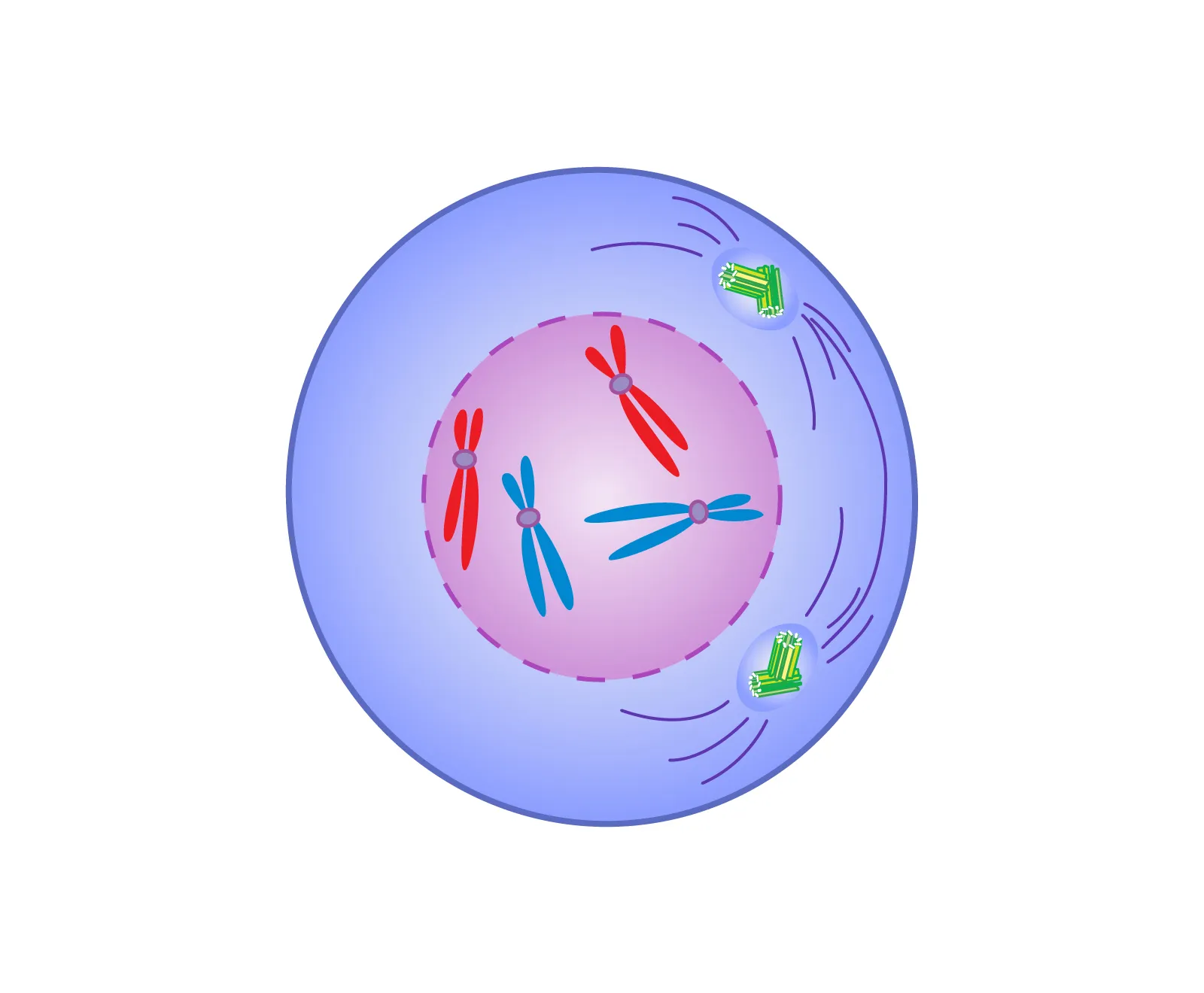
what is occurring in this image?
This image showcases the first stage of mitosis, prophase. during prophase, the DNA tightly coils and forms chromosomes, which consist of two chromatids, joined by a centromere. The nuclear envelope also begins to dissolve, and the centrioles begin to migrate to the opposite ends of the cell. The spindle fibres also start forming.
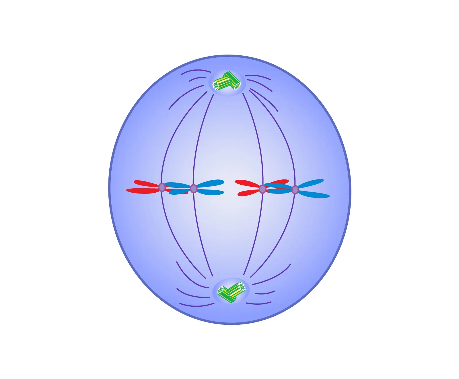
what is occurring in this image?
This image showcases the second stage of mitosis, metaphase. during metaphase, the nuclear envelope completely disappears, and the spindle fibres attach to the centromeres of the chromosomes. The spindle fibres draw the replicated chromosomes to the equator of the cell (metaphase plate).
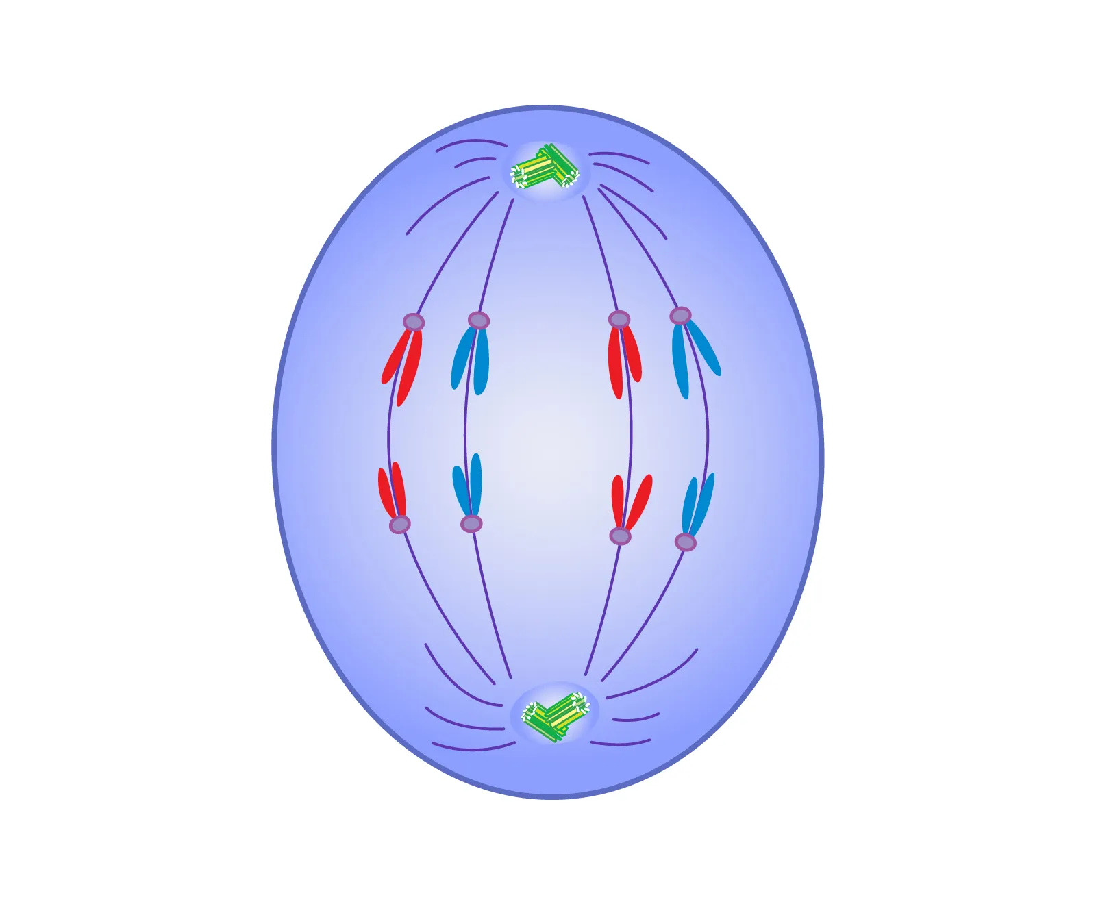
what is occurring in this image?
This image showcases the third stage of mitosis, anaphase. during anaphase, the spindle fibres which were attached to the chromosomes centromeres, pull apart the sister chromatids into polar ends of the cells. This is down to ensure each daughter cell can receive identical, and a complete set of DNA.
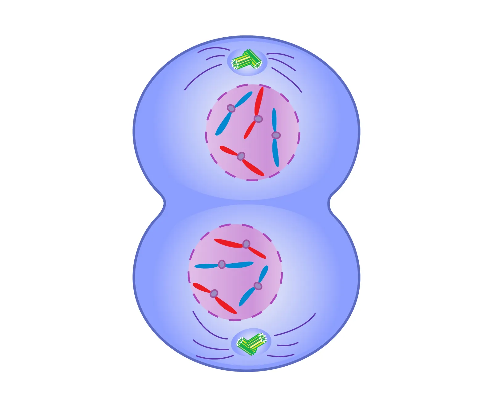
what is occuring in this image?
This image showcases the fourth stage of mitosis, telophase. During telophase, the chromatids arrive at opposite ends of the pole, and the spindle fibres disappear. A new nuclear membrane begins to form around the chromatids, and they begin to uncoil, back into chromatin.
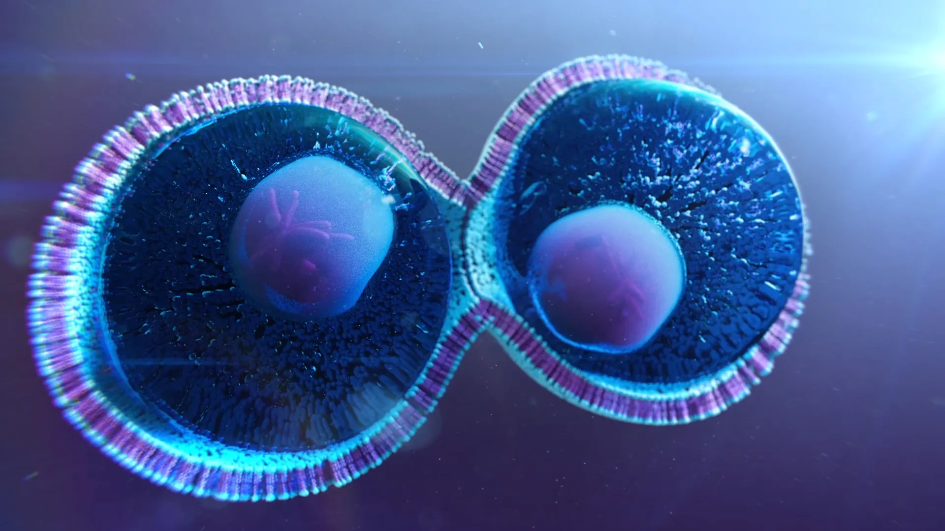
what is occurring in this image?
This image showcases the last stage of the cell cycle, cytokinesis. In animal cells, cytokinesis begins with the formation of a protein contractile ring around the cell. The contractile ring, contracts to form a cleavage furrow, which causes the cell to split into two genetically identical daughter cells. In plant cells, cytokinesis begins with the golgi body releasing vesicle containing cell membrane material. This material is laid down, where the new cell membrane will form.
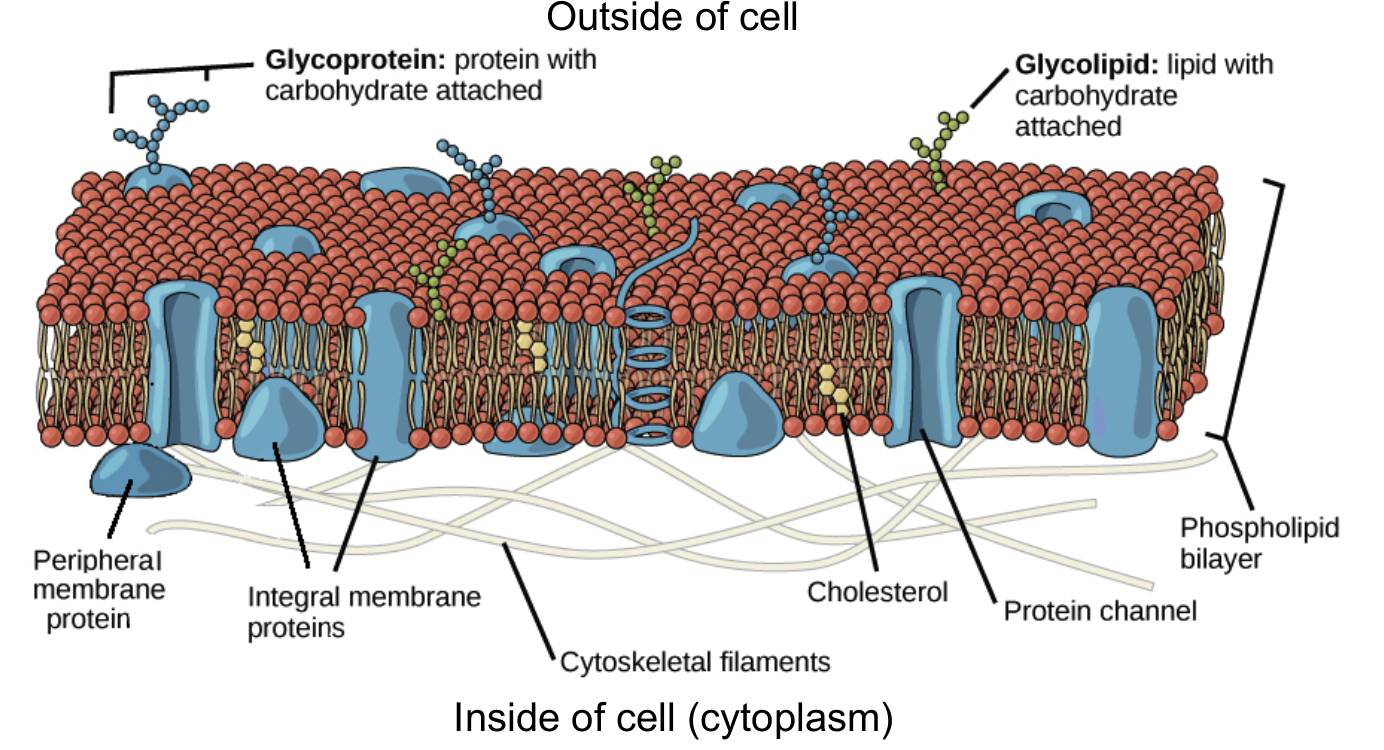
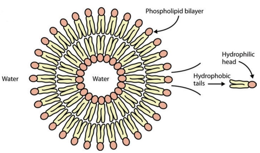
Why is the cell membrane called the fluid mosaic model?
The cell membrane is called the the fluid mosaic model, because of its appearance, and movement. The semipermeable membrane is composed of a phospholipid bilayer, formed by lipids, which have a hydrophobic head that face inwards, and hydrophilic tail which faces towards the intracellular and extracellular space. embedded within this bi-layer are proteins, which can be integral (spanning the membrane), or peripheral (attached to the surface). Another molecule found within the membrane are cholesterol which help regulate the membrane fluidity and carbohydrates which are attached to lipids or proteins. the variety of proteins, cholesterol and carbohydrates on the membrane, give the membrane the mosaic appearance. the cell membrane is also not rigid, and moves laterally, this movement gives the membrane its fluid like quality, therefore making it the fluid mosaic model.
what is the function of the cell membrane?
the semipermeable membrane acts as a selective barrier allowing, and controlling the movement of substances in and out of the cell. the membrane only allows small hydrophobic molecules, such as oxygen and carbon dioxide to enter, while stopping other molecules from entering.
differences between the plant cell and animal cell
Plant cells have a cell wall, chloroplast, and larger vacuole.
describe the structure and function of this organelle
This organelle is called the nucleolus. The nucleolus has no membrane, and is spherical in shape, located inside the nucleus. the main function of the nucleus is to produce ribosomal subunit, and rRNA.
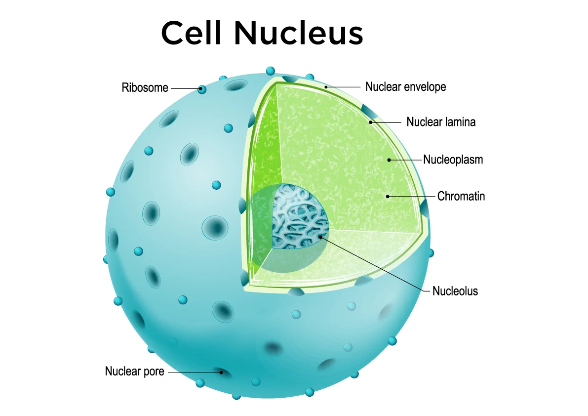
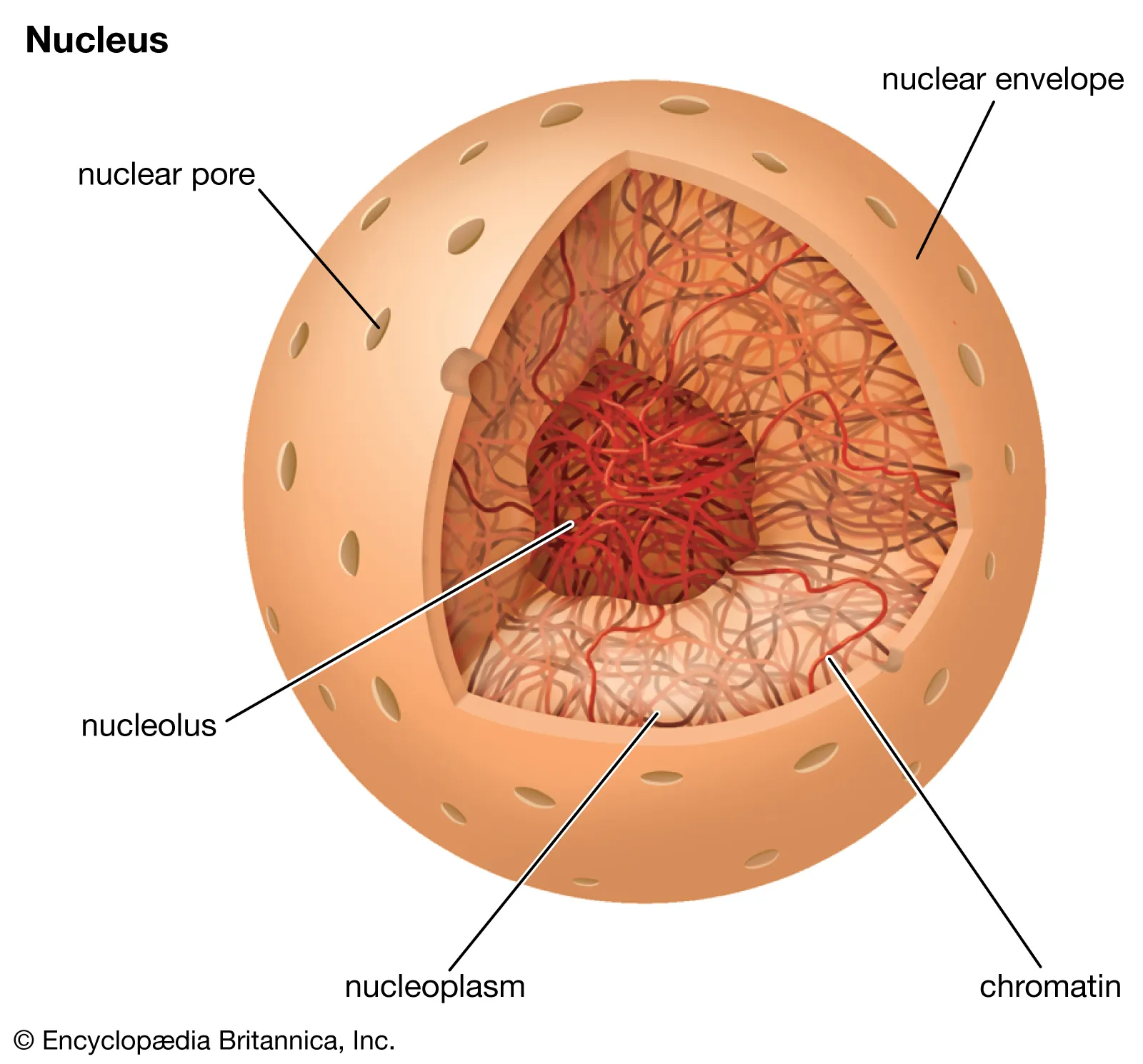
describe the structure and function of this organelle
This organelle is called the nucleus. The nucleus is a single membrane bound organelle, that is spherical in structure. the organelles main function is to store and protect the cells genetic information. The nucleus has a nuclear envelope with pores, to allow DNA to leave the nucleus. The nucleus also hold the nucleolus.
describe the structure and function of this organelle
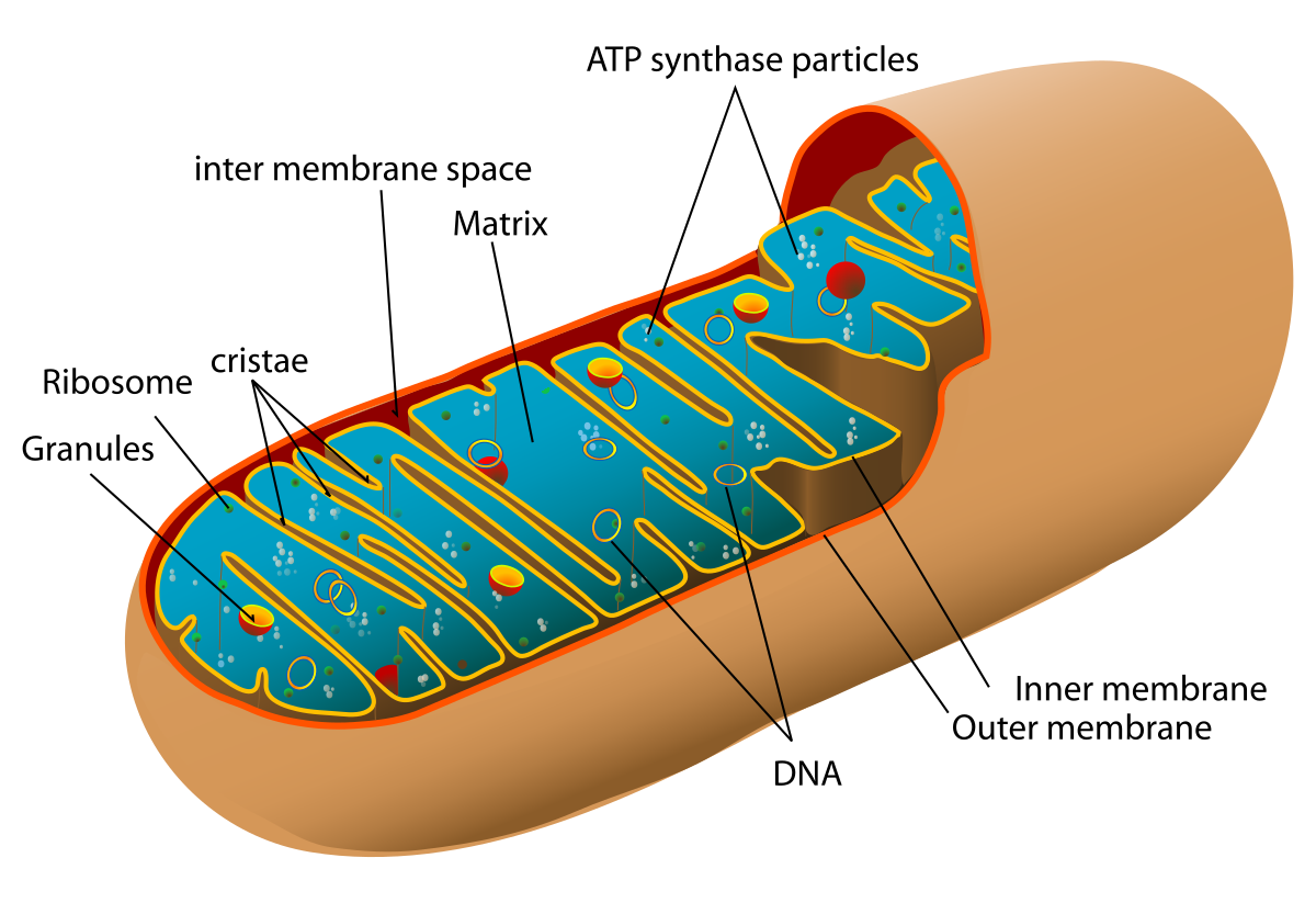
This organelle is called the mitochondria. The mitochondria is a double membrane bound organelle, where its outer membrane is smooth, and inner membrane is highly folded. The mitochondria is the site of aerobic respiration, where the enzymes within the mitochondria break down the glucose molecule. The foldings in the inner membrane is called the cristae. The membrane also has a matrix where, ribosomes and ATP synthase is located.
how does the foldings of the cristae in the mitochondria help its function?
The foldings of the cristae in the mitochondria, increases the surface area of the inner membrane. This provides more space for essential enzymes such as ATP synthase, which are involved in cellular respiration and ATP synthesis. With greater surface area, more ATP synthase enzymes can be embedded, allowing more reactions to take place simultaneously, and thereby increasing the rate of ATP production.
describe the structure and function of this organelle
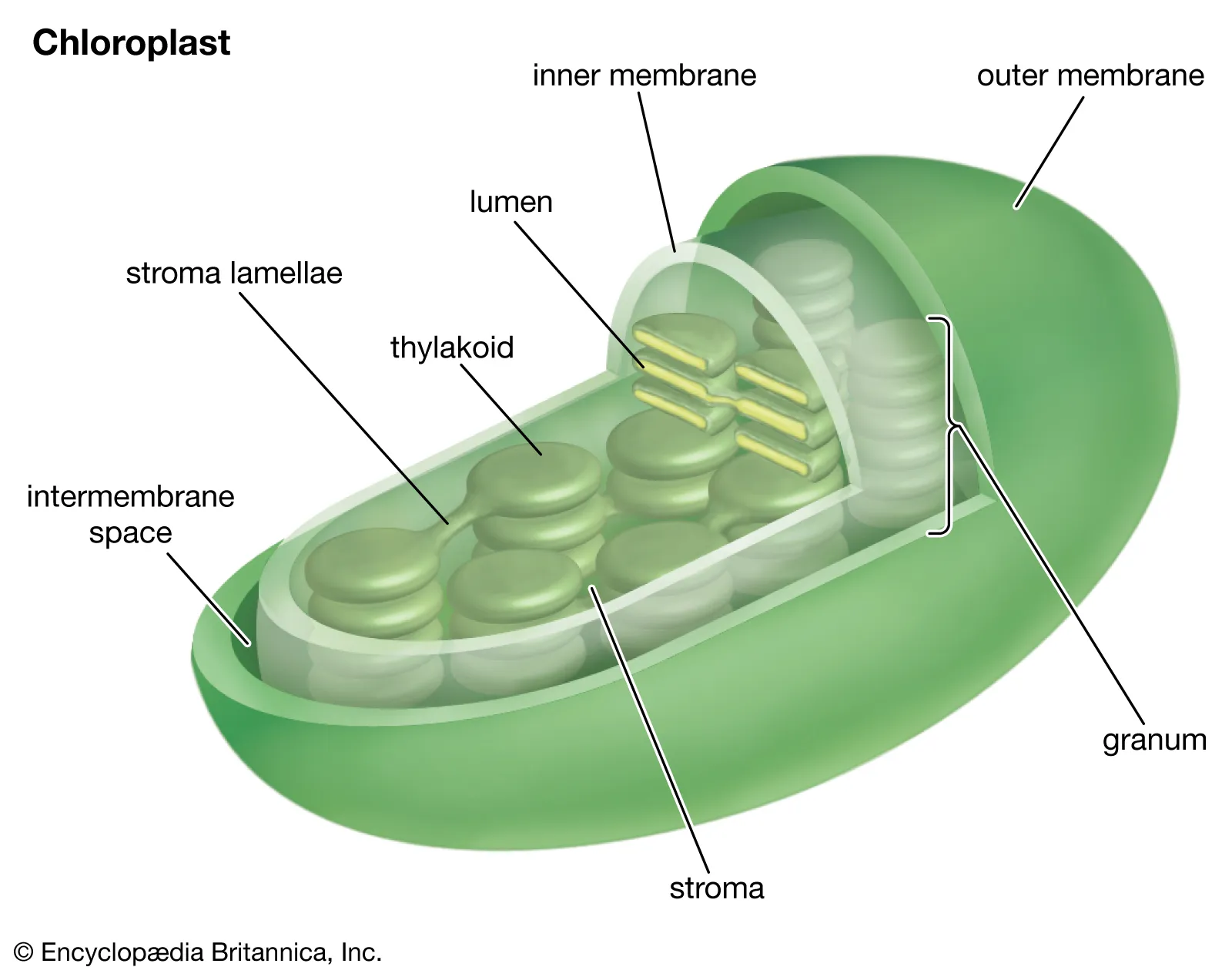
This organelle is the chloroplast, and can only be found in plant cells. chloroplasts have a double membrane, with internal stacked plate-like structures called the thylakoids. these thylakoid contain light trapping pigments called chlorophyll. chlorophyll has the ability to absorb different wavelength of light energy. this ability allows the organelle to convert inorganic material into chemical energy in the form of glucose, through the process of photosynthesis. the main function of chloroplast is to trap energy from sunlight, to build food molecules which can be stored for later use,
How does the stacked nature/structure of the thylakoid help the chloroplasts function?
The stacked structure of the thylakoids increase its surface area. This surface holds important molecules such as chlorophyll, which is needed for the light dependent reaction of photosynthesis. By increasing the surface area of the thylakoids, more light absorbing molecules (chlorophyll), can be packed in, allowing the chloroplast to absorb more sunlight and quicker convert inorganic material into chemical energy in the form of glucose.
describe the structure and function of this organelle
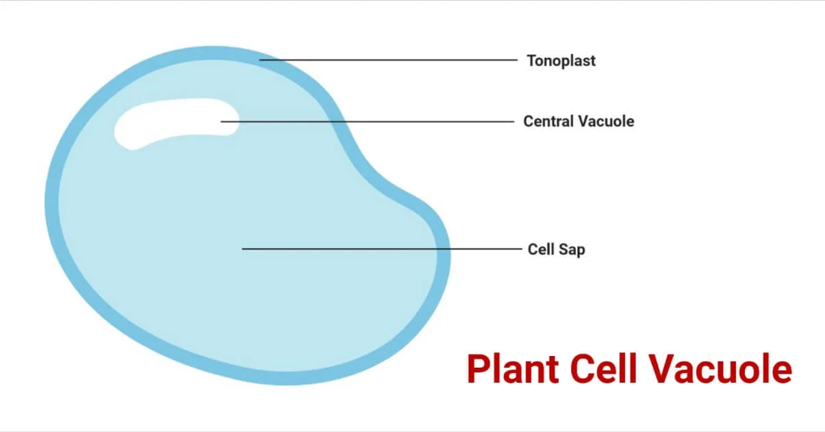
This organelle is called the vacuole. the vacuole is a single membrane bound fluid sac. in animal cells, the vacuole function as storage compartments, used for digestion, and waste removal. in plant cells, vacuole function to maintain the cells shape, and store water and ions.
describe the structure and function of this organelle
This organelle is called the vesicle, and it is a single membrane organelle. the vesicle is composed of a phospholipid bi-layer, with a hydrophobic head, and hydrophilic tail. The vesicle functions to carry and transport material to a specific location within the cell or outside the cell, through exocytosis.
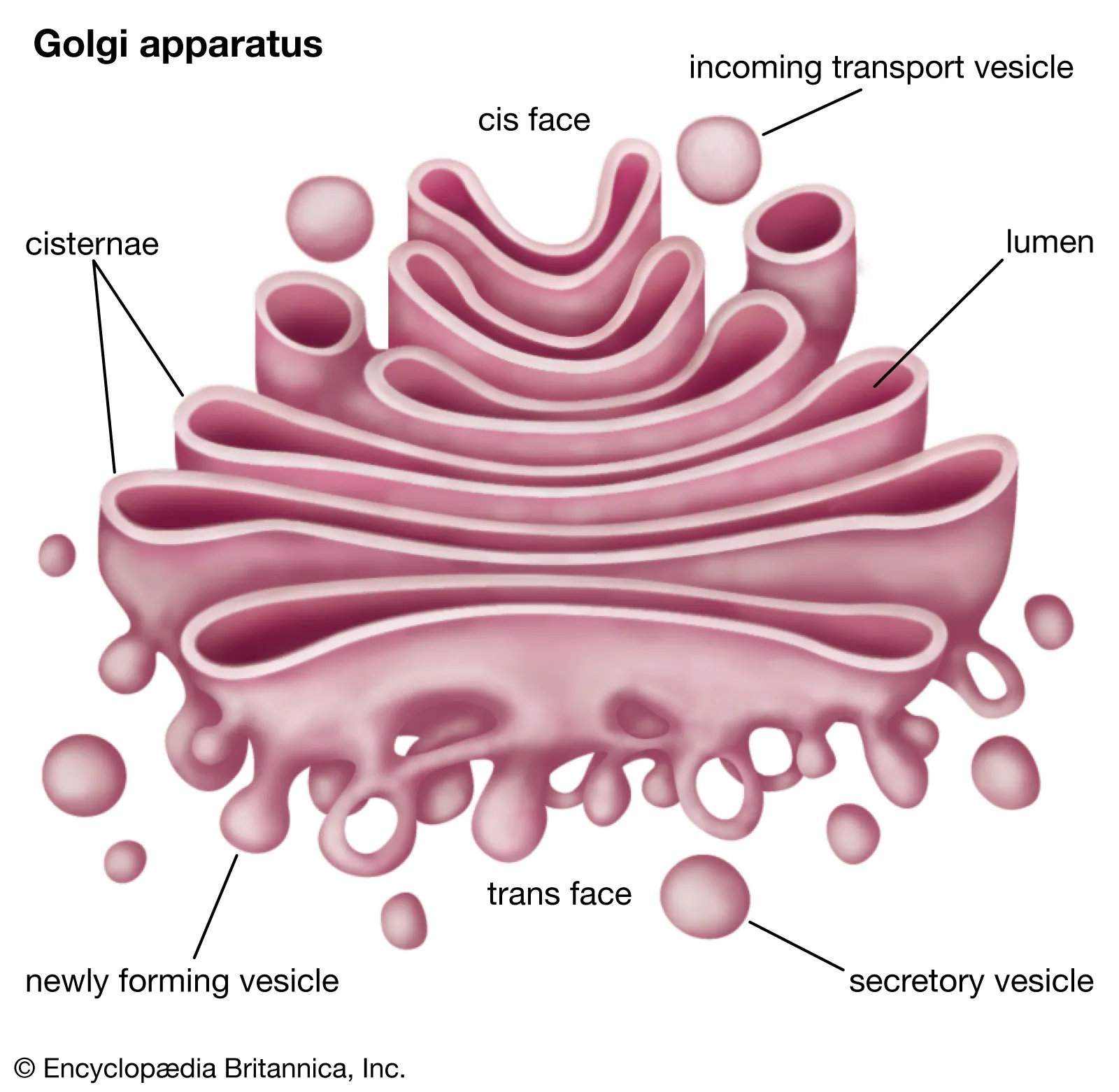
describe the structure and function of this organelle
This organelle is called the golgi body. The golgi body is a single membrane bound organelle, and its structure looks like flattened, disk-shaped sacs. the golgi body can secrete vesicles, a membrane bound sac, which allows the organelle to perform its function, which is to package and transport materials to a specific location.
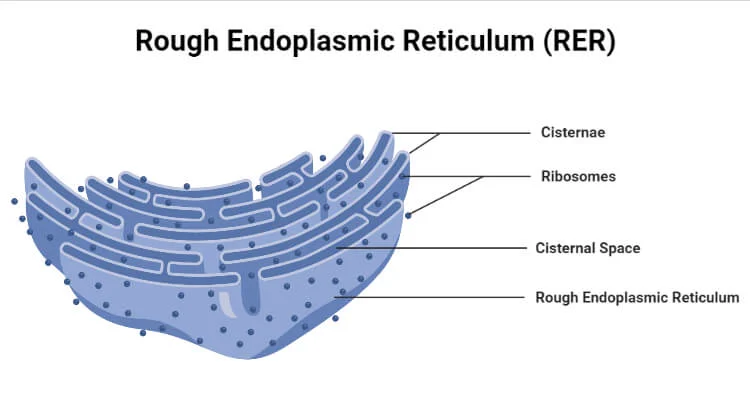
describe the structure and function of this organelle
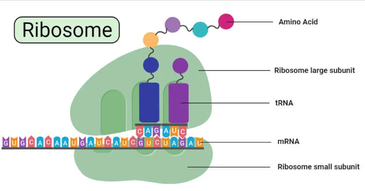
this organelle is called the rough endoplasmic reticulum. the rough er is basically an extension of the nuclear envelope, which is apart of the nucleus. this organelle looks like flattened tubes, with ribosome attached, making it appear '“rough”. Because of its many ribosomes, the rough er is the site of translation, and it functions to synthesize protein.
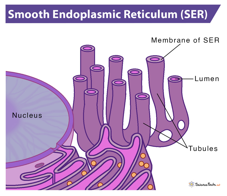
describe the structure and function of this organelle
This organelle is called the smooth endoplasmic reticulum. the smooth er is a single membrane organelle, which connected to the rough er. the smooth er is a series of folded membranes that look like tubes. the smooth er is the site of lipid, and phospholipid synthesis. and it also where the detoxification of some chemicals occur.
describe the structure and function of this organelle
this organelle is called the ribosome, and it is not bounded by a membrane. The ribosome is the site of translation.
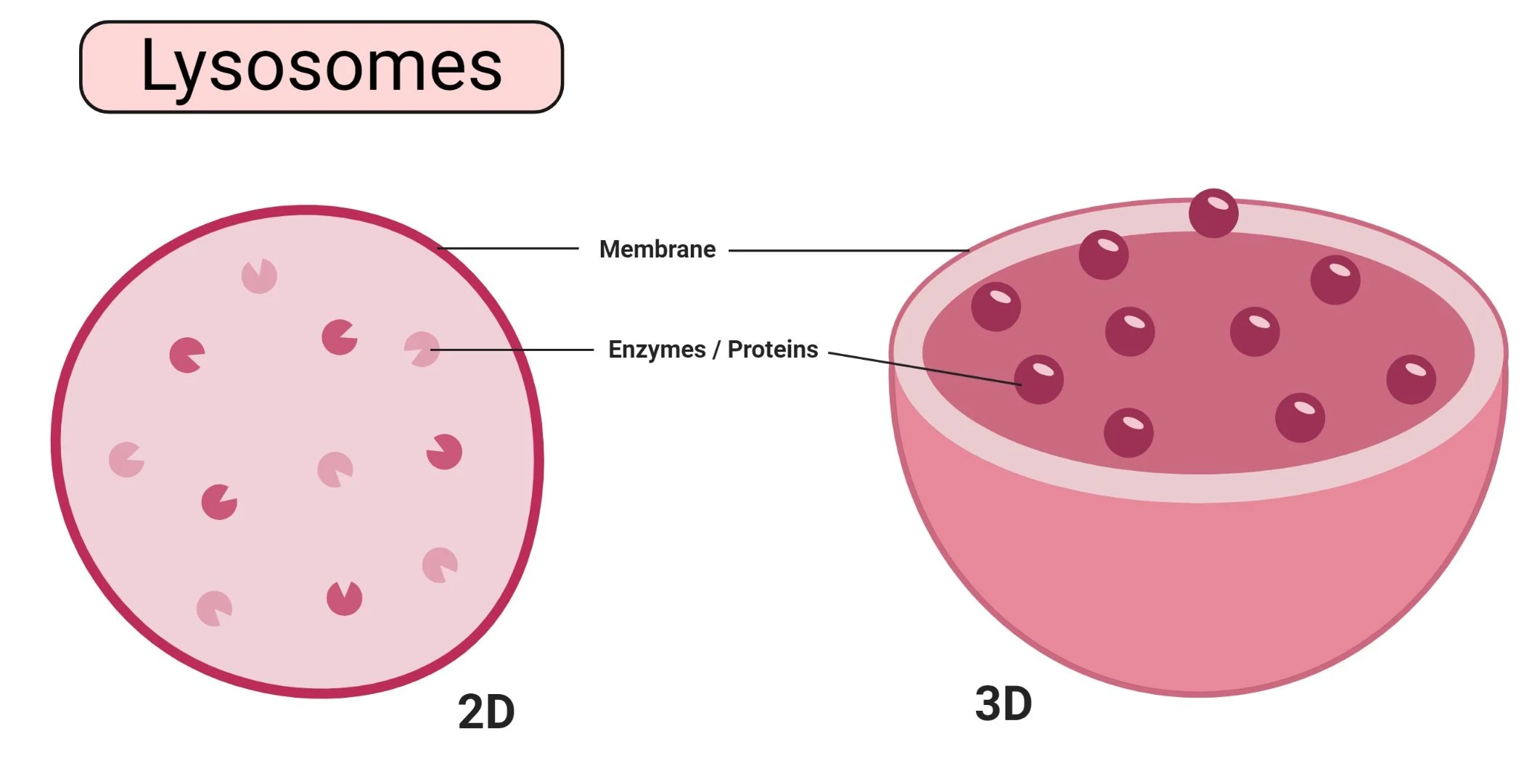
describe the structure and function of this organelle
This organelle is called the lysosome. The lysosome are sphere shaped sacs, filled with digestive enzymes. they function as digestive systems.
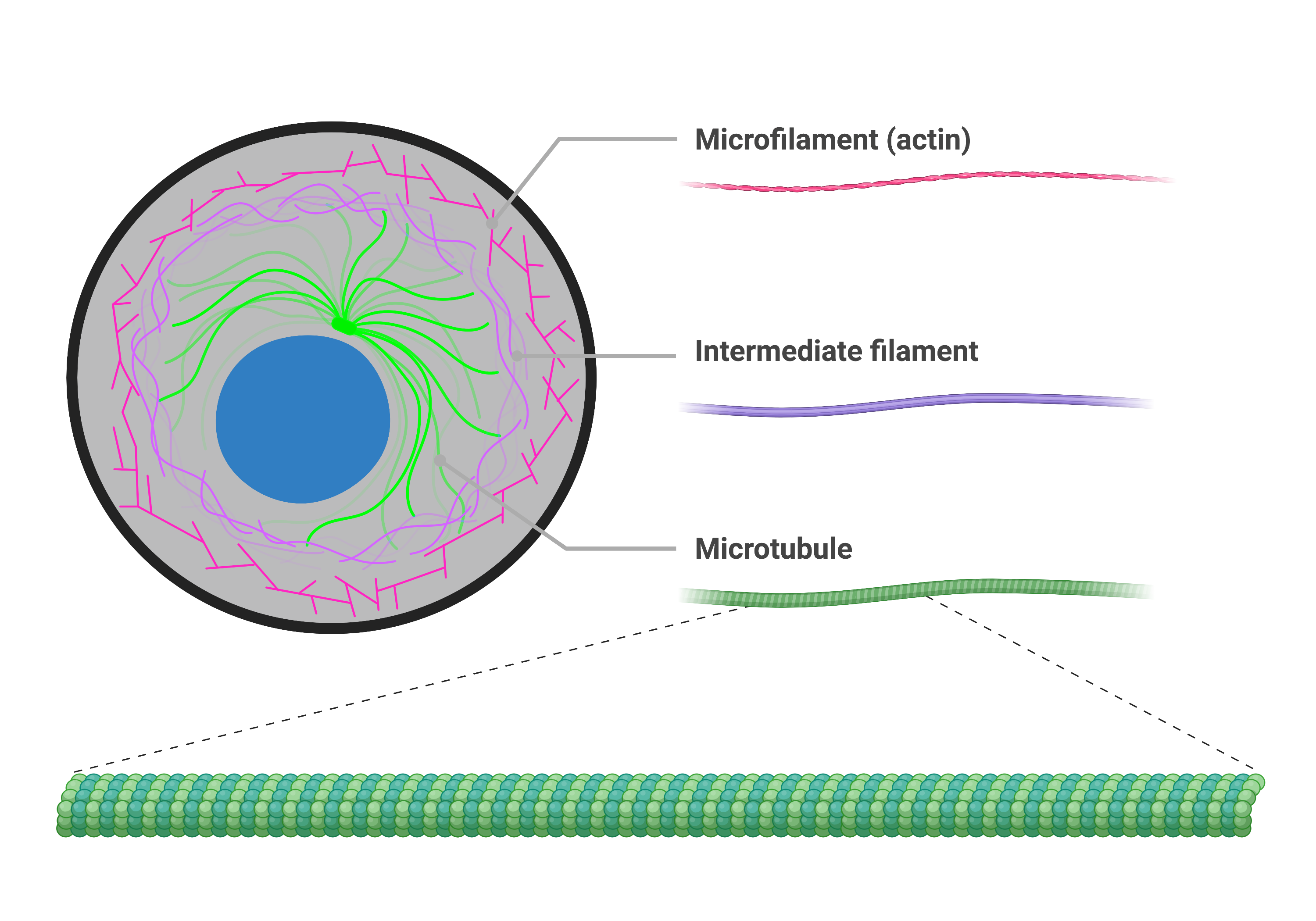
describe the structure and function of this organelle
this organelle is called the cytoskeleton. the cytoksyleenton is a network of protein fibers within cells that provide structural support, and enables movement. the cytoskeleton is composed of three main types of filaments, which work together to maintain cell shape, position organelles, and allow for movement.
Describe the cytoplasmic pathway by which a
secreted protein is produced.
The protein to be produced is transcribed in the nucleus, forming an mRNA strand. This mature mRNA leaves the nucleus through its nuclear pores, and travels to the cytoplasm. In the cytoplasm, ribosomes attached to the rough ER, translate the mRNA into a polypeptide chain which undergoes folding to form a protein. this protein gets packaged in a vesicle and transported to the golgi body. the goligy body repackages the protein into a secretory vesicle, where it then travels to the cell membrane to get excreted through exocytosis.
Distinguish between autotrophs and heterotrophs.
autotrophs are capable of producing their own energy, by converting sunlight energy into chemical energy, specifically to create glucose, through a process known as photosynthesis. heterotrophs are organisms that cannot produce their own food source, so they must rely on other organism for a source of nutrient.
state the chemical formula for photosyntheis
6CO2 + 6H2O —> C6H12O6 + 6O2
how is photosynthesis an energy transformation?
Photosynthesis is an energy transformation because it converts light energy from the sun, into chemical energy stored in glucose.
how is respiration an energy transformation?
Respiration is an energy transformation because it converts chemical energy stored in glucose, into usable energy in the form of ATP.
state the chemical formula for respiration
6O2 (oxygen) + C6H12O6 (glucose)—> 6CO2 (carbon dioxide) + 6H2O (water)
explain anaerobic respiration
anaerobic respiration, is cellular respiration in the absence of oxygen. anaerobic respiration involves the incomplete breakdown of glucose molecules, where the process only reaches the first stage out of the three, with a net ATP production of 2. the first stage is called glycolysis, during glycolysis the glucose molecule is broken down into 2 pyruvate acids. In plant cell, alcohol fermentation occurs, where the pyruvate acids get broken down into ethanol and carbon dioxide. In animal cells, lactic acid fermentation occurs, where the pyruvate acid get broken down forming lactic acid.
explain aerobic respiration in the cytoplasm
The first stage of aerobic respiration, glycolysis, occurs in the cytoplasm. In this stage, glucose molecules are broken down into 2 pyruvate acids, producing 2 ATP and 2 NADH.
explain aerobic respiration in the mitochondria
The second and third stage of aerobic respiration occurs in the mitochondria.
The second stage is called the kreb cycle, and it is located at the mitochondrial matrix. In this stage, a series of enzyme-controlled reactions take place, breaking down the pyruvate molecules produced during glycolysis. This process results in the formation of energy carries, NADH and FADH2.
The third stage is called the electron transport chain, and it takes place in the cristae. In this stage, the NADH and FADH2 produced during the kreb cycle, carry electrons to the electron transport chain. as these electrons move through the chain, energy is released and used to pump hydrogen ions, into the intermembrane space. These hydrogen ion then flow back through proteins called ATP synthase, which uses that energy to produce around 26-28 ATP.
Briefly describe how ATP is generated by aerobic respiration.
ATP is generated by aerobic respiration, through a metabolic process, involving 3 stages.
The first stage of aerobic respiration is glycolysis, which occurs in the cytoplasm of cells. In this stage, glucose is broken down in 2 pyruvate acids, resulting in 2 ATP and 2 NADH.
C6O12H6 —→ 2 pyruvate acids + 2 ATP + 2NADH
In the presence of oxygen, the second stage, the krebs cycle, takes place. In this step, the pyruvate acid is further broken down, in the mitochondrial matrix, producing more ATP, and NADH. The main purpose of this stage is produce energy carrying molecules called NADH and FADH2.
the third stage is the electron transfer chain, which take place in the inner mitochondrial (cristae). in this stage, the NADH and FADH from the krebs cycle carry electrons to the electron transport chain. As the electrons move along the chain, energy is released, which is used to pump hydrogen ions into intermembrane space. These protons then flow back through a protein called ATP synthase, which uses the energy to make ATP.
The overall process of aerobic respiration, creates
state the chemical formula for ATP
ATP —> ADP + Pi
state the chemical formula for lactic acid fermentation
C6H12O6 (glucose) —> 2C3H6O3 (lactic acid) + 2 ATP
state the chemical formula for alcohol fermentation
C6H12O6 (glucose) —> 2C2H5OH (ethanol)+ 2CO2 (carbon dioxide)
Describe the key differences between aerobic
respiration and fermentation (anaerobic respiration)
including the amount of energy released.
The key differences between aerobic respiration and fermentation, lies in their break down of glucose. In aerobic respiration, glucose is completely broken down, resulting in the production of 36-38 ATP. Contrastingly, in anaerobic respiration, glucose is not completely broken down, resulting in only 2 ATP production. In aerobic respiration oxygen is present, unlike anaerobic respiration, this allows the pyruvate acids formed in glycolysis to enter the kreb cycle and move on to the electron transport chain, which occurs in the mitochondria.
Describe the formation of ATP from ADP and Pi.
ATP is formed when ADP gains an inorganic phosphate group, in a process called phosphorylation. This commonly occurs in the mitochondria, where energy released from the breakdown of glucose is used to bond the phosphate group to ADP, to form ATP.
Describe the conversion of ATP to ADP and Pi which
releases energy for some metabolic reactions.
the conversion of ATP into ADP involves the process of hydrolysis. An ATP molecules is composed of an adenine base, ribose sugar, and 3 negatively charged phosphate groups. When the last phosphate group is broken off, releasing energy the ATP is converted into ADP + Pi.
describe endocytosis
endocytosis is a form of active transport. During endocytosis, the cell uses it membrane to surround and engulf large substances from outside the cell, bringing them into the cell inside a vesicle.
this process requires ATP, because it is moving larger molecules or particles against the concentration gradient.
types of endocytosis include
phagocytosis (solids)
pinocytosis (liquids)
receptor mediated endocytosis
describe exocytosis
exocytosis is a form of active transport, where a vesicle containing material for export fuses with the cell membrane to release it content to the extracellular space.
plant cells use exocytosis to export cell wall material
animal cells use exocytosis to secrete hormones
describe facilitated diffusion
facilitated diffusion is a form of passive transport, where molecules move down the concentration gradient, with the help of an embedded protein, called channel proteins. An example of a molecule that requires facilitated diffusion is glucose.,
describe osmosis
osmosis is a specific type of facilitated diffusion (a form of passive transport), that utilses a carrier protein called aquaporins, to help the polar water molecule to move through the concentration gradient, into the area of high solute concentration.
describe how the structure of aquaporins help with its function
Aquaporins have a quaternary structure as they are made up of four identical polypeptide subunits. Each, subunit has a channel which is complementary in shape to that of a water molecule, the inside of the channel is narrow and lined with polar (water-attracting) amino acids, which help the water molecules move through in a single file line. The shape of the channel is specific to a water molecule, inhibiting other substances from going through.
describe active transport
Active transport requires ATP to induce a conformational change in the transporter. active transport utilizes the energy released from the breakdown of ATP molecules.
Explain how the exchange of materials across
membranes is affected by factors including:
surface area to volume ratio (SA/V) of the cell.
Explain how the exchange of materials across
membranes is affected by the physical and chemical nature of the materials being
exchanged (eg. size, charge and relative
hydrophobicity)
explain what the metabolic pathway is
a metabolic pathway is a series of interconnected bio-chemical reactions, each regulated by a specific enzyme. in this pathway, the product of one reaction acts as the substrate for next.
Describe metabolic pathways in terms of energy release.
Explain why energy pathways involve many
regulated steps.
energy pathways involves many regulated steps, because breaking down a large molecule in a single step can release too much energy, which some can transform into heat, this can denature enzymes that catalyse the pathway, stopping the efficiency of the pathway.
Discuss the advantages of having many
regulated steps in biochemical pathways.
the advantages of many regulated steps on a biochemical pathway is that the intermediate compounds formed during the biochemical pathway can be used for various other cellular functions, eg, building amino acids. Additionally, the many regulated steps control the release of energy, which prevent the excessive buildup of heat. the excessive buildup of heat can denature the enzymes involved in the biochemical pathway, reducing the pathways efficiency. having many regulated steps also reduces the activation energy required for the reaction to proceed
what is a homologous chromosome?
chromosome with the same shape, size, and centromerers at the same spot
What occurs in prophase 1?
what occurs in metaphase 1?
what occurs in anaphase 2?
what occurs in telophase 1?
what occurs in prophase 2?
what occurs in metaphase 2?
what occurs in anaphase 2?
what occurs in telophase 2?
State the meaning of haploid. Explain why the
products of meiosis are haploid cells and contain a
single set of chromosomes.
haploid means that the cell has half of it full set of chromosomes, and therefore single set of its two. the products of meiosis (which are sex cells), are haploid cells, because meiosis goes through 2 rounds of division. During meiosis 2, there is no replication of chromosomes, this means that when cell divides, the cell with its full set of chromosomes from the previous round of meiosis, turn into diploid cells with half of its full set.
Explain the importance of crossing-over and
independent assortment in meiosis.
Crossing-over and independent assortment is important in meiosis because it increases the genetic variation in offspring.
Explain the term ‘crossing over’ in prophase 1
crossing over is a process where homologous chromosomes come together to exchange segments of genetic material. this occurs in prophase 1, meiosis 1, when homologous chromosomes pair up to form tetrads. at points called chiasmata, chromatids form chromosomes break and rejoin, swapping segments of DNA. this results in a new combination of alleles on each chromatid, which increases the genetic variation in the gametes.
Explain the term ‘individual assortment’ in metaphase 1
independent assortment is the random arrangement of homologous chromosome pairs along the equator of the cell. this occurs during metaphase 1, meiosis 1, after the homologous chromosomes have paired up during prophase 1. the orientation of each pair is random and independent of others, which means that maternal and paternal chromosomes can be assorted in many different combinations. This results in gametes with different chromosomes, contributing to genetic variation.
Identify the process which involves the fusion of
gametes. State how this process results in a
diploid zygote.
The process which involves the fusion of gametes is called fertilization. gametes are sex cells which have half its full set of chromosomes (therefore a haploid cell), when two gametes fuse during fertilisation, they result in the formation of a diploid zygote with a full set of chromosomes, one set given from the mothers side, and another from the father’s side.
Compare the products of mitotic and meiotic
cell division.
Mitotic cell division results in the formation 2 genetically identical, diploid cells (full set of chromosomes), while in meiotic division, 4 genetically varied haploid cells (half of the full set ) are produced. the products of mitosis is commonly used for tissue repair, and asexual reproduction, while meiotic cell division is used for the production of gametes.
Describe the stages of the cell cycle (including
checkpoints).
the stages of the cell cycle include growth phase 1 (checkpoint), synthesis, growth phase 2 (checkpoint), mitotic division, and cytokinesis.
in G1, the cell grows, preparing itself for DNA replication. in G1, the cell is checked for any DNA damage, it nutrient availability and cell size.
In S, the DNA in the cells replicated, with each each chromosome becoming 2 identical sister chromatids.
In G2, the cells grows in preparation for cell division, and all the organelles get doubled. the cell is also checked to see if the DNA has been replicated correctly.
In M, the cell undergoes mitosis, and is checked again during metaphase.
Explain that hormones (and growth factors) may
regulate cell division.
State two reasons why aerobic respiration must involve a series of small, regulated steps.
Aerobic respiration must involve a series of small, regulated steps because breaking down a large molecule in a single step would release too much energy as heat at once, this can lead to the enzyme being denatured, and losing its function. When the molecule is gradually broken down, over multiple steps, the cell can control the flow energy, and better utilize it to form ATP.
Additionally, the intermediate compounds created during aerobic respiration can be used for various cellular functions, beyond energy production, for example amino acids and nucleotide formation.
How does ATP turn into ADP?
ATP is made out of an adenine base, ribose sugar and three negatively charged phosphate groups. when the cell needs energy, ATP undergoes hydrolysis, where the bond between the last phosphate group is broken forming ADP and Pi. This reaction releases energy which can be used for cellular processes.
how does ADP turn into ATP?
To regenerate ATP, ADP must undergo phosphorylation, where a phosphate group is bonded to the ADP molecule through the energy released from the break down of glucose.