Human Anatomy & Physiology Basics
 Call Kai
 Learn
Learn Practice Test
 Spaced Repetition
 Match
Match Flashcards
 Knowt Play
1/92

Earn XP
Description and Tags
Flashcards covering human anatomy basics including directional terms, planes and axes of motion, osteokinematic actions, body cavities, and anatomical regions.
Name | Mastery | Learn | Test | Matching | Spaced | Call with Kai | Chat |
|---|
No analytics yet
Send a link to your students to track their progress
93 Terms
Anatomical Position
The standard universal reference position used by clinicians to describe body parts and positions, with the body standing erect, facing forward, arms at sides, and palms facing forward.
Anterior/Ventral
Pertaining to the front surface or towards the front of the body; also referred to as ventral.
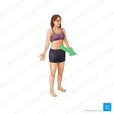
Posterior/Dorsal
Pertaining to the back surface or towards the back of the body; also referred to as dorsal.

Proximal
Located closer to the trunk of the body or point of origin/attachment.

Distal
Located farther from the trunk of the body or point of origin/attachment.

Median Plane
The single vertical plane dividing the body centrally into equal right and left halves.

Medial
Positioned closer to the median plane or midline of the body.

Lateral
Positioned farther away from the median plane or midline of the body.

Superior
Positioned higher or towards the head end of the body.

Inferior
Positioned lower or towards the feet end of the body.

External/Superficial
Located on or near the outer surface of the body

Internal/Deep
Located farther inside or away from the body surface

Frontal Bone
The anterior cranial bone that forms the forehead and upper rims of the orbits.

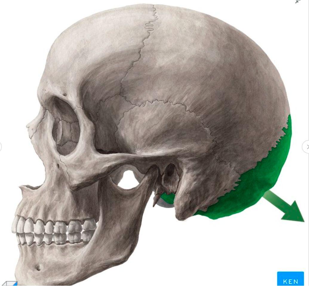
Occipital Bone
The posterior cranial bone forming the back and lower base of the skull.
Body Planes
Imaginary flat 2D surfaces that section or cut through the human body in anatomical position.
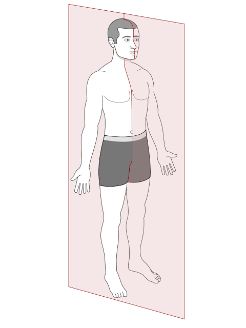
Sagittal Plane
A vertical plane dividing the body into right and left sections.
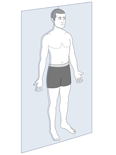
Coronal Plane
A vertical plane dividing the body into anterior (front) and posterior (back) sections.
Transverse Plane
A horizontal plane dividing the body into superior (upper) and inferior (lower) sections.

Anatomical Axes
Perpendicular imaginary lines passing through the body around which joint rotation occurs.
Horizontal Axis (X-axis)
medial to lateral.

Vertical Axis (Y-axis)
superior to inferior.
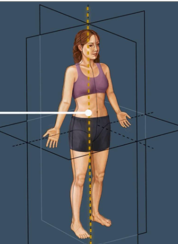
Sagittal Axis (Z-axis)
anterior to posterior.
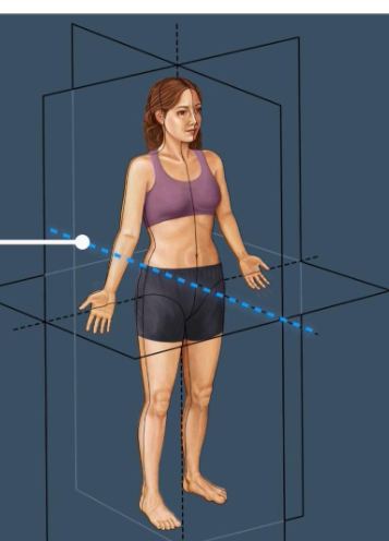
Osteokinematic Actions
Gross movements of bones relative to one another at joints.
Flexion
A joint movement that decreases the angle between two articulating bones (e.g., angle <90× or <90o).

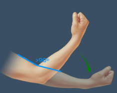
Extension
A joint movement that increases the angle between two articulating bones (e.g., angle >90o).
Lateral Flexion
A sideways bending movement of the trunk or spine away from the midline.

Abduction
Movement of a limb or body structure away from the midline of the body.

Adduction
Movement of a limb or body structure towards the midline of the body.
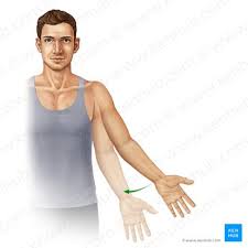
External Rotation
Rotational movement of a limb region around its axis away from the midline (lateral rotation).
Internal Rotation
Rotational movement of a limb region around its axis towards the midline (medial rotation).
Protraction
Anterior or forward movement of a body structure across a plane, such as pushing shoulders forward.
Retraction
Posterior or backward movement of a body structure across a plane, such as pulling scapulae together.
Elevation
Movement of a body part in a superior (upward) direction.
Depression
Movement of a body part in an inferior (downward) direction.
Circumduction
A circular movement combining flexion, extension, abduction, and adduction at ball-and-socket or condyloid joints.
Supination
Forearm movement that rotates the palm anteriorly or upward.
Pronation
Forearm movement that rotates the palm posteriorly or downward.
Ulnar Deviation
Wrist movement bending the hand medially toward the ulna.
Radial Deviation
Wrist movement bending the hand laterally toward the radius.
Opposition
Movement bringing the pad of the thumb across the palm to contact other digits, enabling grip.
Reposition
Movement returning the thumb from opposition back to its anatomical stance.
Plantarflexion
Ankle movement pointing the foot or toes downward toward the ground.

Dorsiflexion
Ankle movement lifting the foot or toes upward toward the shin.

Inversion
Movement turning the plantar surface (sole) of the foot inward toward the midline.

Eversion
Movement turning the plantar surface (sole) of the foot outward away from the midline.

Agonist
The prime mover muscle primarily responsible for producing a specific motion (e.g., Biceps brachii in elbow flexion).
Antagonist
The muscle that opposes the action of an agonist muscle (e.g., Triceps brachii in elbow flexion).
Simple Joints
Uniaxial hinge joints that accommodate basic flexion and extension (e.g., elbows, knees, interphalangeal joints).
Compound Joints
Complex joint combinations involved in movement, such as the intervertebral joints of the neck and spine.
Body Cavities
Fluid-filled internal spaces that function to protect organs, compartmentalize systems, and limit the spread of infection.
Dorsal Body Cavity
The posterior fluid-filled cavity consisting of the cranial cavity and the vertebral canal.
Ventral Body Cavity
The anterior body space encompassing the thoracic, abdominal, and pelvic cavities.
Cranial Cavity
The cavity inside the skull that encloses and protects the brain.
Neurocranium
The protective bony braincase consisting of 8 bones: 1 ethmoid, 1 sphenoid, 1 frontal, 2 parietal, 2 temporal, and 1 occipital.
Ethmoid Bone
A single cranial bone forming part of the nasal cavity walls, neurocranium, and superior border of the anterior cranial fossa.
Sphenoid Bone
An unpaired wedge-shaped bone at the base of the skull forming central neurocranial structures.
Parietal Bones
A pair of bones forming the curved roof and superior side walls of the skull.
Temporal Bones
A pair of bones forming the lower sides and base of the skull.
Calvaria
The roof of the skull (skullcap) composed of the frontal bone, parietal bones, and occipital bone.
Foramen Magnum
The large opening in the base of the occipital bone allowing the spinal cord to exit the cranial cavity.
Anterior Cranial Fossa (Fossa cranii anterior)
The shallowest cranial fossa bounded by the orbital plate of the frontal bone, superior ethmoid bone, and lesser wings of the sphenoid bone.
Middle Cranial Fossa (Fossa cranii media)
The middle cranial depression formed by the sphenoid bone, temporal bones, and parietal bones.
Posterior Cranial Fossa (Fossa cranii posterior)
The deepest cranial fossa bounded by the temporal bones, parietal bones, and occipital bone.
Cerebrum
The major superior brain structure housed inside the cranial cavity.
Cerebellum
The posterior brain structure situated in the posterior cranial fossa controlling motor coordination.
Brainstem
The central trunk structure of the brain housed within the cranial cavity connecting the cerebrum to the spinal cord.
Vertebral Canal (Canalis vertebralis)
The bony canal extending through the vertebral column that houses the spinal cord and spinal nerves.
Spinal Cord
The primary cylindrical nerve trunk contained within the vertebral canal.
Spinal Nerves
Paired nerve roots issuing laterally from the spinal cord through intervertebral spaces.
Cauda Equina
The collection of lumbar and sacral nerve roots extending downward beyond the end of the spinal cord within the vertebral canal.
Thoracic Cavity
The upper division of the ventral cavity bounded by the ribcage, sternum, spine, and respiratory diaphragm.
Superior Border of Thoracic Cavity
Anatomical border formed by the body of T1 vertebra, the first pair of ribs, and the manubrium of the sternum.
Inferior Border of Thoracic Cavity
The anatomical floor of the thoracic cavity formed by the respiratory diaphragm.
Contents of Thoracic Cavity
Key internal organs consisting of the trachea, bronchi, lungs, oesophagus, heart, and great vessels.
Pleural Cavity
The serous space enclosing each lung lined by pleural membranes.
Parietal Pleura
The outer fibrous membrane lining the internal surface of the thoracic cavity wall.
Visceral Pleura
The inner delicate membrane adhering tightly to the surface of the lung.
Intrapleural Pressure
The pressure inside the pleural cavity, which must remain lower than intrapulmonary pressure to prevent lung collapse.
Pericardial Cavity (Cavitas pericardiaca)
The fluid-filled serous space surrounding the heart situated in the inferior mediastinum.
Fibrous Pericardium
The tough external fibrous connective sac enclosing the heart and pericardial cavity.
Parietal Pericardium
The outer serous layer lining the interior aspect of the fibrous pericardium.
Visceral Pericardium
The inner serous layer that directly covers the epicardial surface of the heart.
Pericardial Fluid
Serous fluid contained in the pericardial space that reduces friction during heart contractions.
Abdominal Cavity
The ventral body cavity region situated between the diaphragm and pelvis housing digestive and metabolic organs.
Greater Omentum
A large apron-like fold of visceral peritoneum extending downward from the stomach over the abdominal viscera.
Mesentery
A double-layered fold of peritoneum attaching the intestines to the posterior abdominal wall.
Pelvic Cavity
The lowermost division of the ventral body cavity situated within the bony pelvis.
Female Pelvic Cavity Contents
Encloses the sigmoid colon, rectum, ureters, urinary bladder, vagina, uterus, and Fallopian tubes.
Male Pelvic Cavity Contents
Encloses the rectum, distal end of the sigmoid colon, bladder, seminal vessels, and prostate.
Right Upper Quadrant (RUQ)
The superior-right division of the abdominal wall in a 4-quadrant anatomical layout.
Subcostal Plane
The upper horizontal grid line passing beneath the costal margins in the 9-region abdominal breakdown.
Intertubercular Plane
The lower horizontal grid line intersecting the iliac tubercles in the 9-region abdominal breakdown.
Epigastric Region
The upper central abdominal region located superior to the umbilical region and between hypochondriac regions.
