Heart Anatomy and Function
 Call Kai
 Learn
Learn Practice Test
 Spaced Repetition
 Match
Match Flashcards
 Knowt Play
1/104

There's no tags or description
Looks like no tags are added yet.
Name | Mastery | Learn | Test | Matching | Spaced | Call with Kai | Chat |
|---|
No analytics yet
Send a link to your students to track their progress
105 Terms

Base
1

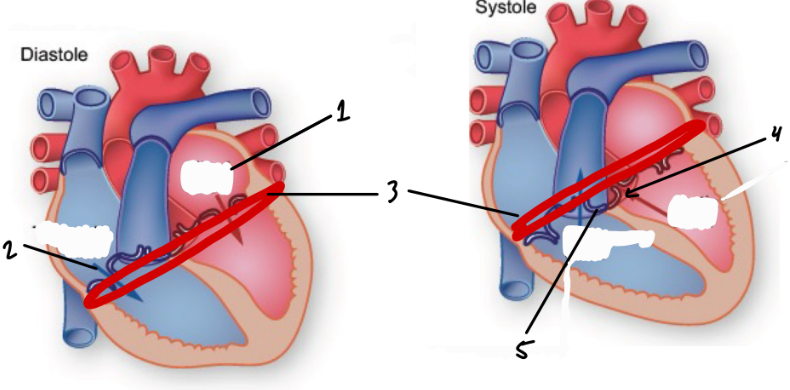
Bicuspid Valve
1


Apex
2
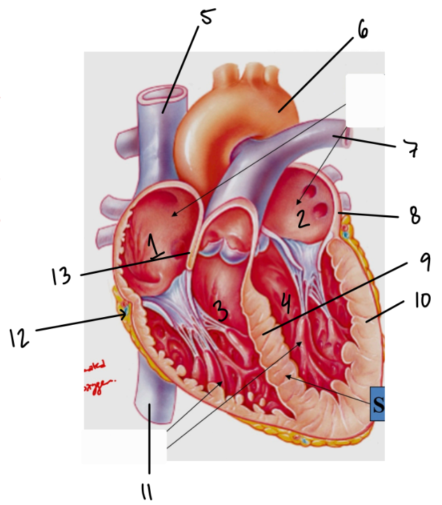
Superior Vena Cava
5
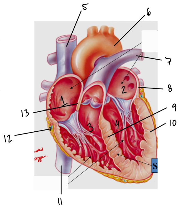

Aorta
6
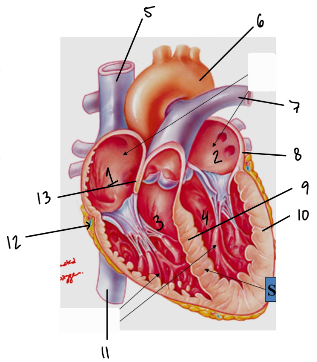

Pulmonary Trunk
7


Ventricular Wall
10
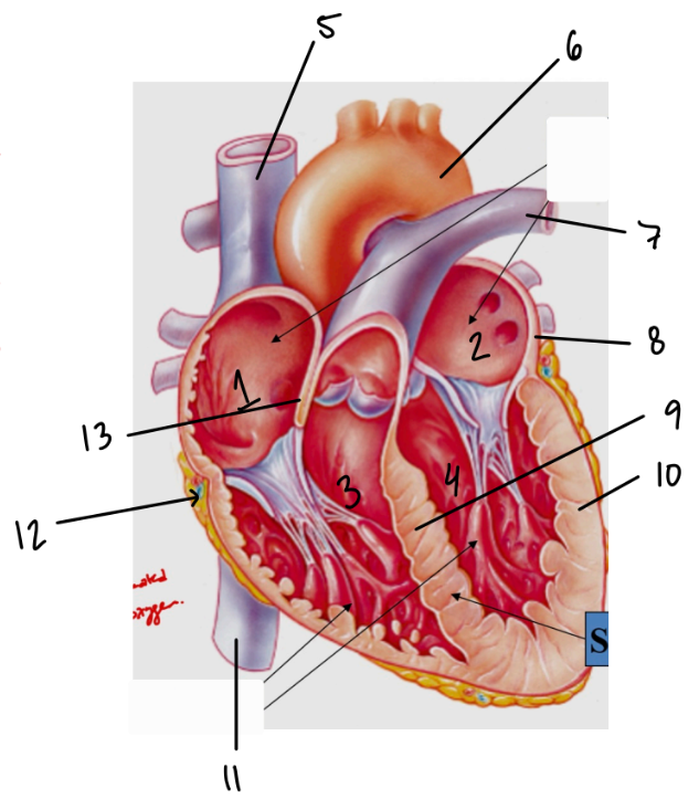
Septum
9

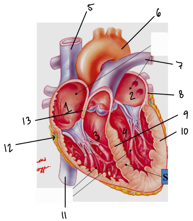
Interatrial Septum
13
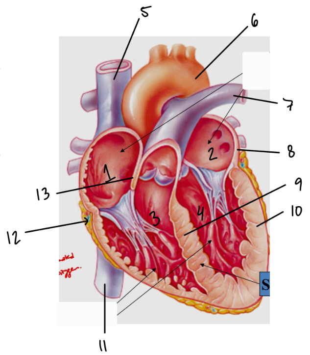
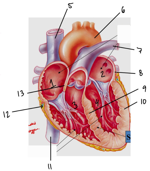
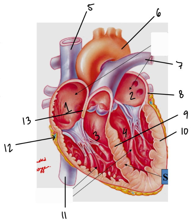
Interventricular Septum
9


Inferior Vena Cava
11

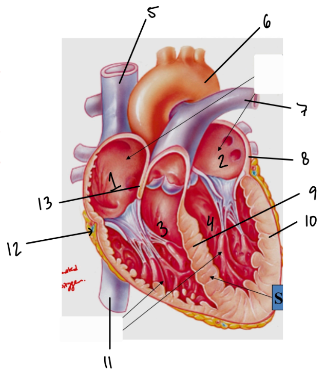
Coronary Sinus
12

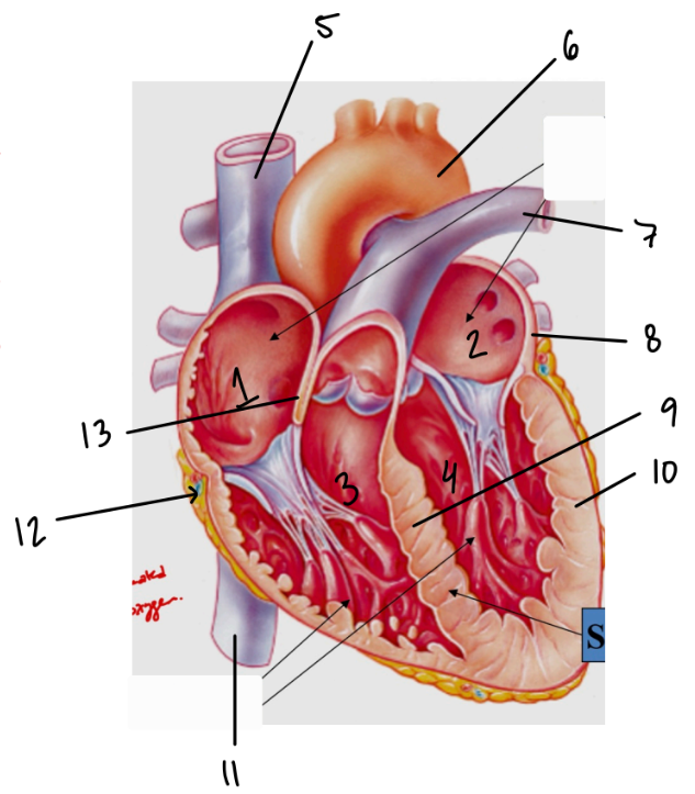
Atrial Wall
8


Auricles
1


Anterior Interventricular Sulcus
2


Left Pulmonary Veins
2
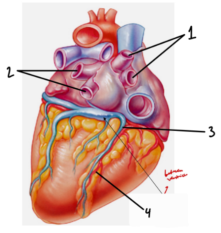

Right Pulmonary Veins
3


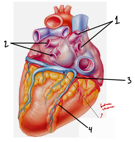
Posterior Interventricular Sulcus
4


Coronary Sulcus
3
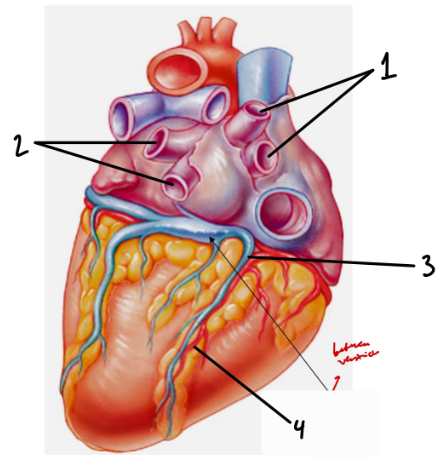

Atrioventricular Groove
3
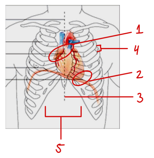
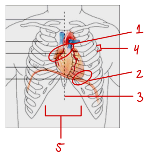
Mediastinum
5
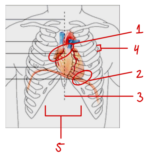

Intercostal Space
4
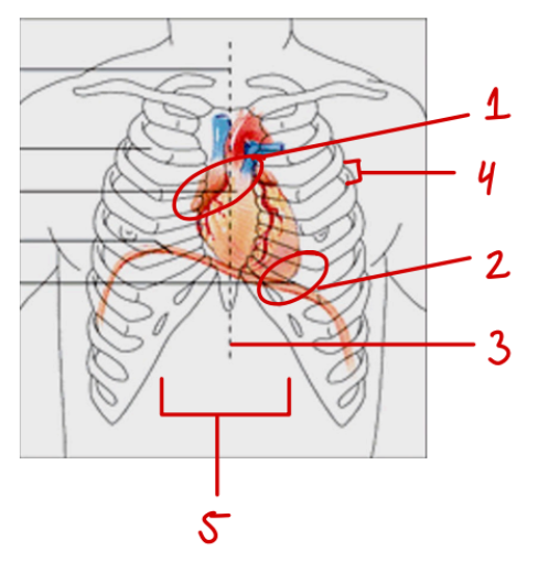

Midsternal Line
3

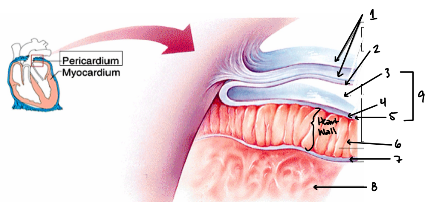
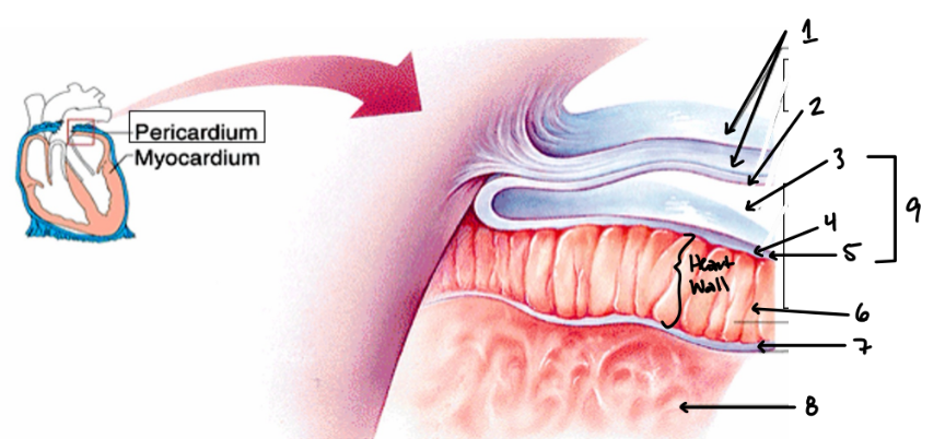
Pericardium
9

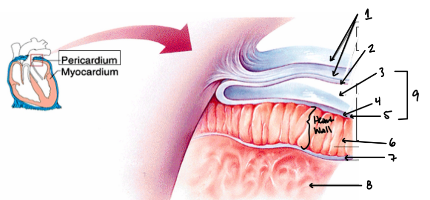
Fibrous Pericardium
1


Parietal Layer of Serous Pericardium
2
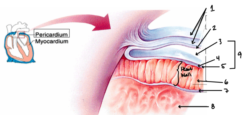

Pericardial Cavity
3

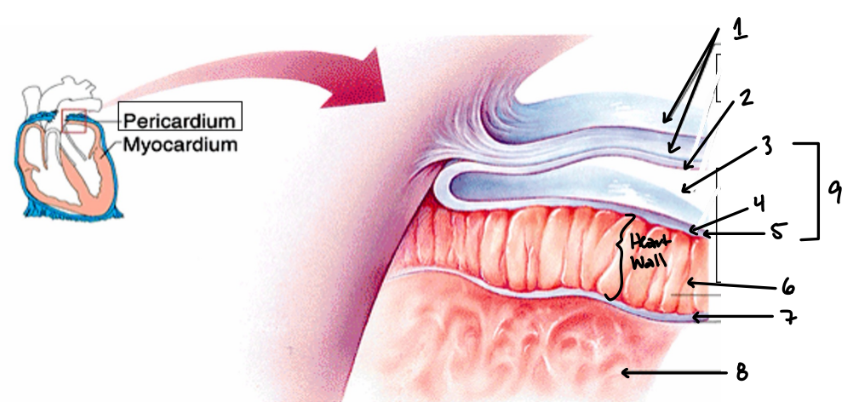
Pericardial Fluid
Inside 3


Visceral Layer of Serous Pericardium
4

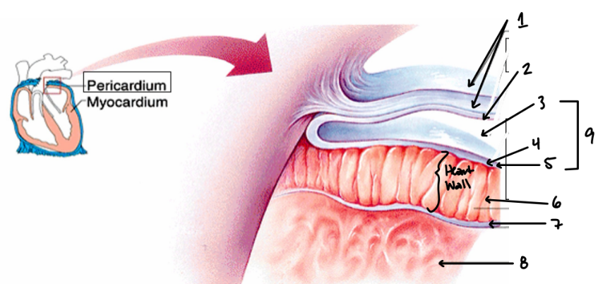
Epicardium
5

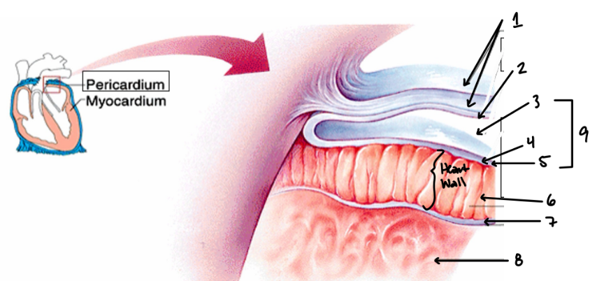
Myocardium
6
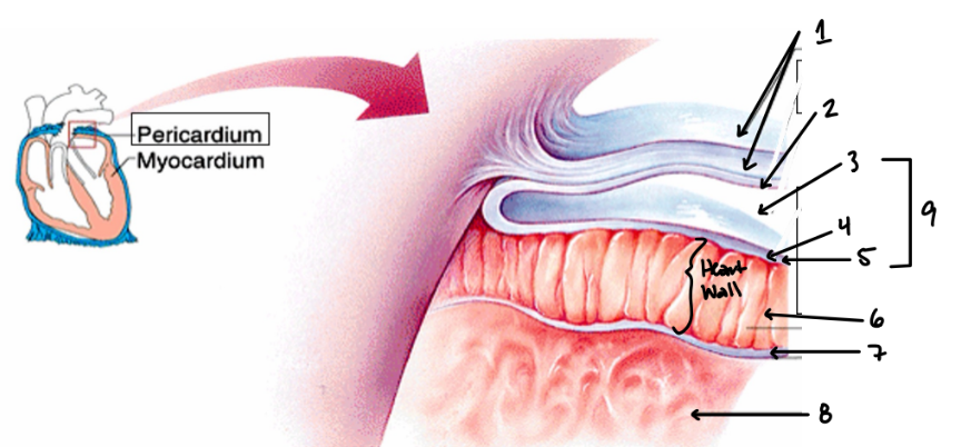

Endocardium
7

Pectinate Muscle
3
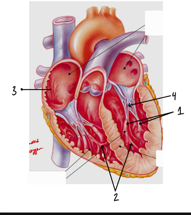

Papillary Muscles
2
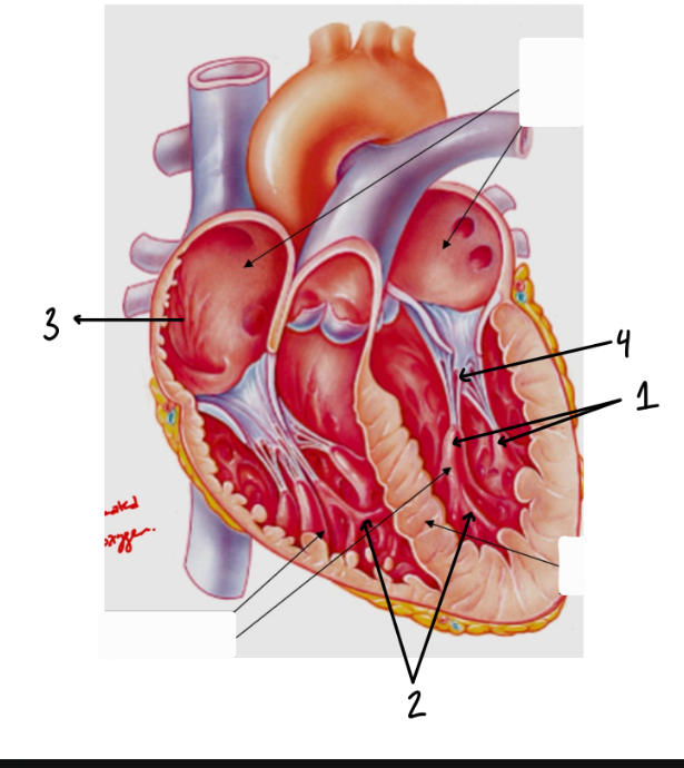

Trabeculae Carnae
1

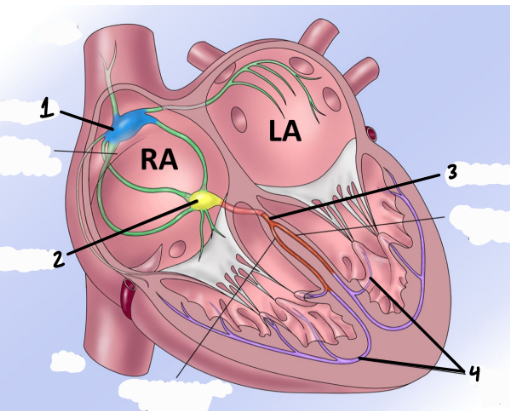
Sinoatrial Node
1


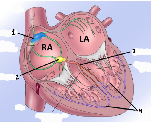
Atrioventricular Node
2

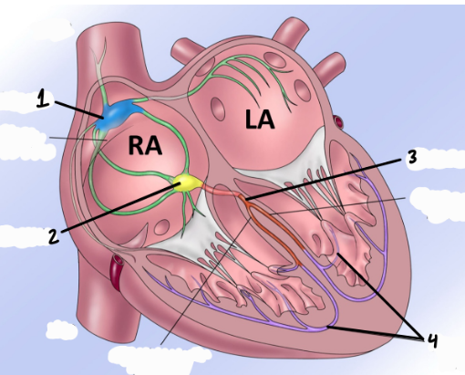
His Bundle
3


Purkinje Fibers
4
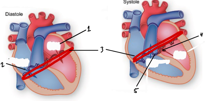
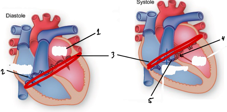
Atrioventricular Valves
1/2

Cusps
Thin flaps of tissue in the valves of heart
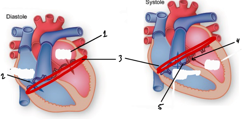
Mitral Valve
1

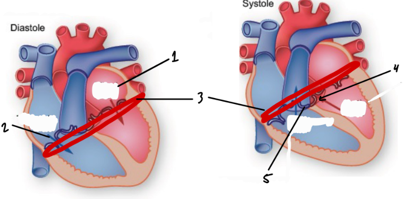
Bicuspid Valve
Between LV/LA

Tricuspid Valve
2


Fibrous Skeleton
3

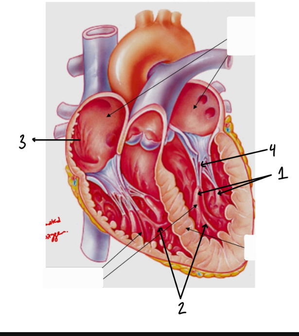
Chordae Tendoneae
4


Semilunar Valves
4/5
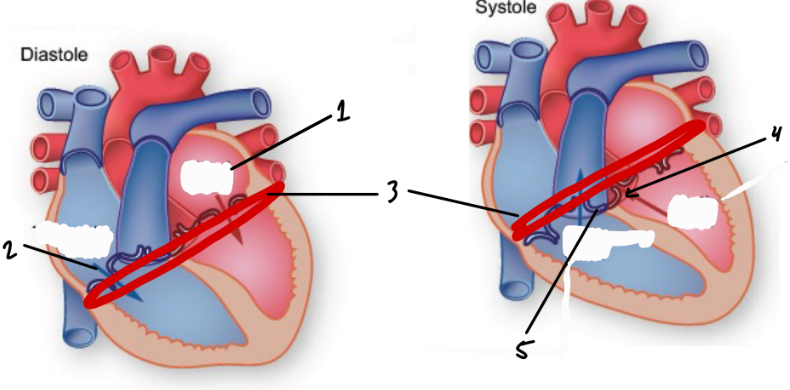

Aortic Valve
4

Pulmnary Valve
5

Superior Vena Cava - Function
Brings deoxygenated blood from superior to the diaphragm into the RA
Left Atrium - Function
Pumps oxygenated blood into the LV
Right Atrium - Function
Pumps deoxygenated blood into the RV
Aorta - Function
Brings oxygenated blood out of the LV to the rest of the body
Pulmonary Trunk - Function
Brings deoxygenated blood from RV to the lungs
Ventricular Wall - Function
3x thicker than atrial wall due to more required force
Septum - Function
Membrane between left and right sides of heart
Interarterial Septum - Function
Separates atria and provides support for pumping
Interventricular Septum - Function
Separates ventricles and provides support for pumping
Left Ventricle - Function
Pumps oxygenated blood into the aorta for the rest of the body
Right Ventricle - Function
Pumps deoxygenated blood into pulmonary trunk for the lungs
Inferior Vena Cava - Function
Brings deoxygenated blood from inferior to the diaphragm into the RA
Coronary Sinus - Function
Brings deoxygenated blood from coronoary circulation into the RA
Atrial Wall - Function
Auricles - Function
Small appendages that store a small volume of blood; expand to decrease pressure during diasole
Anterior Interventricular Sulcus - Function
Groove separating LV and RV
Left Pulmonary Veins - Function
Bring oxygenated blood from left lung to the LA
Right Pulmonary Veins - Function
Bring oxygenated blood from right lung to the LA
Posterior Interventricular Sulcus - Function
Division between LV and RV on the posterior side
Coronary Sulcus - Function
Divides atria and ventricles; carries blood vessels that supply the myocardium; AKA Atrioventricular groove
Atrioventricular Groove - Function
Divides atria and ventricles; carries blood vessels that supply the myocardium; AKA Coronary Sulcus
Myocardium - Function
Muscle of the heart
Mediastinum - Function
Space between the lungs
Pericardium - Function
Three walled sac that protects the heart; Fibrous pericardium, Serous pericardium, Pericardial cavity
Fibrous Pericardium - Function
Dense connective tissue; protects heart, anchors heart, prevents overfilling
Parietal Layer of Serous Pericardium - Function
Adheres to the internal surface of the fibrous pericardium
Pericardial Cavity - Function
Contains pericardial fluid
Pericardial Fluid - Function
Non-compressible fluid to protect heart and lubricate movement
Visceral Layer of Serous Pericardium - Function
Encapsulates the organ; AKA Epicardium
Endocardium - Function
Encapsulates the organ; AKA Visceral Layer of Serious Pericardium; Lined with endothelial cells
Myocardium
The heart muscle cells
Endocardium - Function
Lines the chambers and valves of the heart; Lined with endothelial cells
Pectinate Muscle - Function
Increase contraction power in the atria
Papillary Muscles - Function
Anchor the AV valves in the ventricles and control their opening/closing
Trabeculae Carnae
Ridges of muscle that increase contraction power in the ventricles
Sinoatrial Node - Function
Main pacemaker for the heart; Electrical impulse begins here
Atrioventricular Node - Function
Transition point for the electrical impulse to spread to the ventricles from the atria
His Bundle - Function
Transmits the electrical impulse into the ventricles; Branches into Purkinje fibers
Purkinje Fibers - Function
Spread the electrical impulse through the ventricles
Steps of Conduction - Function
SA node -> Atria -> AV node -> Bundle of His -> Purkinje fibers -> Ventricles
Vagus Nerve - Function
Parasympathetic nerve that slows the heart rate
Sympathetic NS - Function
Nervous system that accelerates the HR
Parasympathetic NS - Function
Nervous system that deccelerates the HR
Coronary Circulation - Function
Supplies heart muscle with blood; supplied by aorta; drained by coronary sinus
Right/Left Coronary Arteries - Function
Bring oxygenated blood to the right/left sides of the heart
Atrioventricular Valves - Function
Separate the ventricles and atria; Prevent backflow of blood into the atria during contraction
Cusps - Function
Flaps of endocardium that fill with blood and block valves
Mitral Valve - Function
Separates LA/LV; Has two cusps; AKA Bicuspid
Bicuspid Valve - Function
Separates LA/LV; Has two cusps; AKA Mitral Valve
Tricuspid Valve - Function
Separates RA/RV; Has three cusps
Atrialventricular Groove - Function
Dense connective tissue between atria and ventricles; Anchors valve cusps; Prevents overdilation; Blocks direct spread of electrical signals from atria to ventricles