Structures of brain (CNS) development
1/8
There's no tags or description
Looks like no tags are added yet.
Name | Mastery | Learn | Test | Matching | Spaced |
|---|
No study sessions yet.
9 Terms
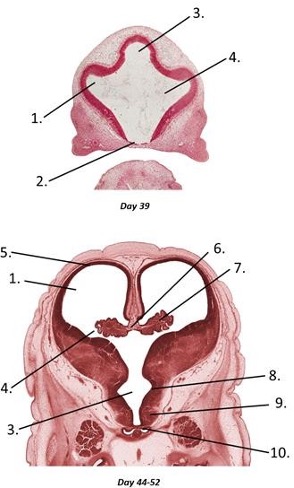
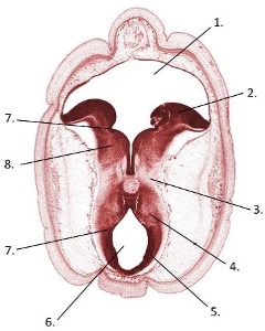
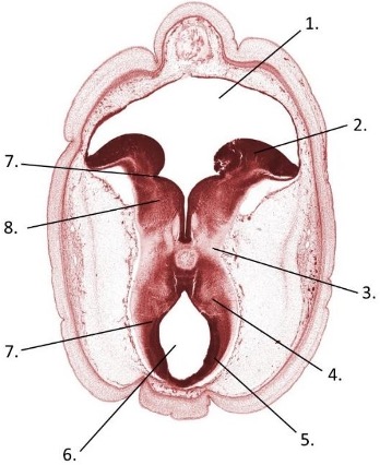
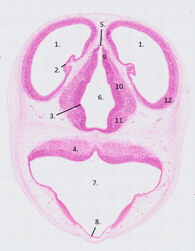
Transverse sections of the telencephalon and diencephalon of a human embryo of 39 days and between 49 and 52 days old, respectively
lateral ventricles
lamina terinalis
third ventricle
interventricular foramen
cerebral hemisphere
choroid fissure
choroid plexus
hypothalamic sulcus
hypothalamus
neurohypophysis
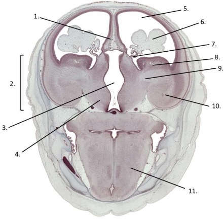
Transverse section of a 56-day old human embryo in which the telencephalon, diencephalon and myelencephalon are visible
hippocampus
corpus striatum
third ventricle
hypothalamus
lateral ventricle
chroid plexus
choroid fissure
caudate nucleus
internal capsule
lentiform nucleus
myelencephalon
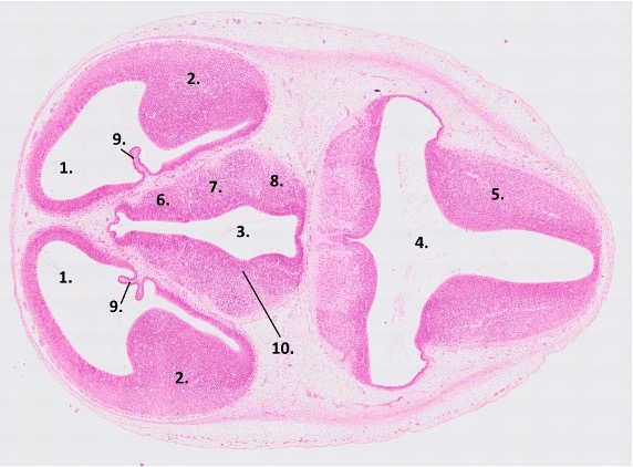
Transverse section of a 15-days old rat embryo. The myelencephalon, diencephalon and telencephalon are visible
lateral ventricle
telencephalon
third ventricle
fourth ventricle
myelencephalon
epithalamus
thalamus
hypothalamus
choroid plexus
hypothalamus plexus
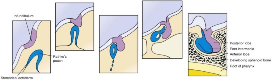
Transverse section through an 11-days old mouse embryo in which the telencephalon, diencephalon and myelencephalon are visible. The cut-out shows the two main structures of the developing pituitary in more detail
telencephalon
lateral ventricles
diencephalon
third ventricles
myelencephalon
fourth ventricle
A. infundibulum
B. Rathke’s pouch
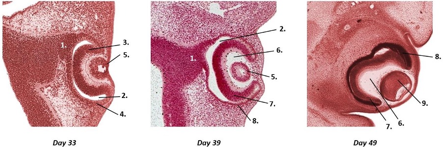
Three transverse sections of the developing human eye at days 33, 39 and 49 post-fertilization.
optic stalk
intraretinal space
inner layer of the optic cup
outer layer of the optic cup
lens vesicle
optic cup cavity
neural layer of retina
pigmented layer of retina
primary lens fibers
Transverse section of the mesencephalon and metencephalon of a human embryo of between 49 and 52 days old.
fourth ventricle
rhombic lip
cerebral preduncle
tegmentum
tectum
aqueduct of sylvius (mesocoel)
sulcus limitans
metencephalon
Transverse section of the mesencephalon and metencephalon of a human embryo of between 49 and 52 days old.
fourth ventricle
rhombic lip
cerebral peduncle
tegmentum
tectum
aqueduct of Sylvius
sulcus limitans
metencephalon
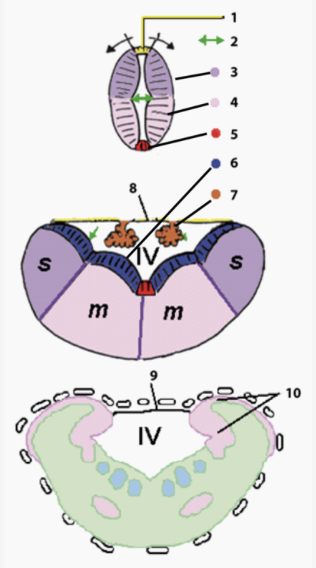
The illustrations represent three different stages in metencephalon development.
roof plate
sulcus limitans
alar plate
basal plate
floorplate
ventricular/ependymal zone
choroid plexus
ependymal roof
ependymal roof
rhombic lips
Shown is a transverse section of a 14-day old rat embryo. The telencephalon, diencephalon and myelencephalon are visible. Name the numbered structures.
lateral ventricle
choroid plexus
hypothalamic sulcus
myelencephalon
future epiphysis
third ventricle
fourth ventricle
ependymal roof
epithalamus
thalamus
hypothalamus
telencephalon