Rad Bio Exam 1
 Call Kai
 Learn
Learn Practice Test
 Spaced Repetition
 Match
Match Flashcards
 Knowt Play
1/46

There's no tags or description
Looks like no tags are added yet.
Name | Mastery | Learn | Test | Matching | Spaced | Call with Kai | Chat |
|---|
No analytics yet
Send a link to your students to track their progress
47 Terms
Acute tissue change
Typically inflammatory
Changes are the result of cells dying in the tissues within the radiation field
Two possible outcomes
Regeneration- may be complete or partial
Replacement- original cell population replaced by a different population. usually fibroblasts
Chronic tissue change
Changes manifest after healing process
ex) loss of vascular supply to a tissue such as the intestine after mucosal regeneration has occurred
Factors in radiation response
Volume of tissue irradiated
Oxygenation at the cellular level
Presence of some chemicals
Dose rate
Cellular kenetics
Cellular kinetics
Cell type
Hemopoietic (blood and lymph)
The parenchymal cells of the bone marrow and the circulating blood
Red blood cells are the most resistant cell in mammals to radiation injury
Skin and oral mucosa
Erythema w/ early and late effects at 10 gray and above
Digestive system
Early effects are mucosal depopulation
late effects are repopulation, epithelial metaplasia (loss of cunftion), scarring, and stricture (obstruction of GI tract)
Male reproductive system
Adult sperm are FPM cells - resistant
but chromosomal damage may be passed onto fetus
Germinal cells are very sensitive
don’t shoot the balls
Female reproductive system
6.25Gy to both ovaries - expect sterility
Can affect hormonal function
Decreased or lost above 25 Gy
Follicular cells are more susceptible to harm from radiation
Cardiovascular system
Can cause thrombosis and possible hemorrhage\
the heart is considered resistant but effects may be seen years later
Liver and Kidneys
Whole organ doses of 30 Gy are lethal
Central nervous system
Considered radioresistant in adults
Excitation
Electron of an atom is raised to a higher energy state but not ejected
Ionization
Energy imparted is great enough to cause an electron to be ejected
X-ray characteristics
EM radiation
No charge
Frequency * wavelength= velocity of the wave
Produced
Brenstrahlung
Characteristic
Considered a photon
Alpha particles
2 protons and 2 neutrons
2+ charge
short range cause they fat bastards
Directly ionizing
Charged particles can directly disrupt atomic structure (nucleus or electrons)
Cause damage
Indirectly ionizing (photons)
Don’t directly cause damage
Set in motion charged particles (electrons) which may cause damage
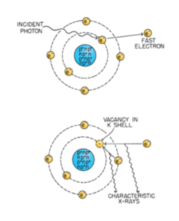
Photo-electric
< 100 KeV
Proportional to Z³
Secondary emissions
PE ejected electron
Characteristic X-rays
Auger electrons
Compton scattering
25 KeV to 25 MeV
Most fall into this category
Secondary emissions
CS photon
CS electron

Direct action of radiation
Cause damage or biological effect by direct excitation or ionization
Dominant process for high linear energy transfer (LET)
Responsible for 1/3 of damage
Indirect action of radiation
Radiation interacts with atoms and produces free radicals
Free radicals can cause excitation and ionization
Represents 2/3 of damage
Free radicals
Radiation interacts w/ water
High degree of chemical reactivity
Atom with unpaired orbital electron in outer shell
Mitosis steps
Prophase
Metaphase
Anaphase
Telophase
Prophase
DNA organizes into identifiable chromosomes
Metaphase
DNA aligns with centromeres on equatorial plate
Anaphase
DNA separates and moves to opposite ends of cell
Telophase
Cell cytoplasm divides at equatorial plate
Functional Death
Loss of a specific function
Cells that do not proliferate (nerve, muscle, secretory)
Reproductive Death
Cell can still be present
Cell can still function
Cell may be able to synthesize DNA
Cell may have 1-2 mitoses but cannot sustain indefinitely
Mitotic Death
Death when attempting to divide
Apoptosis
Programmed cell death
Destroy cell function in non-proliferating systems
100 Gy
Loss of reproductive capacity
2Gy
Plating Efficiency PE
PE = (colonies counted/cells seeded) * 100
Surviving fraction
SF = (Colonies counted/ Cells seeded) * PE
D1
Based on initial slope of survival curve
Reciprocal of initial slope
Dose required to reduce fraction of surviving cells to 37% of previous value
D0
Based on the final slope resulting from multi-event killing
Reciprocal of the final slope
Dose required to reduce fraction of surviving cells to 37% of previous value
Dq
Quasi-Threshold dose
Measure of the shoulder of the curve
Dose at which the straight portion of the survival curve extrapolated backward intercepts the Y-axis
Threshold dose: Dose below which there is no effect
Extrapolation Number
Measure of width of shoulder
If n > 10 (brads shoulder)
If n is equal to 1.5-2.0 (narrow shoulder)
Linear Quadratic Model
S = e- (alphaD-betaD²)
S= surviving fraction
alpha= component associated with non repairable component (double-strand break)
Beta= component associated with repairable component (single-strand break)
D= dose
Bystander effect
Death of (some) cells that are next to other cells who have been irradiated
Up to 30% of bystander cells may be killed
Mitotic Death
Most common type of cell death
Prodromal Radiation Syndrome
> 100 Gy
Time onset within 5-15 minutes
Symptoms may reach max at 30 min
Cerebrovascular (CNS) Syndrome
Dose:100 Gy
Death: 24-48 hours
Neurological breakdown
Cardiovascular breakdown
All organ systems are damaged
Increase of fluid in brain leading to a build-up of pressure
GI Syndrome
Dose: 5 Gy-12 Gy
Death: 3-10 days
Destruction of GI Mucosa
Bone Marrow (Hematopoietic) Syndrome
Dose: 2.5 Gy- 5 Gy
Death: Weeks to months
Effect on blood forming organs
Bone Marrow Syndrome
Dose: 250 Gy to 500 Gy
Death: very lethal
Mitotically active precursor cells are sterilized
Blood count decreased
Latent period of 1 to 3 weeks after prodromal syndrome