Exocrine Pancreas and Peritoneum: Systemic Path Lab
1/15
Earn XP
Description and Tags
Year 2 Semester 1
Name | Mastery | Learn | Test | Matching | Spaced |
|---|
No study sessions yet.
16 Terms
2 possible morphologic diagnosis
Exocrine pancreatic hypoplasia
Exocrine pancreatic atrophy
Tissues from a 6-month-old dog. Histologic examination reveals small pancreatic lobules that are infiltrated by moderate numbers of lymphocytes. Acinar structures are reduced in number, and acinar cells lack zymogen granules. Scattered cells contain lipofuscin. Name of condition? Predisposed breed of dog?
Exocrine pancreatic atrophy (juvenile pancreatic atrophy)
German Shepherd (rough collie, Eurasian dog, Greyhound)
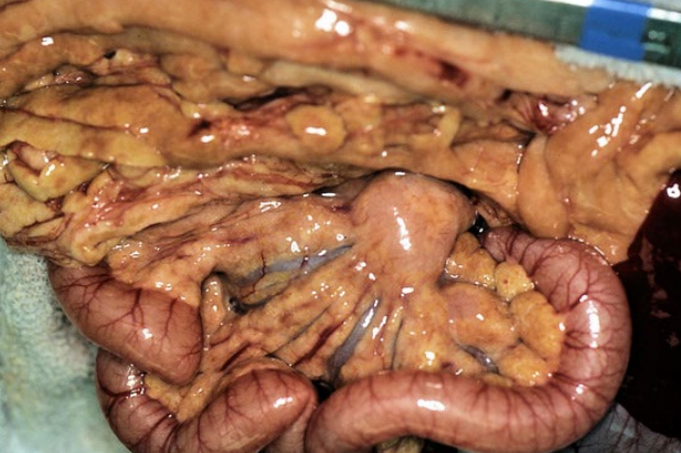
Pancreas from a dog. Clinical history of having a high-fat meal 3 days ago (ate pork scraps). Presented with anorexia, vomiting, depression, and a fever. Abdominal ultrasound revealed a lack of detail in the cranial abdomen (edema and/or local peritonitis); no mass is observed. Morphological diagnosis?
Pancreas: severe, diffuse, acute, pancreatic necrosis (necrotizing pancreatitis)
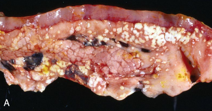
Tissues from a 5-year-old, spayed female dog. Clinical history: The dog ate a pound of butter and an entire rhubarb pie. Morphological diagnosis? Two possible long-term sequelae (if chronic and recurrent)
Pancreas: moderate, multifocal, acute, pancreatic necrosis with extensive peripancreatic fat necrosis (and saponification of fat)
Exocrine pancreatic insufficiency
Diabetes Mellitus
Pancreas from a 5-yearold, spayed female, Domestic Short Hair cat. Clinical history: The cat presented with a history of anorexia, vomiting, and diarrhea. AFAST (an ultrasound technique) revealed peripancreatic free fluid. Spec fPLI yielded a positive test result (feline pancreatic lipase immunoreactivity >5.4 g/L). Morphological diagnosis?
Severe, diffuse, acute, pancreatic necrosis (necrotizing pancreatitis)
Pancreas from an 8-year-old, spayed female, mixed breed cat. The cat has diabetes mellitus which you have been managing well for the past year. She presented with a history of hyporexia progressing to anorexia, lethargy, vomiting, and diarrhea. Morphological diagnosis?
Mild, diffuse, chronic, lymphoplasmacytic interstitial pancreatitis, with exocrine pancreatic nodular hyperplasia
Pancreas from an 8-year-old, spayed female, mixed breed cat. The cat has diabetes mellitus which you have been managing well for the past year. She presented with a history of hyporexia progressing to anorexia, lethargy, vomiting, and diarrhea. What conditions might this cat have?
Inflammatory bowel disease
lymphocytic cholangitis
exocrine pancreatic insufficiency
hepatic lipidosis
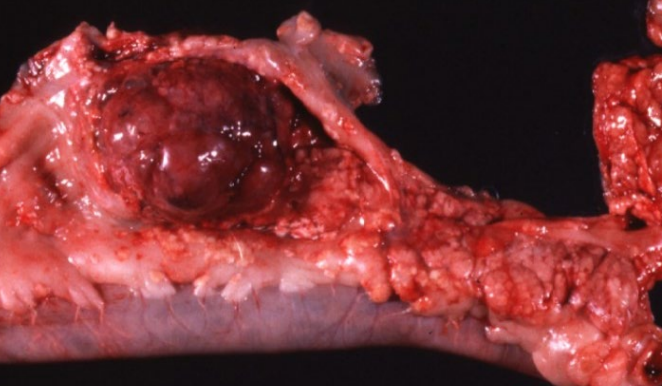
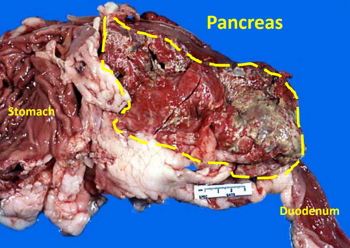
Tissue from a dog. Presented with anorexia, vomiting, depression, and a fever. Abdominal ultrasound revealed a cranial abdominal mass. Morphologic diagnoses for the exocrine pancreatic mass? What is the expected biological behaviour of this mass?
Exocrine pancreatic carcinoma
Malignant tumor with metastasis often present at diagnosis
Tissue from a 3-year-old, MC, DSH cat. Clinical history: The cat has been on an all-tuna diet for the past 6 months. Abdominal palpation revealed that the subcutaneous and visceral fat stores felt firmer and more nodular than expected. Morphologic diagnosis? Name of condition?
Severe, chronic, diffuse, granulomatous mesenteric steatitis (+ granulmotous panniculitis indicated in the clinical history)
Nutritional pansteatitis (= Yellow Fat Disease)

Tissue from a 3-year-old, MC, DSH cat. Clinical history: The cat has been on an all-tuna diet for the past 6 months. Abdominal palpation revealed that the subcutaneous and visceral fat stores felt firmer and more nodular than expected. what kind of diet would you expect this pateint to be on?
unusual diet that is high in unsaturated fatty acids, low in vitamin E or both. Classic history: all fish diets, such as all tuna diet
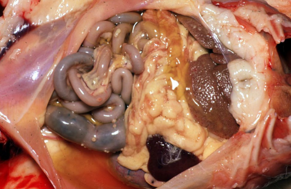
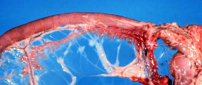
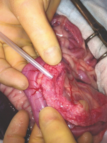
Abdomen as a whole (peritoneal contents) from a 3-month-old, male, purebred Himalayan cat. Clinical history: The cat was recently rehomed. Ocular exam revealed anterior uveitis. The cat has a potbellied appearance with a palpable fluid wave (intra-abdominal fluid). Morph? Name of disease? Cause?
Morph - Severe, multifocal, chronic, pyogranulomatous peritonitis
Disease - Feline infectious peritonitis (FIP)
Cause - Feline infectious peritonitis virus (FIPv), a mutated feline enteric coronavirus
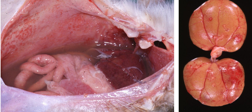
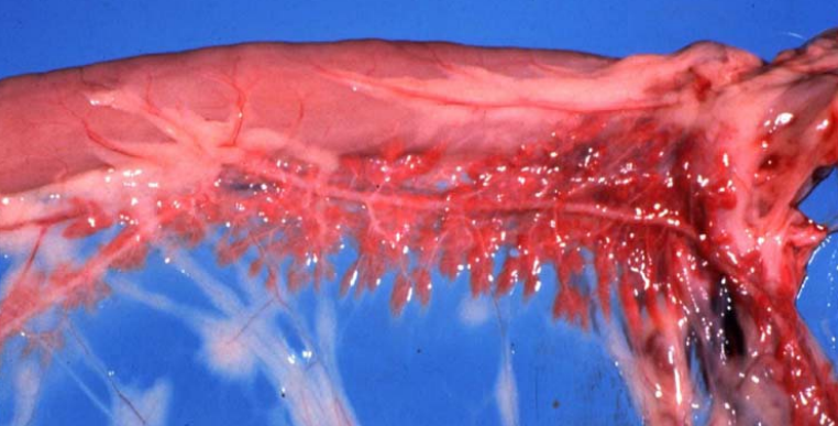
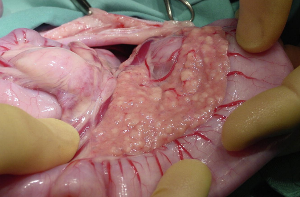
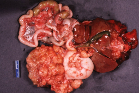
Abdomen as a whole (peritoneal contents and kidneys) from a 6-month-old, male, purebred Bengal cat. Clinical history: The cat was in a breeding facility. Clinical signs included failure to gain weight, lethargy, pot belly with a fluid wave, and intermittent diarrhea. Morph? Name of disease? Cause?
Morph - severe, multifocal, chronic, pyogranulomatous peritonitis (+ nephritis)
Disease - Feline infectious peritonitis (FIP)
Cause - Feline infectious peritonitis virus (FIPv), [mutated feline enteric coronavirus]
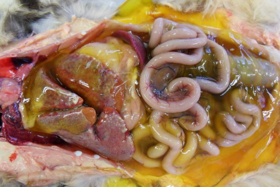
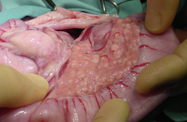
Abdomen as a whole from a 9-month-old, male, purebred Siamese cat. Clinical history: The cat was smaller than normal (“runty”). Serum biochemistry revealed mild anemia, lymphopenia, hyperbilirubinemia, and hypergammaglobulinemia with a high albumin to globulin ration (A:G ratio). Morph? Name of disease? Cause?
Severe, multifocal, chronic, pyogranulomatous peritonitis
Feline Infectious Peritonitis (FIP)
Feline Infectious Peritonitis virus (FIPv) [mutated feline enteric coronavirus]
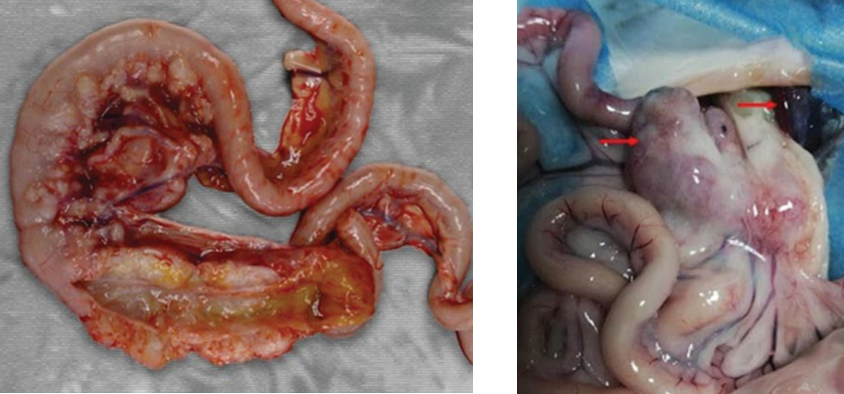
Tissues from a 9-month-old, male, purebred Siamese cat with severe ascites. Necropsy findings: The small intestinal walls are multifocally thickened by dense, white to cream, plaques and nodules and the mesenteric lymph nodes are markedly enlarged. Two most likely differential diagnoses for these findings? Most likely diagnosis?
Feline infectious peritonitis and intestinal lymphosarcoma
FIP
Intra-abdominal contents from a 13-year-old FS, DSH cat with a history of chronic weight loss. Digital imaging revealed a napkin-ring like area of ileal wall thickening (beside the ileocecal junction) with luminal stenosis. A full-thickness biopsy of area of thickening of the ileal wall revealed transmural infiltration of the ileum by neoplastic epithelial cells embedded in fibrous stroma (desmoplasia). morph?
Intestinal adenocarcinoma with carcinomatosis