Week #2 Readings - 11.1, 11.4, 11.5 (MITOSIS and CELL CYCLE)
1/39
There's no tags or description
Looks like no tags are added yet.
Name | Mastery | Learn | Test | Matching | Spaced | Call with Kai |
|---|
No study sessions yet.
40 Terms
Cell Division
Cell division is the process by which a single cell produces two daughter cells.
Must be large enough to divide in two and contribute sufficient nuclear and cytoplasmic components to each daughter cell
Key cellular components are duplicated -DNA
What does Cell Division Occur For?
Growth (Single to Multi)
Cell Replacement
Healing (Ex. Wound → New skin cells)
Reproduction (Asexual and Sexual)
Cell Cycle
The series of steps that take place as a eukaryotic cell
Grows
Replicates its DNA
Divides to produce daughter cells
Life cycle of a cell
Prokaryotic vs Eukaryotic Cell Division

Chromosomes
Packaged genetic material
Consist of a single DNA molecule and associated proteins
Prokaryotic → Organized as a single, relatively small, circular chromosome
Eukaryotic → Larger and is organized into one or more linear chromosomes
Binary Fission
The process by which cells of bacteria and archaea divide to form two daughter cells
A cell replicates its DNA, increases in size, and divides into two daughter cells
Each daughter cell receives one copy of the replicated parental DNA.
Red - Origin of Replication
Important gene - FtsZ encodes a protein that forms a ring at the site of constriction where the new cell wall forms between the two daughter cells

Eukaryotic Cell Cycle
More complicated
Two distinct phases
M Phase → Divides into two daughter cells
First divide the nucleus by mitosis (chromosomes separated into two nuclei)
Then divide the cytoplasm into two daughter cells by cytokinesis
Interphase occurs between two
successive M phases
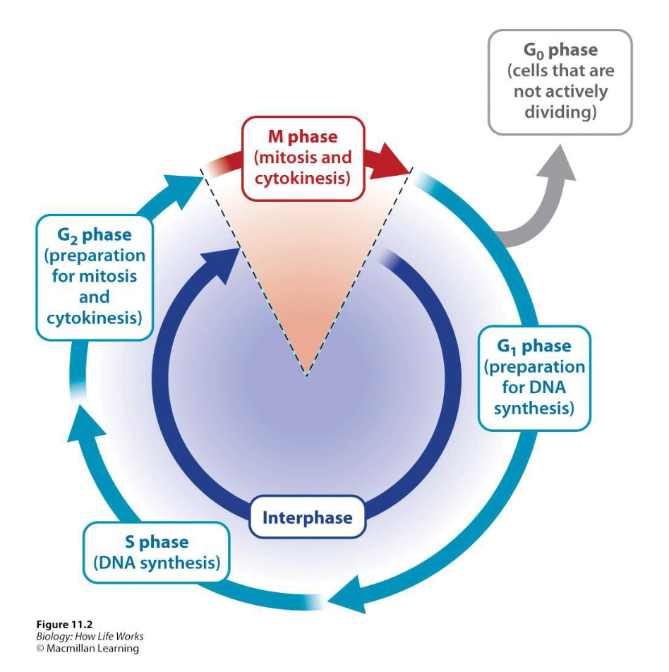
What occurs during Interphase?
Preparations for division.
DNA replication and cell growth.
DNA in the nucleus first replicates so that each daughter cell receives a copy of the genetic material.
Cell then increases in size so that each daughter cell receives enough of cytoplasmic and membrane components to allow it to survive on its own.
What are the Three (4?) Phases of Interphase?
G1 Phase (Gap 1) → Cell growth and expression of regulatory proteins
Ex…Kinase, activate enzymes that synthesize DNA
S Phase (Synthesis)→ Replication of DNA
G2 Phase (Gap 2) → Size and protein content of the cell increase in preparation for M-phase
S phase does not immediately precede or follow mitosis but is separated from it by two gap phases
G0 Phase (“Fourth Phase”)
The gap phase of the cell cycle in which cells pause in the cell cycle between M phase and S phase; it may last for periods ranging from days to more than a year.
These cells are said to be quiescent.
Ex. A neuron, axon, and dendrites would not re-enter the cell cycle
Ex. Liver remains in G0 for a year than re-enters
Still perform their specialized functions
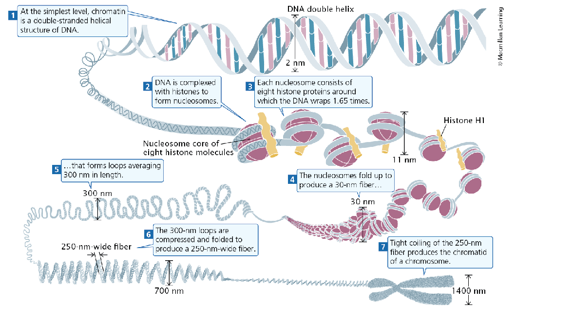
How is DNA stored?
Length of DNA 1-2 meters → Heavily condensed!
In eukaryotic cells, DNA is organized with histones and other proteins into chromatin, which can be looped and packaged to
form chromosomesFurther condensed during cell division so it does not tangle as it separates into daughter cells
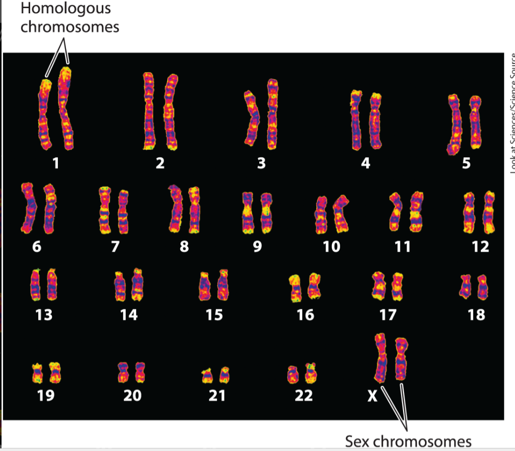
Karyotype
A standard arrangement of chromosomes, showing the number and shapes of the chromosomes representative of a species.
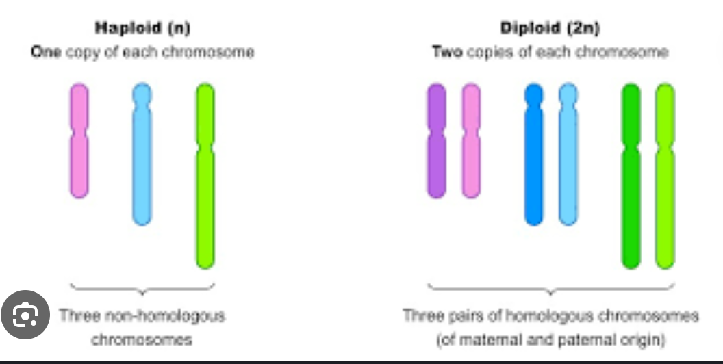
Most cells of the human body has 46 chromosomes (23 pairs).
There are 22 pairs of homologous chromosomes, 1 pair of sex
chromosomes.Homologous chromosomes carry the same set of genes + match in size and appearance
One from the mother and one from the father.
The sex chromosomes are X and Y
Two X chromosomes is a female
An X and a Y chromosome is a male.
Sets of Chromosomes
The number of complete sets of chromosomes in a cell is known as its ploidy.
One complete set of chromosomes is haploid (n)
Two complete sets of chromosomes is diploid (2n)
Plants can have polyploids! (4n)
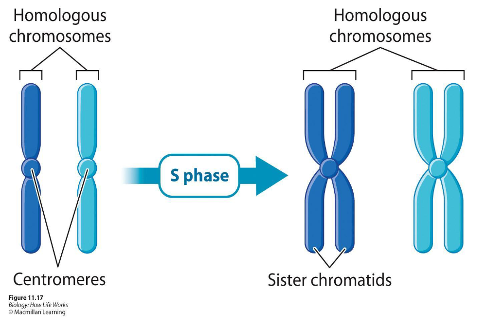
S Phase and Sister Chromatids
Duplication occurs during S phase
Every chromosome in the parent cell must be duplicated so that each daughter cell receives a full set of chromosomes
Sister chromatids – The two identical copies that are made after duplication
Do not separate
Are held together at the
centromere.Cell still contains 46 chromosomes, count each pair as 1!
Stages of Mitosis
Is divided into 5 phases
Prophase
Prometaphase
Metaphase
Anaphase
Telophase
These 5 stages are followed by Cytokinesis
Each of the five stages of mitosis can be determined using a microscope depending on the position of the chromosomes.
Please Pass Me A Taco, Chef
Prophase
Characterized by the appearance of visible chromosomes
Chromosome Condensation → The progressive coiling of the chromatin fiber, an active, energy-consuming process requiring the participation of several types of proteins.
Chromosomes change from long, thin, threadlike structures to short, dense forms (visible under microscope)
Centrosomes radiate microtubules
Migrate to opposite poles
What happens to the cytoskeleton in this phase?
Microtubules assemble into the mitotic spindle, a structure that pulls the chromosomes to opposite ends of the dividing cell.
These spindles radiate from the centrosome, a compact structure that is the microtubule organizing center for animal cells.
These centrosomes define the future daughter cell poles and organize the microtubules that guide chromosome movement.
Prometaphase
Nuclear envelope breaks down
The microtubules of the mitotic spindle attach to chromosomes
Attach to the chromosomes at their centromeres
Kinetochores → Protein complexes that are sites for spindle attachment
Ensures that each sister chromatid travels to an opposite pole
Metaphase
Once each chromosome is attached to the mitotic spindles from both poles of the cell, the microtubules of the mitotic spindle lengthen or shorten to move the chromosomes to the middle of the cell.
There the chromosomes are lined up in a single plane that is roughly equidistant from both poles of the cell.
Anaphase
The centromere holding a pair of sister chromatids together splits, allowing the two sister chromatids to separate from each other.
After separation, each chromatid is considered to be a full-fledged chromosome.
Anaphase ensures that one chromatid from each pair of sister chromatids goes to opposite poles of the cell
Telophase
A complete set of chromosomes is now at each pole of the cell.
The microtubules break down completely and the nuclear envelope re-forms.
Chromosomes decondense, marking the end of telophase and mitosis.
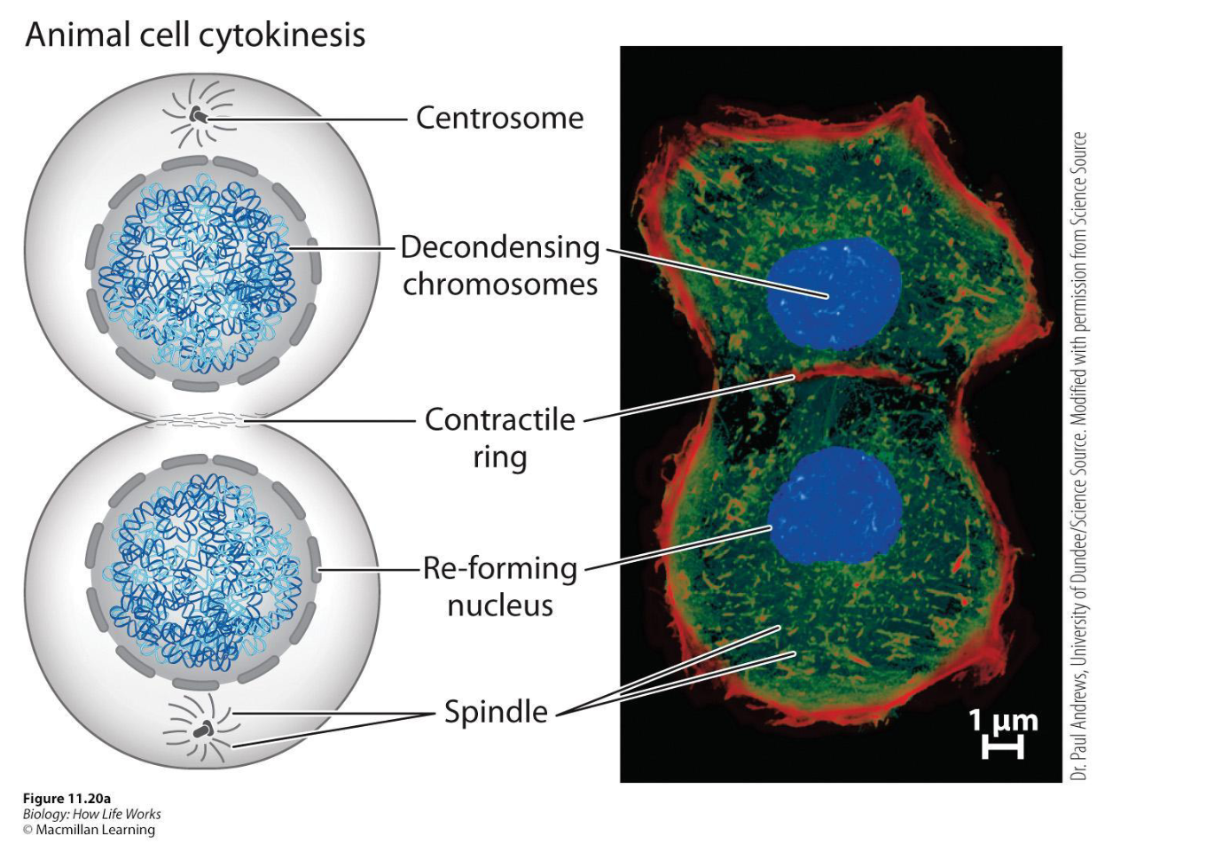
Cytokinesis: In Animal Cells
As mitosis ends, cytokinesis begins when actin filaments form a contractile ring at the equator of the cell, perpendicular to the spindle
Motor proteins (FtsZ) cause the ring to constrict like a drawstring, pinching the cytoplasm and dividing the parent cell into two daughter cells, each with its own nucleus, which then enter G₁ phase.
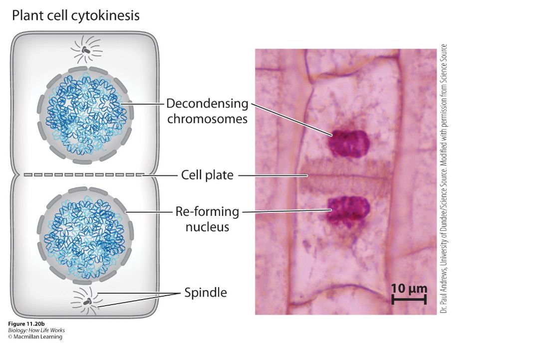
Cytokinesis: In Plant Cells
Since plant cells have a cell wall, the cell divides in two by constructing a new cell wall.
During telophase, a phragmoplast of microtubules guides vesicles to the cell’s center, where they fuse to form a cell plate that grows outward and joins the original cell wall, separating the cell into two daughter cells.
How do cells know when to divide?
In response to external signals
When internal requirements are met - does not divide unless ready
DNA replication is complete
The cell is large enough to divide into two
Uncontrolled division is dangerous and can lead to cancer
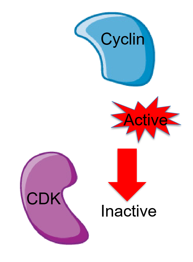
Regulation of the Cell Cycle
Progression through the cell cycle is
controlled by cyclinsActivate kinases (cyclin-dependent kinases (CDKs), which target proteins that promote cell division
Are always present within the cell but are active only when bound to the appropriate cyclin.
KINASE- Enzymes that phosphorylate other
molecules
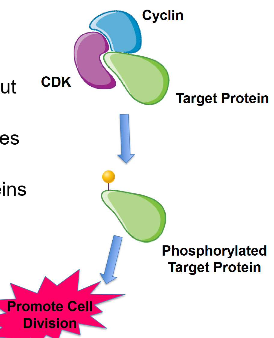
CDK/Cyclin: Regulation of the Cell Cycle
Cyclins appear and disappear throughout the cell cycle.
Cyclins activate cyclin-dependent kinases (CDKs).
Active CDKs phosphorylate target proteins
The phosphorylated target proteins
promote cell division and progression through the stages of the cell cycle.
Cyclin-CDK complexes and Different stages of the cell cycle
There are 3 specific time points that are regulated.
G1 → S
S → G2
G2 → M
Different cyclins and CDKs act at these specific steps of the cell cycle.
The G1/S cyclin–CDK complex
Prepares the cell for S phase (Cyclin D and E rise)
Promotes the expression of histone proteins needed to package DNA
Activate transcription factors that lead to the expression and activation of DNA polymerase and other enzymes
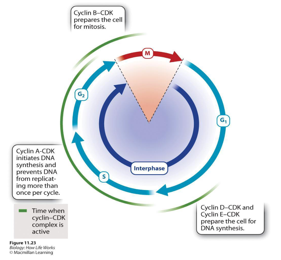
The S cyclin–CDK complex
Initiation of DNA synthesis (Cyclin A rise and activate specific CDKs)
Inhibit the activity of DNA synthesis enzymes once replication is over (prevent replication proteins from reassembling)

The M cyclin–CDK complex
Cyclin B binds to CDKs that activate enzymes that initiate multiple events associated with mitosis
The breakdown of the nuclear
envelope during prophaseThe formation of the mitotic spindle

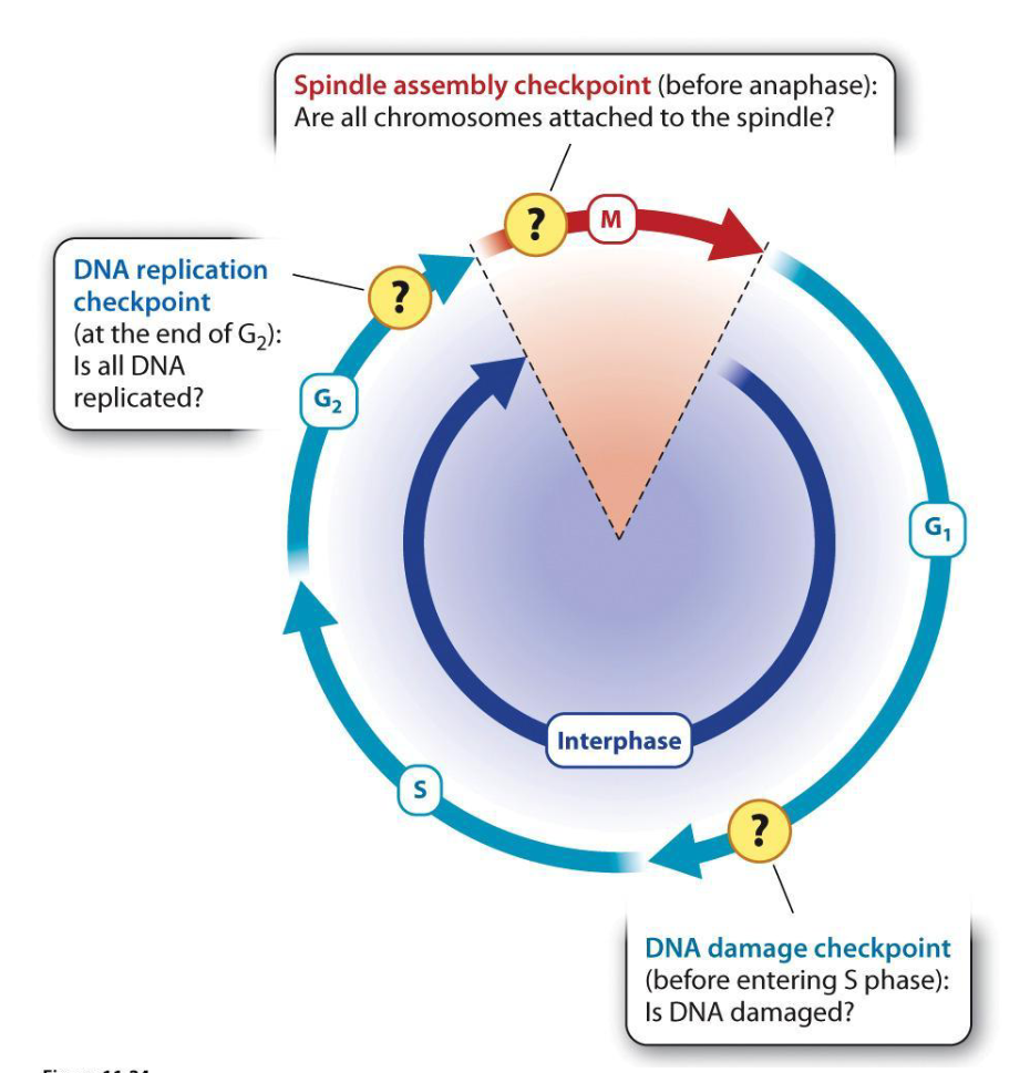
Cell Cycle Checkpoints
Checkpoints in the cell cycle ensure
the cell is prepared at different stages to proceed to the next stage.Mechanisms that block the cyclin–CDK activity required for the next step, pausing the cell cycle until preparations are complete or the damage is repaired
What are the Three Major Checkpoints?
1. DNA damage checkpoint
Presence of damaged DNA arrests the cell at the end of G1 before DNA synthesis
Ex. Damaged DNA (radiation) → double-stranded breaks → checkpoint delays progression until repaired
2. DNA replication checkpoint
Presence of unreplicated DNA arrests the cell at the end of before the cell enters mitosis
3. Spindle assembly checkpoint
Abnormalities in chromosome attachment to the spindle arrest the cell in early mitosis
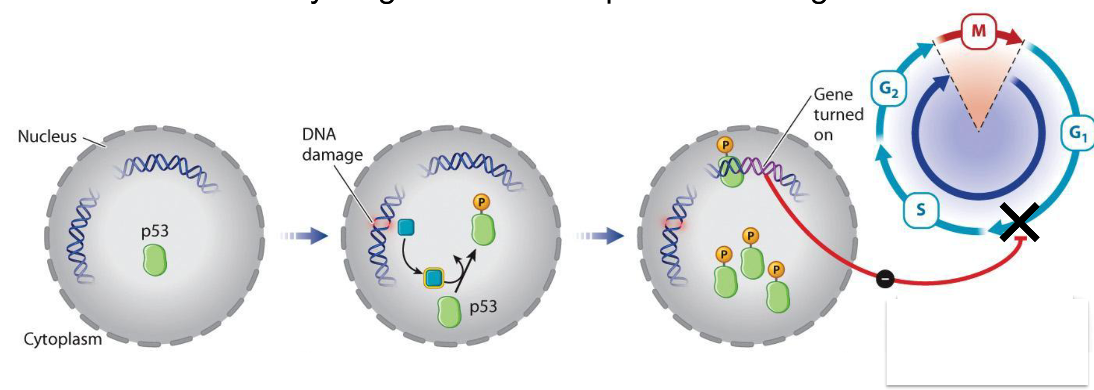
DNA Damage Checkpoint: p53 Regulation
When DNA is damaged by radiation, a protein kinase is activated that phosphorylates a protein called p53
p53 activates the synthesis of
proteins that block G1/S Cyclin-CDK complexp53 arrests the cell at the G 1 /S transition → time to repair DNA
“Guardian of the genome”
How does Apoptosis Occur?
When DNA is damaged, phosphorylated p53 increases transcription of Bax and represses Bcl-2.
This shifts the balance from Bax/Bcl-2 dimers to Bax/Bax complexes
Trigger apoptosis, a controlled and orderly form of programmed cell death.
Responsible for eliminating cells that are unneeded, damaged, or harmful

Cancer
Characterized by uncontrolled cell division
Occurs when mechanisms that promote cell division are activated inappropriately or the usual checks on cell division are lost
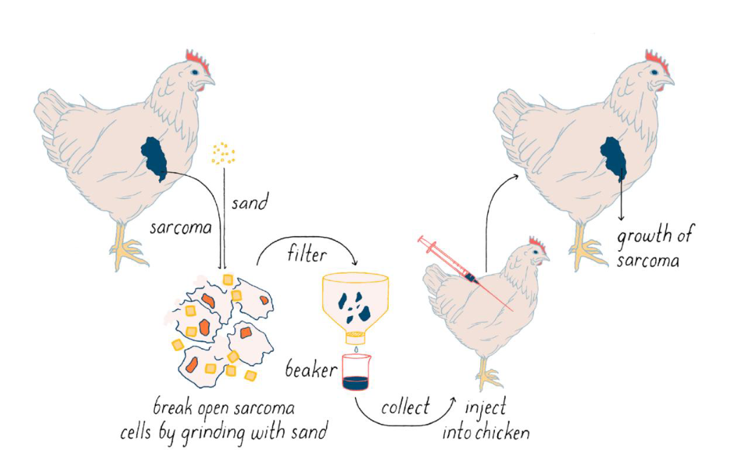
Peyton Rous and His Research
Studied cancers called sarcomas in chickens
Discovered oncogenes; cancer-causing genes
First discovered in viruses → discovery that they weren’t just in viruses
Ex. Rous Sarcoma Virus has a gene that promotes uncontrolled cell division
Encodes an overactive protein kinase that functions as a signal
This gene is an example of an oncogene
Proto-Oncogenes
Normal genes important in cell division that have the potential to become cancerous if mutated
Various proteins that performs roles in signalling cascades leading to cell division can be a product of a protooncogene
Growth factors
Cell surface receptors
G proteins
Protein kinases
If mutated, any of these can become an oncogene
Environmental agents (cigarate smoke) can damage and mutate DNA
p53 and Cancer
Cycle checkpoints that halt the cell cycle until the cell is ready to divide → p53 (stops due to DNA damage)
When the p53 protein is mutated or its function is inhibited, the cell can divide before the DNA damage is repaired.
The p53 protein is mutated in many types of human cancer, highlighting its critical role in regulating the cell cycle.
Tumor Suppressors and Proto-Oncogenes
Tumor suppressors: genes that encode proteins whose normal activities inhibit cell division
Ex. p53
Act in opposition to proto-oncogenes
Proto-oncogenes must be turned on and tumor suppressors must be turned off for a cell to divide
Two counterbalancing systems that must be in agreement before the cell divides
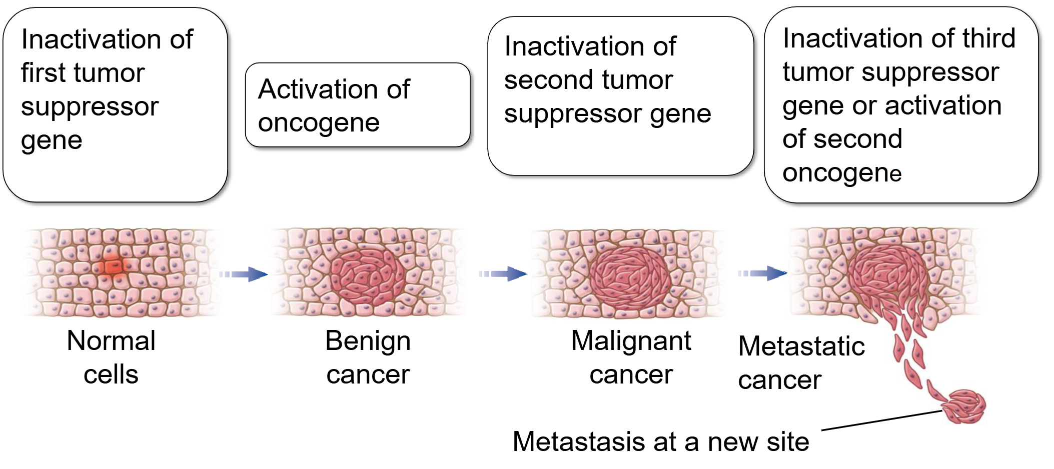
Multiple Mutation Model for Cancer Development
Require the accumulation of mutations in multiple genes (noy just one)
When several different cell cycle regulators fail, leading to both the overactivation of oncogenes and the loss of tumor suppressor activity, cancer will likely develop
Benign: relatively slow growing and does not invade the surrounding tissue
Malignant: grows rapidly and invades surrounding tissues
Ex. Colon Cancer → Tumor cells contain at least one overactive oncogene and several inactive tumor suppressor genes
A cancer cell is one that no longer plays by the “rules” of a stable cellular community.