Chapter 4: Form and Function
1/58
Earn XP
Description and Tags
Name | Mastery | Learn | Test | Matching | Spaced | Call with Kai |
|---|
No study sessions yet.
59 Terms
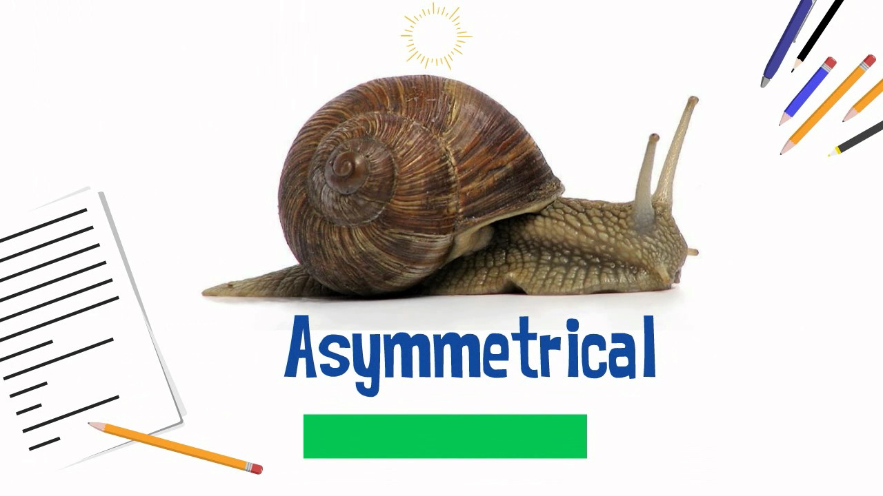
Asymmetrical Animals
Animals that have no symmetry and therefore no specific form, such as sponges.
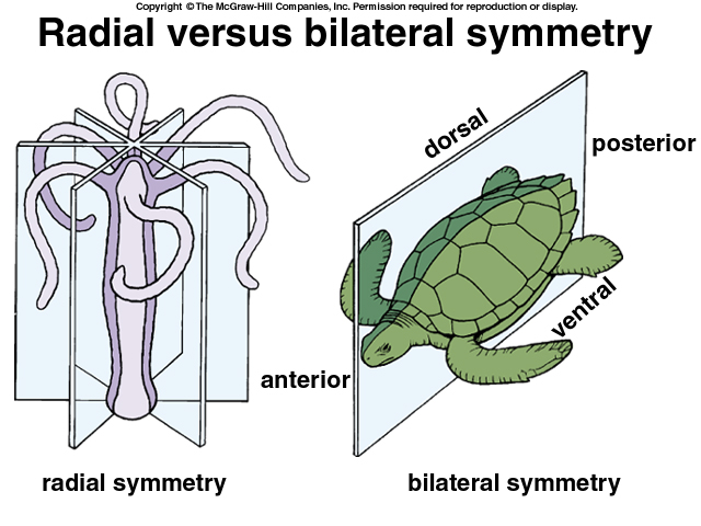
Symmetrical Animals
Animals that can be classified into two forms based on symmetry: radial and bilateral.
Example: Humans have bilateral symmetry
Sagittal Plane
A body plane that divides the body into right and left portions.
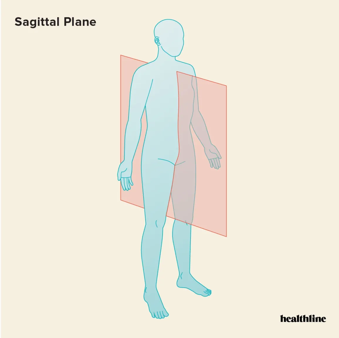
Midsagittal Plane
A body plane that divides the body exactly in the middle into equal right and left halves.
Parasagittal Plane
A body plane that divides the body into left and right portions, but not exactly in the middle.
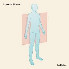
Frontal (Coronal) Plane
A body plane that separates the front from the back.
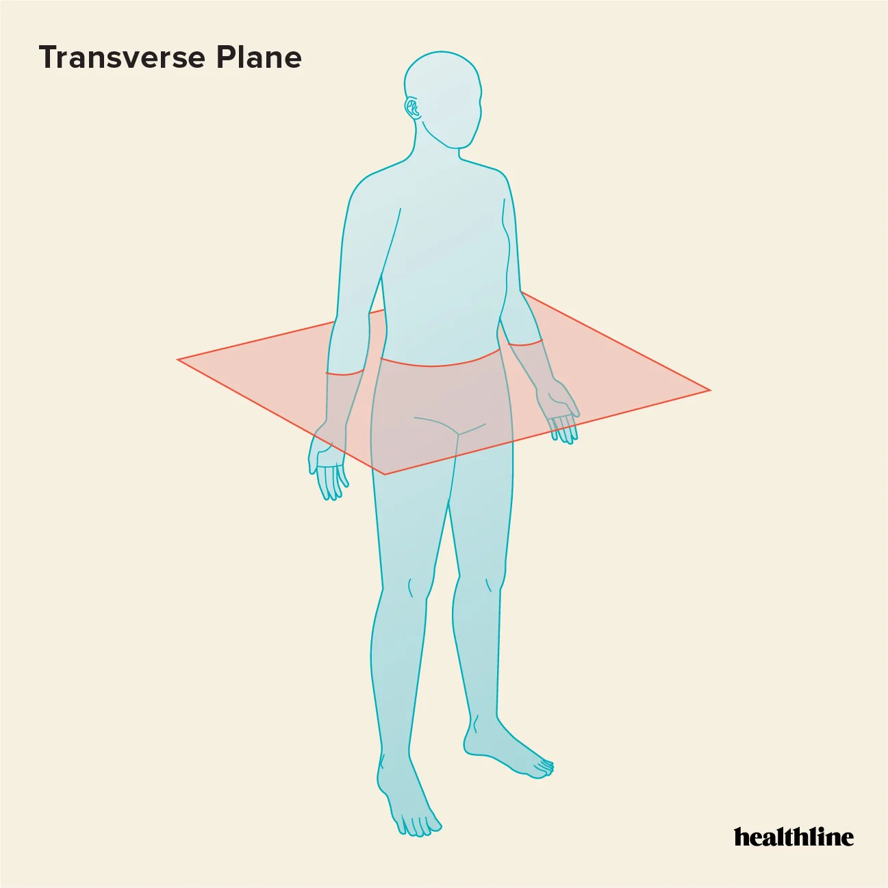
Transverse (Horizontal) Plane
A body plane that divides the body into upper and lower portions, also known as a cross section.
Dorsal Cavity
The cavity located at the back of the body.
Includes the cranial, spinal, and pelvic cavity.
Ventral Cavity
The cavity located at the front of the body.
Includes the thoracic, abdominal, and abdominopelvic cavity.
Tissue
A group of cells with similar functions.
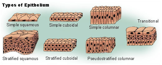
Epithelial Tissue
Tissue that covers the body and lines cavities, classified by shape and layers.
Shape: Squamous (flat), cuboidal, columnar
Layers: simple, stratified (more than 1 layer), or pseudostratified (appear to be layered but only consist of 1)
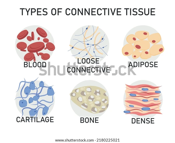
Connective Tissue
Tissue that holds other tissues together, composed of cells and a matrix.
Types: Fibrous (loose or dense), supportive (cartilage or bone), and fluid (blood or lymph)
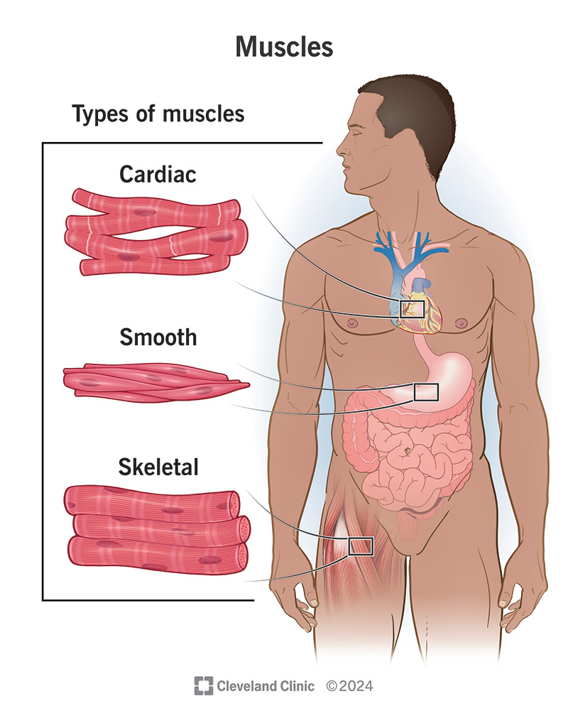
Muscle Tissue
Tissue that can contract and shorten to provide movement, with three types: skeletal, smooth, and cardiac.
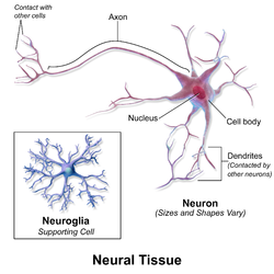
Nervous Tissue
Excitable tissue found in neurons (functional cells) and neuroglia (supportive cells).
Homeostasis
The process by which an organism maintains internal stability while adjusting to external conditions.
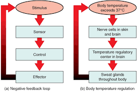
Negative Feedback Loops
Mechanisms that limit stimuli to maintain homeostasis, such as sweating.
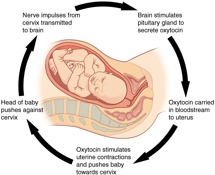
Positive Feedback Loops
Mechanisms that enhance stimuli, pushing the organism further out of homeostasis, such as during childbirth.
Endotherms
Organisms that maintain a constant body temperature, often referred to as warm-blooded.
Have a higher metabolic rate
Example: Humans are endotherms
Ectotherms
Organisms that change their temperature based on the environment, often referred to as cold-blooded.
Example: Reptiles are ectotherms
Endoskeleton
A skeletal system consisting of hard, mineralized structures located within soft tissue, as seen in humans.
The human body as 206 bones.
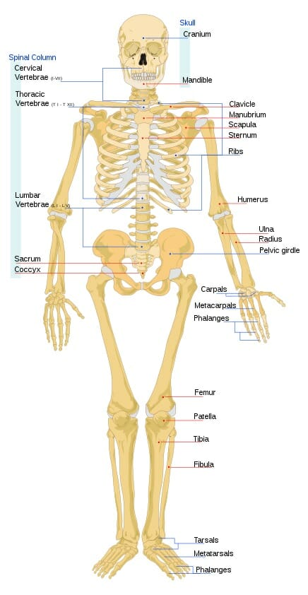
Axial Skeleton
The part of the skeletal system that includes the skull, vertebral column, rib cage, and sternum.
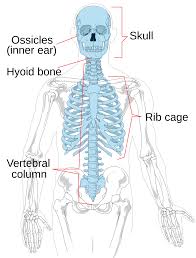
Appendicular Skeleton
The part of the skeletal system that includes the shoulder, upper limb, pelvis, and lower limb.

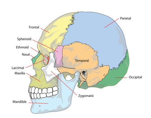
Skull
The bony structure that supports the face and protects the brain, consisting of 22 bones.
Cranial Bones: Frontal, parietal temporal, occipital, sphenoid, and the ethmoid bone (8)
Facial Bones: Nasal, maxillary, zygomatic, palatine, vomer, lacrimal, inferior nasal conchae, and the mandible (14)
Vertebral Column
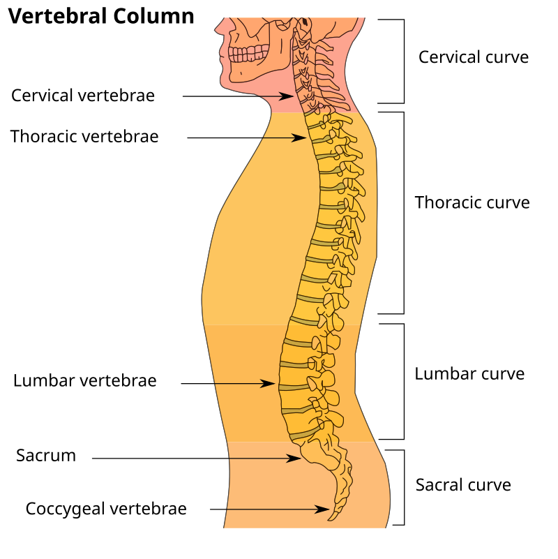
An S-shaped column composed of 33 vertebrae that houses and protects the spinal cord.
Divided into 7 cervical vertebrae, 12 thoracic, vertebrae, and 5 lumbar vertebrae
Intervertebral Discs
These discs lie between adjacent vertebral bodies from the second cervical vertebra to the sacrum.
Each disk is part of a joint that allows for some movement of the spine and acts as a cushion to absorb shocks from movement such as walking and running.
Thoracic Cage
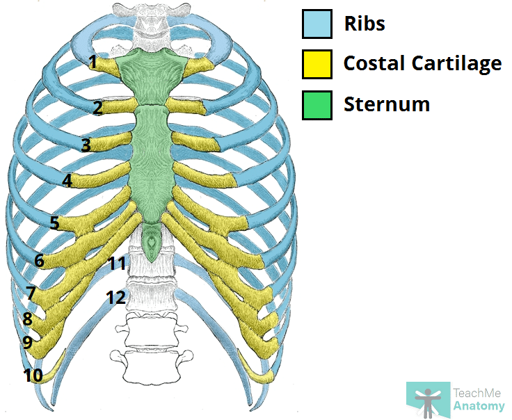
Also known as the ribcage, it consists of ribs, sternum, and thoracic vertebrae
Protects the heart and lungs.
Provides support for the shoulder girdles and upper limbs.
Serves as the attachment point for the diaphragm, muscles of the back, chest, neck, and shoulders
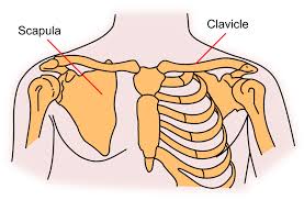
Pectoral Girdle
The structure that connects the upper limbs to the axial skeleton, consisting of the clavicle (collarbone) and scapula (shoulder blades).
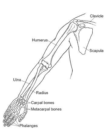
Upper Limb
Contains 30 bones in three regions: the arm (shoulder to elbow), the forearm (ulna and radius), the wrist (carpals), and hand (metacarpals and phalanges).
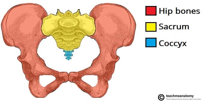
Pelvic Girdle
The structure that attaches the lower limbs to the axial skeleton, responsible for weight-bearing and locomotion.
Attached to the axial skeleton by strong ligaments.
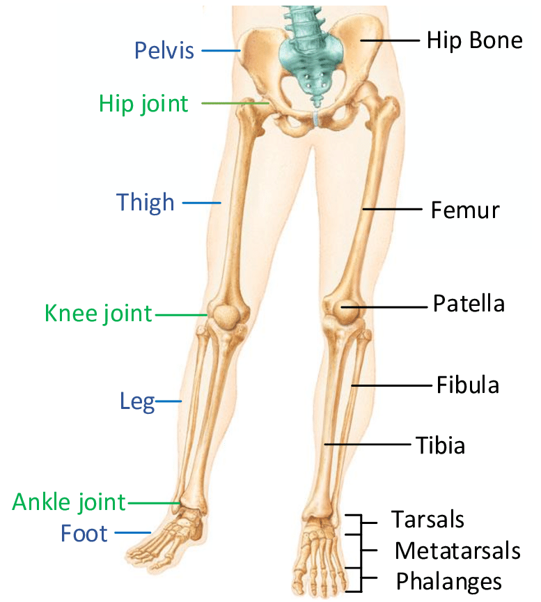
Lower Limb
Consist of the thigh, leg, and foot.
The bones of the lower limbs are thicker and stronger than the bones of the upper limbs because of the need to support the entire body weight.
Bone (Osseous Tissue)
A solid connective tissue that makes up the endoskeleton.
It contains specialized cells and a matrix of mineral salts (hydroxyapatite) and collagen fibers.
Calcification
The process of deposition of mineral salts on the collagen fiber matrix that crystallizes and hardens tissue.
Only occurs in the presence of collagen fibers.
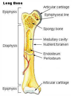
Long Bones
Bones that are longer than they are wide, such as the femur and tibia.
Diaphysis - contains bone marrow in a medullary cavity.
Epiphyses - covered with articular cartilage and are filled with red bone marrow, which produces blood cells.
Short (Cuboidal) Bones
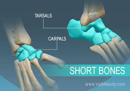
These bones are the same width and length, giving them a cube-like shape.
Example: The bones of the wrist (carpals) and ankle (tarsals) are short bones.
Flat Bones
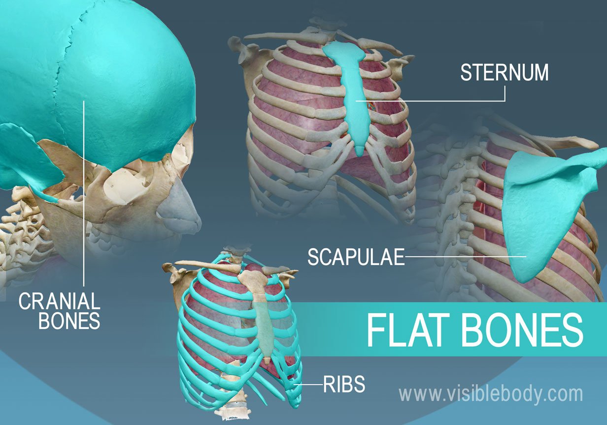
These bones are thin and relatively broad that are found where extensive protection of organs is required or where broad surfaces of muscle attachment are required.
Example: The sternum (breast bone), ribs, scapulae (shoulder blades), and the roof of the skull are flat bones.
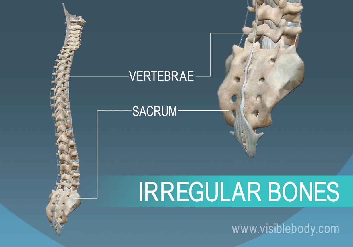
Irregular bones
These bones have complex shapes; they may have short, flat, notched, or ridged surfaces.
Example: The vertebrae, hip bones, and several skull bones.
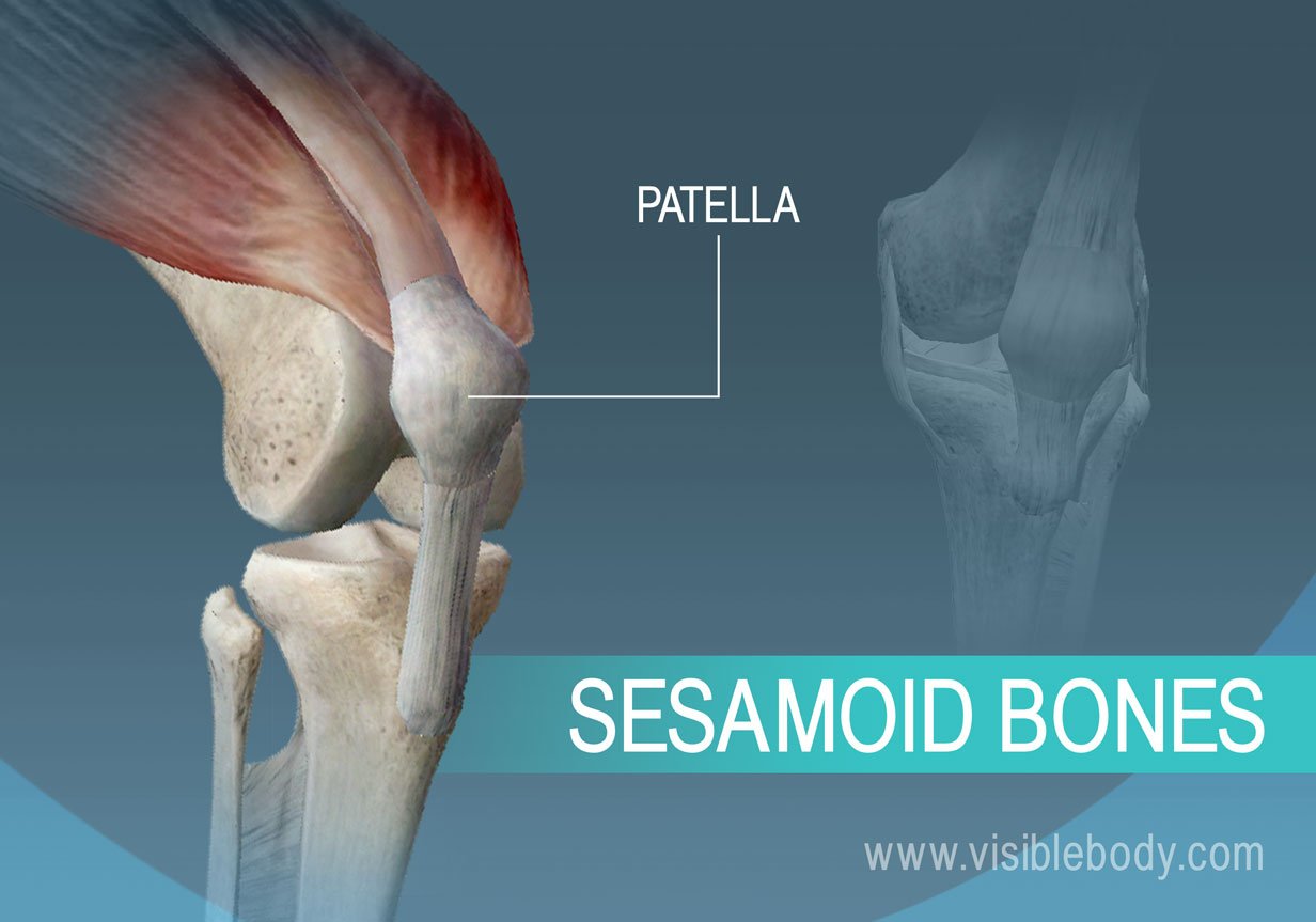
Sesamoid bones
Small, flat bones that are shaped like a sesame seed.
They develop inside tendons and may be found near joints at the knees, hands, and feet.
Example: The patellae
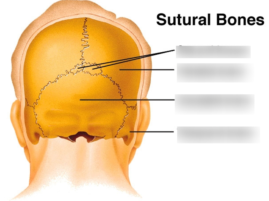
Sutural bones
Small, flat, irregularly shaped bones.
They may be found between the flat bones of the skull.
They vary in number, shape, size, and position.
Compact (Cortical) Bone
The hard external layer of bones that provides strength and protection.
Prominent in areas of bone at which stresses are applied in only a few directions.
Consists of osteons or Haversian systems
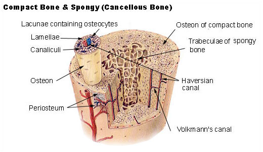
Spongy Bone
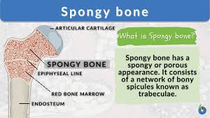
The inner layer of bones that consists of trabeculae and contains red bone marrow.
Prominent in areas of bones that are not heavily stressed or where stresses arrive from many directions.
Osteoblasts
Bone cells that are responsible for bone formation
Blast = build
Osteoclasts
Bone cells that remove bone structure by releasing lysosomal enzymes and acids that dissolve the bony matrix.
Help regulate calcium concentrations in blood.
Clast = destroy
Osteocytes
The mature cells of bone tissue that maintain normal bone structure by recycling mineral salts in the bony matrix.
They cannot divide.
Osteoprogenitor cells
Squamous stem cells that divide to produce daughter cells that differentiate into osteoblasts.
Important for repairing of fractures.
Progenitor = special (differentiate)
Ossification (Osteogenesis)
The process of bone formation that begins in the embryo and continues until about age 25.
Intramembranous Ossification
The process of bone development from fibrous membranes, forming flat bones of the skull.
Endochondral Ossification
The process of bone development from hyaline cartilage, forming most bones in the body.
Epiphyseal Plate
The growth plate responsible for the lengthwise growth of long bones.
Appositional Growth
The increase in the diameter of bones through the addition of bony tissue at the surface.
Bone Remodeling
The process of replacing old bone tissue with new bone tissue, involving osteoblasts and osteoclasts.
Joints
Structures where bones meet, allows for the motion of bones
Classified by structure or function (extent of mobility provided by the joint)
Function: Synarthroses (immovable), amphiarthrosis (slightly movable), diarthroses (freely movable)
Structure: Fibrous (tend to be immovable), synovial (tend to be freely movable), and cartilaginous (exhibit a range of mobilities)
Fibrous Joints
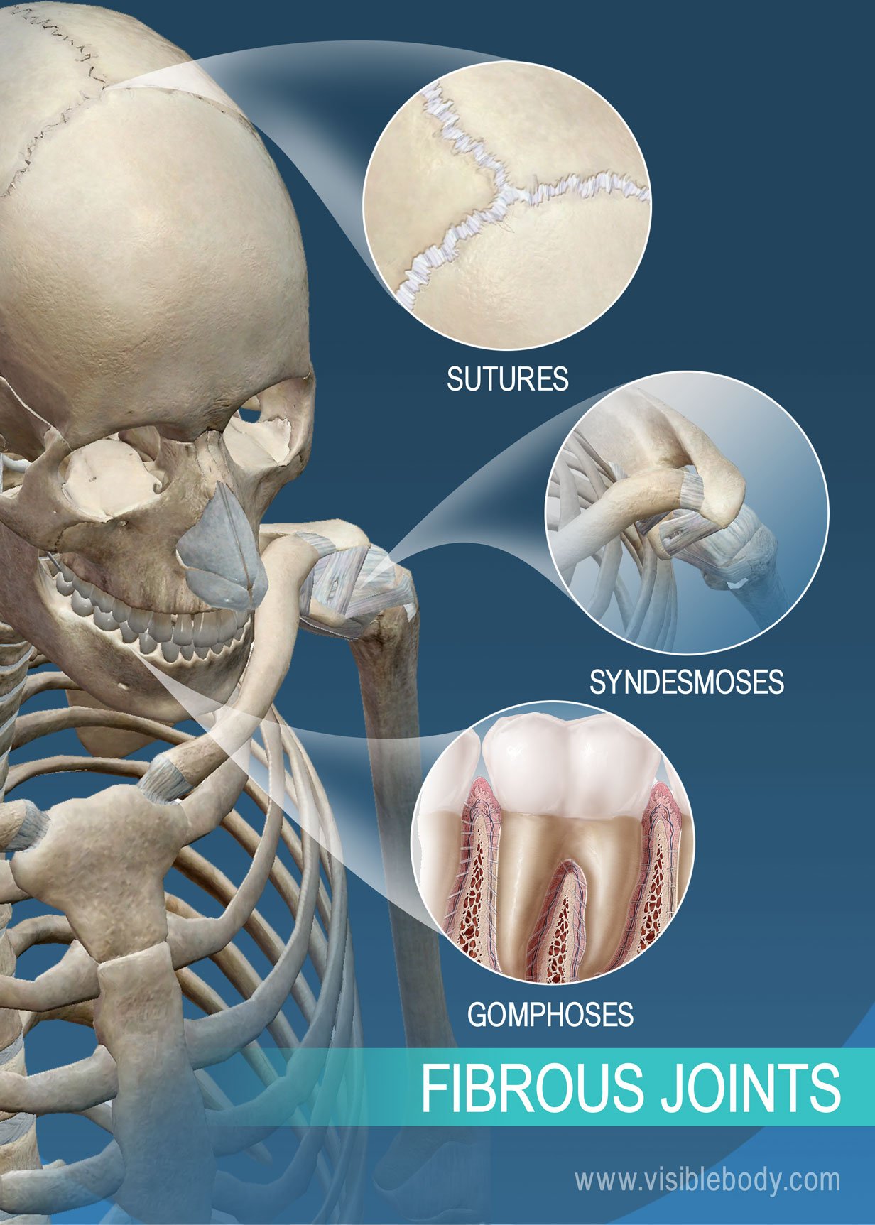
Contains lots of dense, fibrous connective tissue
No joint cavity
Connect bones that don’t require a lot of movement
Three types: sutures (found only in skull), syndesmoses (found where bones are connected only by ligaments), and gomphoses (found only in mouth)
Cartilaginous Joints
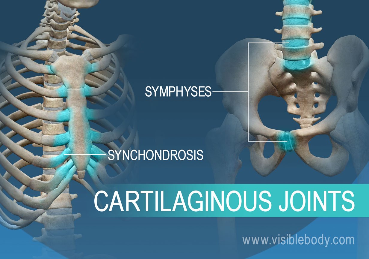
Bones are connected by cartilage
Lack a joint cavity
Not particularly movable
Two Types: synchondroses (contain hyaline cartilage, sympheses (contain fibrocartilage, compressible)
Synovial Joints
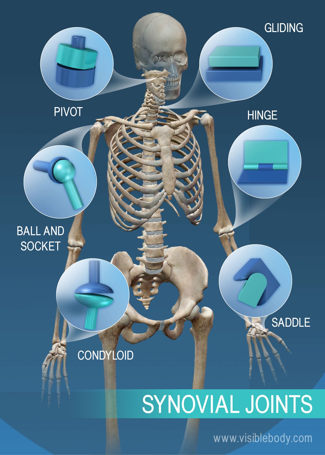
Joints that contain a cavity filled with fluid, allowing for a wide range of motion.
Most joints are this type, especially the ones in limbs.
Types of Motion
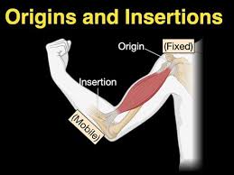
Muscles have an origin attached to an immovable bone, and an insertion attached to a movable bone.
When muscles contract around joints, it causes movement.
Synovial Joint Movement
Nonaxial movement - slipping movement
Uniaxial movement - movement in one plane
Biaxial movement - movement in two planes
Multiaxial movement - movement in all three planes
Gliding movement - when one flat bone surface slips over another
Occurs at the ankles and wrists
Angular movement - when the angle between two bones changes
Flexion - decreases the angle of the joint (i.e. bending the head forward)
Extension - increases the angle of the joint (i.e. straightening your neck)
Hyperextension - goes beyond extension (i.e. bending your head back)
Abduction - motion of a limb away from the midline plate of the body (i.e. moving arms up and away from your side)
Adduction - motion of a limb toward the midline (i.e. bringing arms down to your side)
Circumduction - making circles with a limb
Rotation - turning a bone around its own axis
Special Movements
Supination and pronation - refers to the radius moving around the ulna
Dorsiflexion and plantar flexion - refers to movements in the foot
Protraction and retraction - refer to movements in the mandible
Skeletal (Striated) Muscle
This muscle is used to move the skeleton.
They are under the direct control of the nervous system and can produce contractions ranging from quick twitches to powerful sustained tension.
Structure of Skeletal Muscle
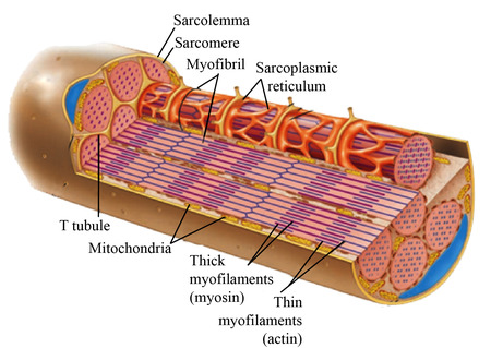
Muscle fibers - among the largest cells of the human body (10 to 100 mm), run the entire length of the muscle.
Sarcolemma - outer membrane that covers a muscle fiber.
Myofibrils - individual contractile subunits that make up each muscle fiber, extending from one end of it to the other.
Sarcoplasmic reticulum (SR) - a complex of membranes forming a network of interconnected hollow tubes, surrounding each myofibril.
Contains fluid rich in calcium ions.
T (Transverse) Tubules - deep indentations of the muscle cell membrane that extend down into the muscle fiber, passing very close to portions of the SR.
Crucial in controlling muscle contraction.
Sarcomeres - subunits of myofibrils that are made up of precise arrangements of actin and myosin filaments.
Z-lines - the junction points of where sarcomeres are attached end to end throughout the length of the myofibril.
Strands of actin and two accessory proteins are attached to them.
Suspended between the thin filaments are thick filaments composed of myosin.
Cross bridges - small arms that extend from the strands of biosin and contact the thin filaments
Each subunit of actin in thin filaments has a binding site for a myosin cross bridge
