19.1 Pulmonary and Systemic Cicuits / 19.2 Gross Anataomty of the Heart, The Chambers + The Valves
 Call Kai
 Learn
Learn Practice Test
 Spaced Repetition
 Match
Match Flashcards
 Knowt Play
1/35

There's no tags or description
Looks like no tags are added yet.
Name | Mastery | Learn | Test | Matching | Spaced | Call with Kai | Chat |
|---|
No analytics yet
Send a link to your students to track their progress
36 Terms
Cardiovascular system
Heart and blood vessels
Heart is the muscular pump keeping the blood flowing through vessels
Vessels deliver blood to all the body’s organs and then return to the heart
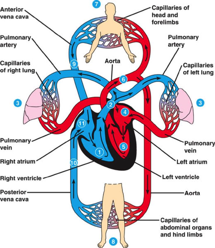
What are the two cardiovascular systems
Pulmonary and systemic
Pulmonary circuit
Carries blood to the lungs for gas exchange and then returns to the heart
Right half of the heart supplies this circuit, receives blood that has circulated through the body, unloaded its oxygen and nutrients and picked up a load of carbon dioxide and other waste
It then pumps oxygen poor blood into large artery known as pulmonary trunk which then divides into right and left pulmonary arteries
The arterories tranport blood into air sacs (alveoli) of the lungs, where carbon dioxide is unloaded and oxygen is picked up
Oxygen rich blood then flows through pulmonary veins to the left side of the heart
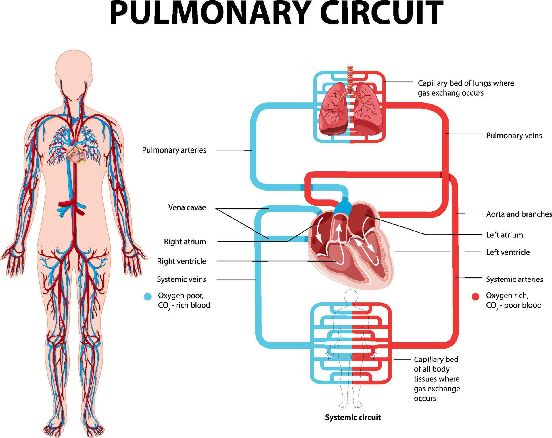
Systemic Circuit
Supplies blood to every organ in the body, including parts of the lungs and walls of the heart
Left side of the heart supplies this circuit
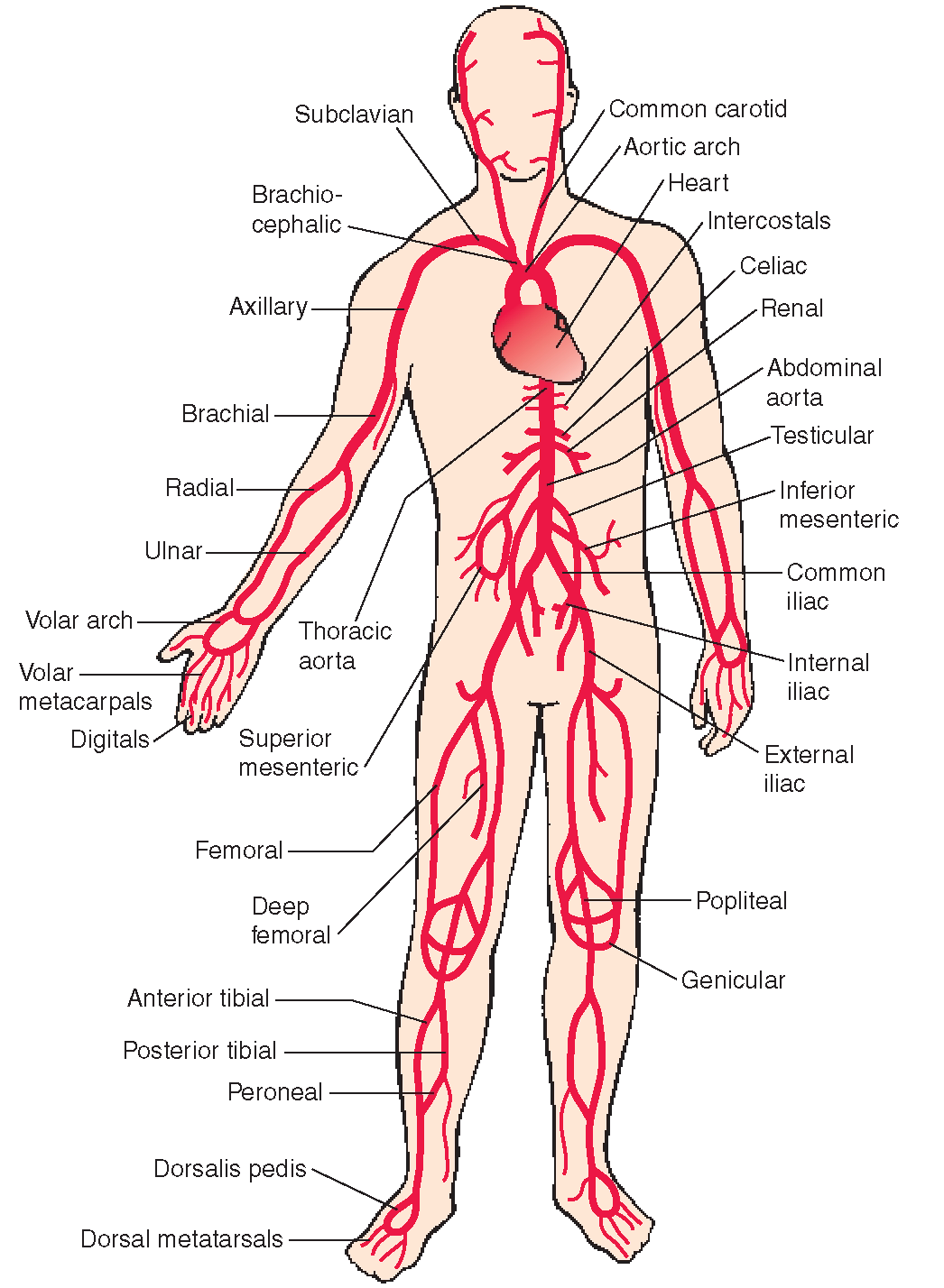
Blood leaves by large artery known as the aorta - the aorta is like an inverted U (aortic arch) and passes downward posterior to the heart
The arch gives off arteries that supply the head, neck, and upper limbs, then travels through thoracic and abdominal cavities and issues smaller arteries to the other smaller organs, before branching into lower limbs
After circulating the whole body, the deoxygenated blood systemic blood returns to the right side of the heart by two veins the superior vena cava (draining of upper body) and inferior vena cava (draining everything below the diaphram)
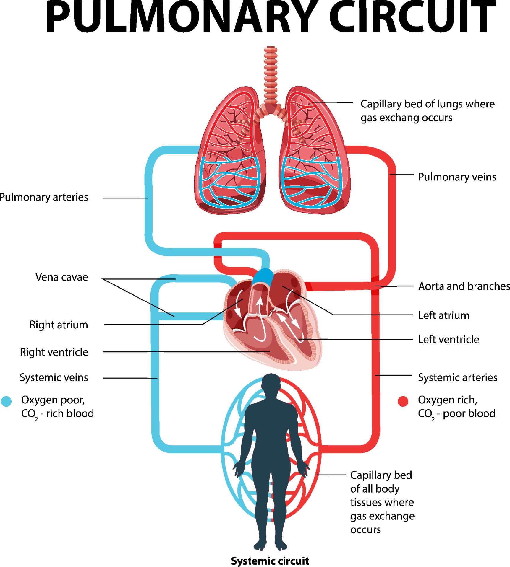
What are the great vessels?
The major arteries and veins entering and leaving the heart
They are called that because they have relatively large diameters
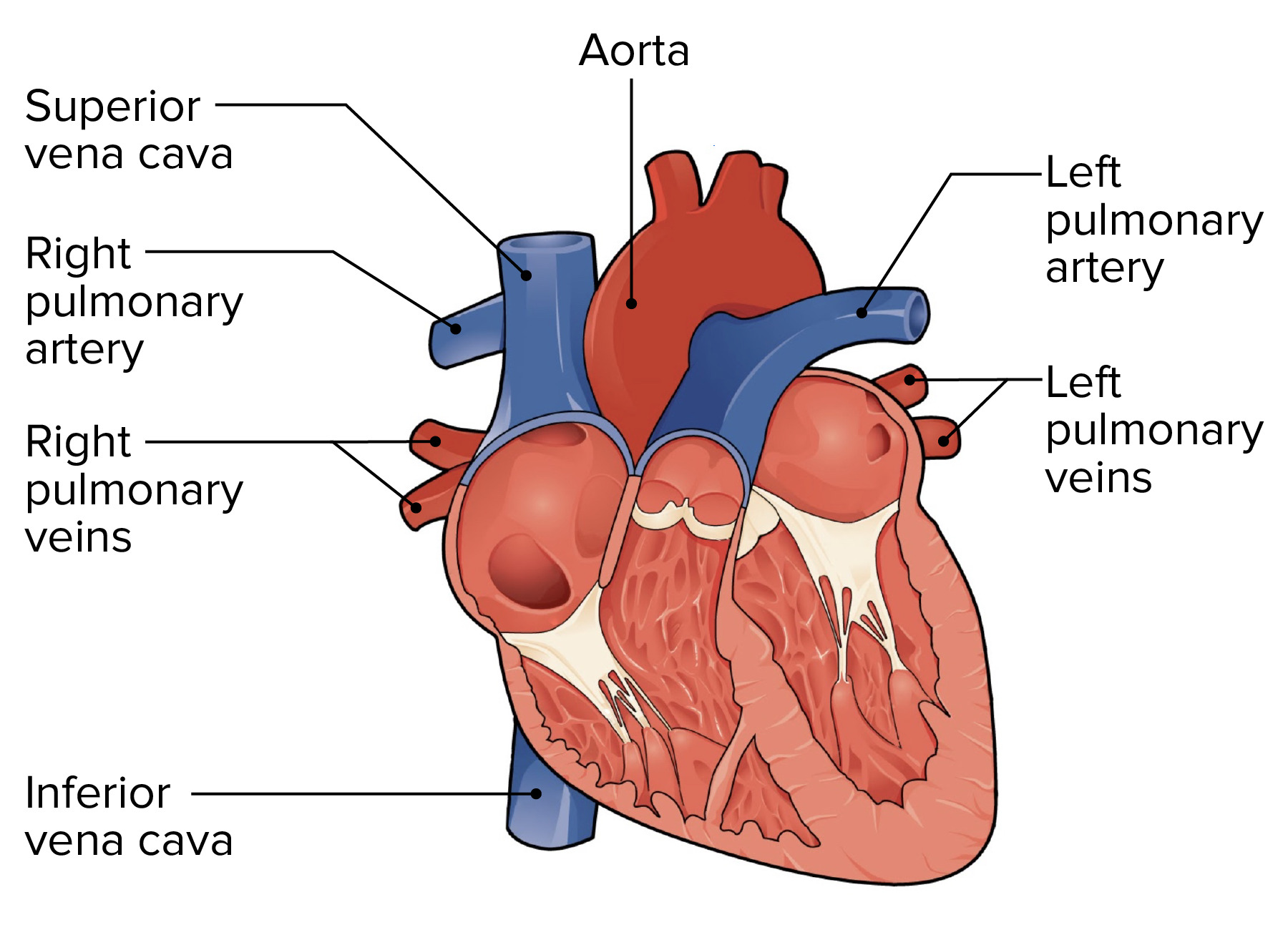
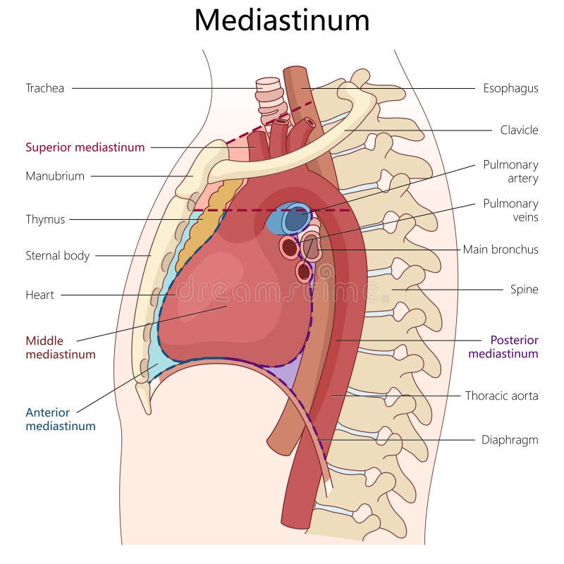
What is the mediastinum?
The thick partition between the two lungs where the heart lives
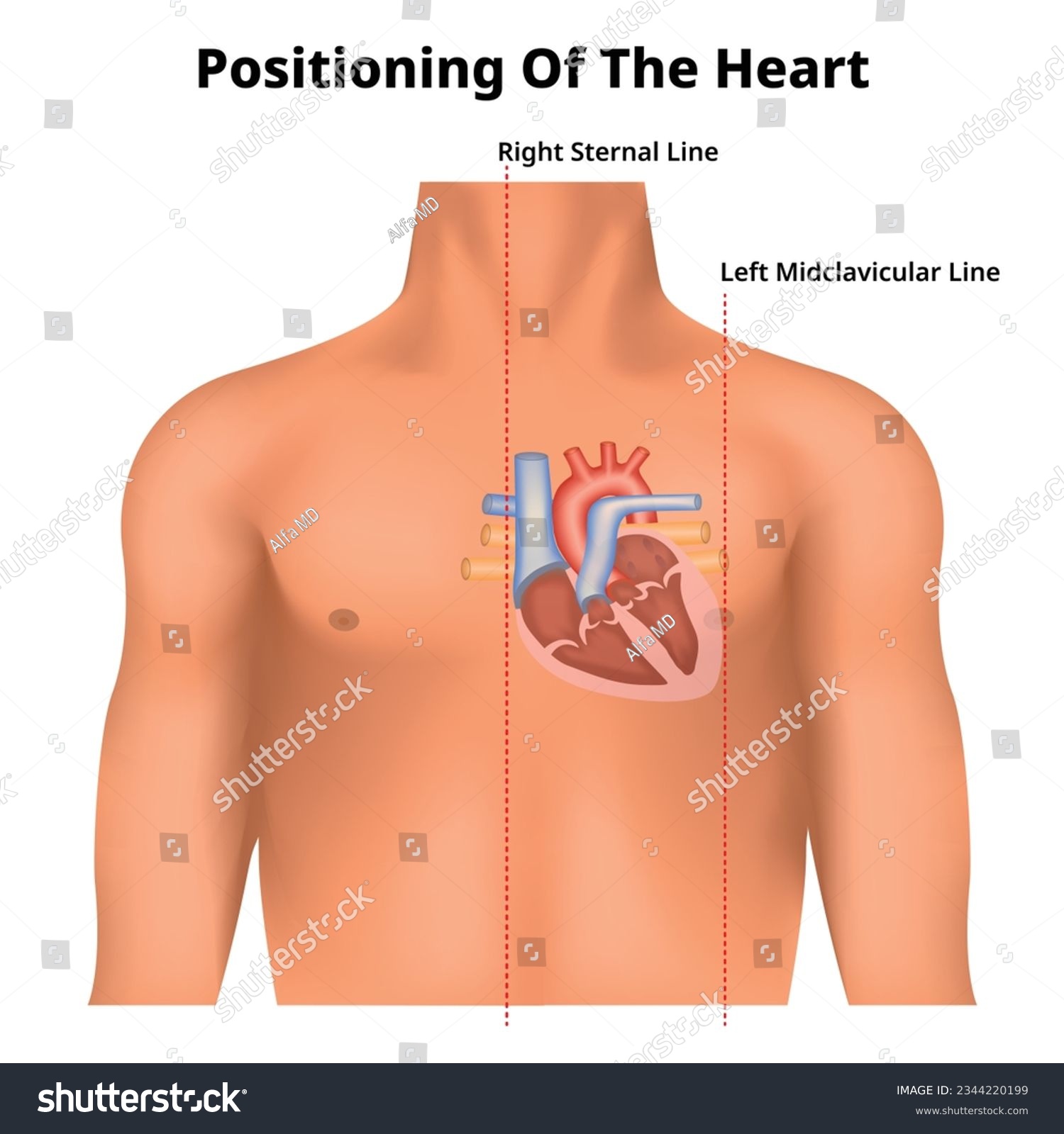
How is the heart shaped and positioned?
It has a base at the very beginning, where the great vessels attach
And apex which is bluntly pointed at the lower end, just above the diaphram
It tilts towards the left from the base to apex, so that more than half the heart is on the left side of the median plan
The heart is approximately 3.5” wide at the base, 5” long from base to apex, and 2.5” wide anterior to posterior
Roughly the size of your hand
10 ounces in weight
What is the pericardium?
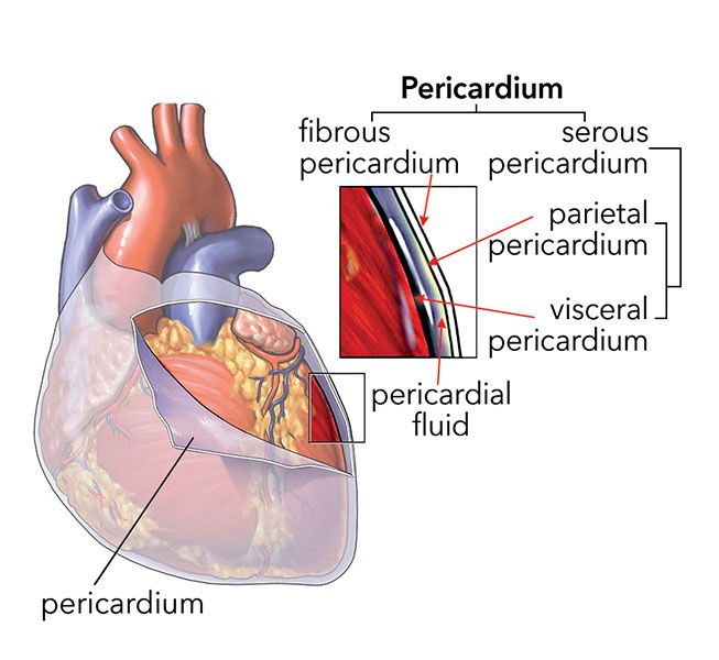
Double walled sac that encloses the heart
Isolated the heart from other thoracic organs, anchors it within the thorax
Gives the heart room to expand but resists excessive expansion
What is the outermost layer of the pericardium?
Fibrous Pericardium
Tough and fibrous
Surround the heart but isn’t attached
Is anchored by ligaments to the diaphram below and the sternum which is anterior to it
Loosely anchored by fibrous connective tissue to the mediastinal tissues posterior to the heart
What is deep to the fiberous pericardium?
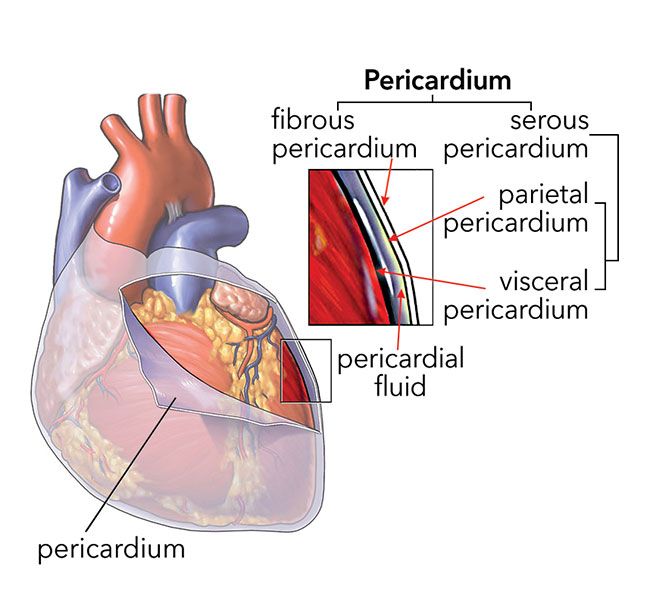
Serous pericardium, has two layers
Parietal layer, lines the inside of the fibrous pericardium
Visceral layer, adheres to the heart surface and form outermost layer of the heart itself, the epicardium
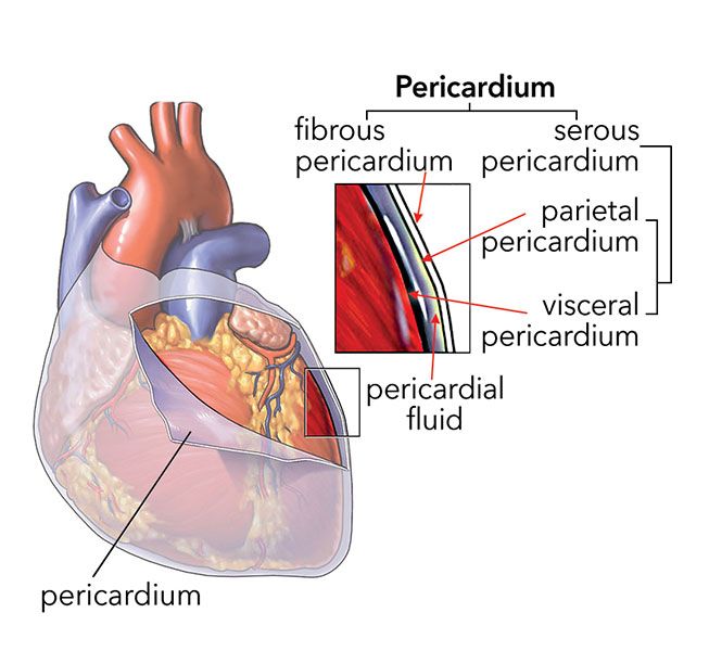
Pericardial Cavity
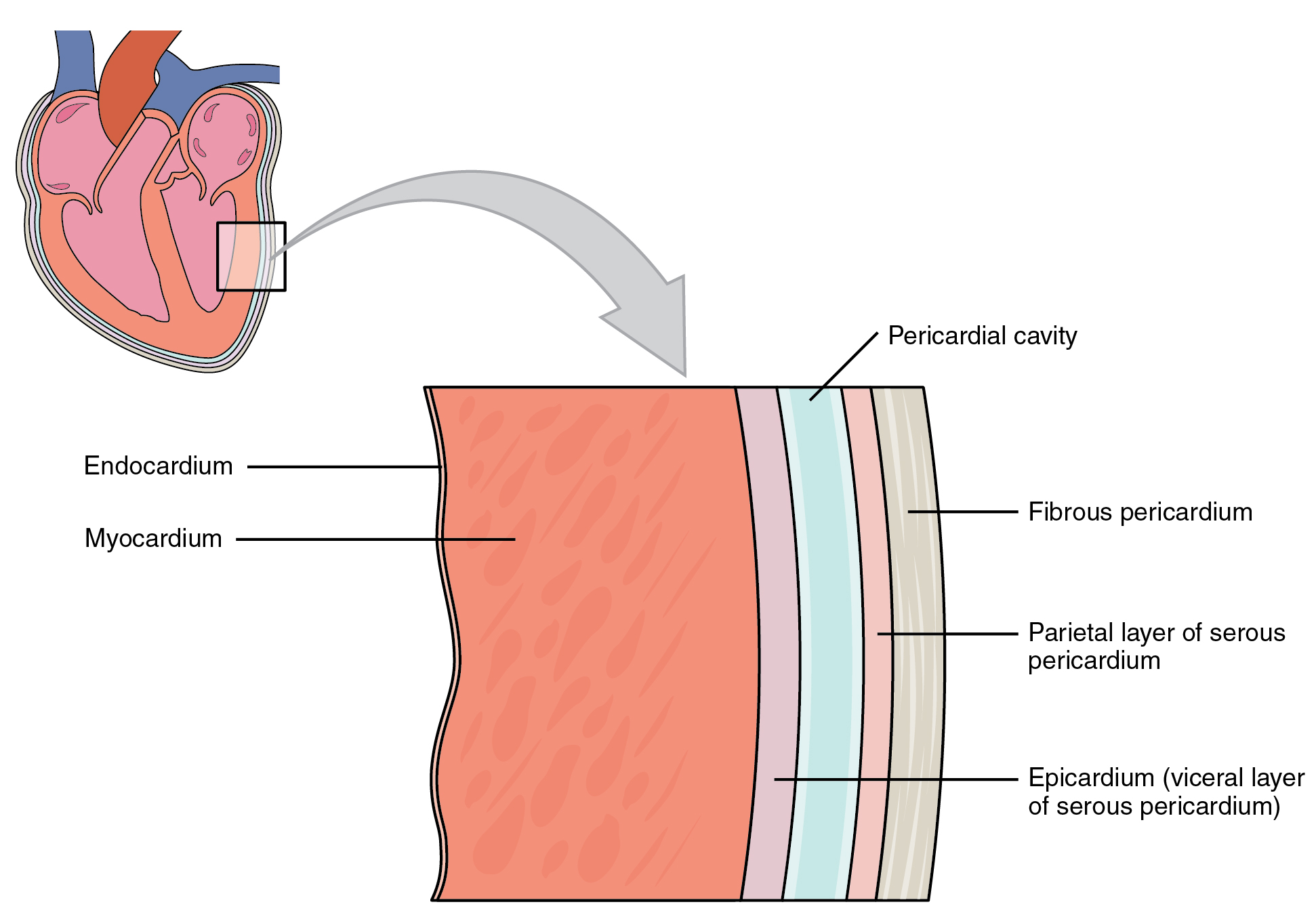
Space between the parietal and visceral layers of serous pericardium
Heart isn’t inside the pericardial cavity but is enfolded by it
An analogy is that of a fist pushed into a underinflated balloon: balloon surface in contact with the fist = epicardium, outer balloon = parietal layer, air space between = pericardium cavity
What is pericardial fluid?
Inside the pericardial cavity
Contains 5 to 30 mL
Is excuded by the serous pericardium
Fluid lubricates the membranes and allows heart to beat with minimal friction
What is pericarditis
Inflammation of the pericardium, membranes can become roughened and produce painful, friction rub with each heartbeat
The heart wall: epicardium
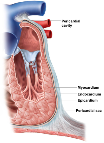
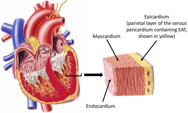
Visceral layer of serous pericardium, serous membrane external to hearts surface
Simple squamous epithelium, overlaying thin layer of areolar tissue
In some places, it also had thick layer of adipose tissue
Other areas, fat free and translucent, and the myocardium (heart tissue/cells) shows through
Largest branches of the coronary blood vessels travel through ehre
The heart wall: endocardium
lines interior of hearts chambers
simple squamous epithelium overlying thin areolar tissue
no adipose tissue
covers valve surface and is continuous with endothelium of blood vessels
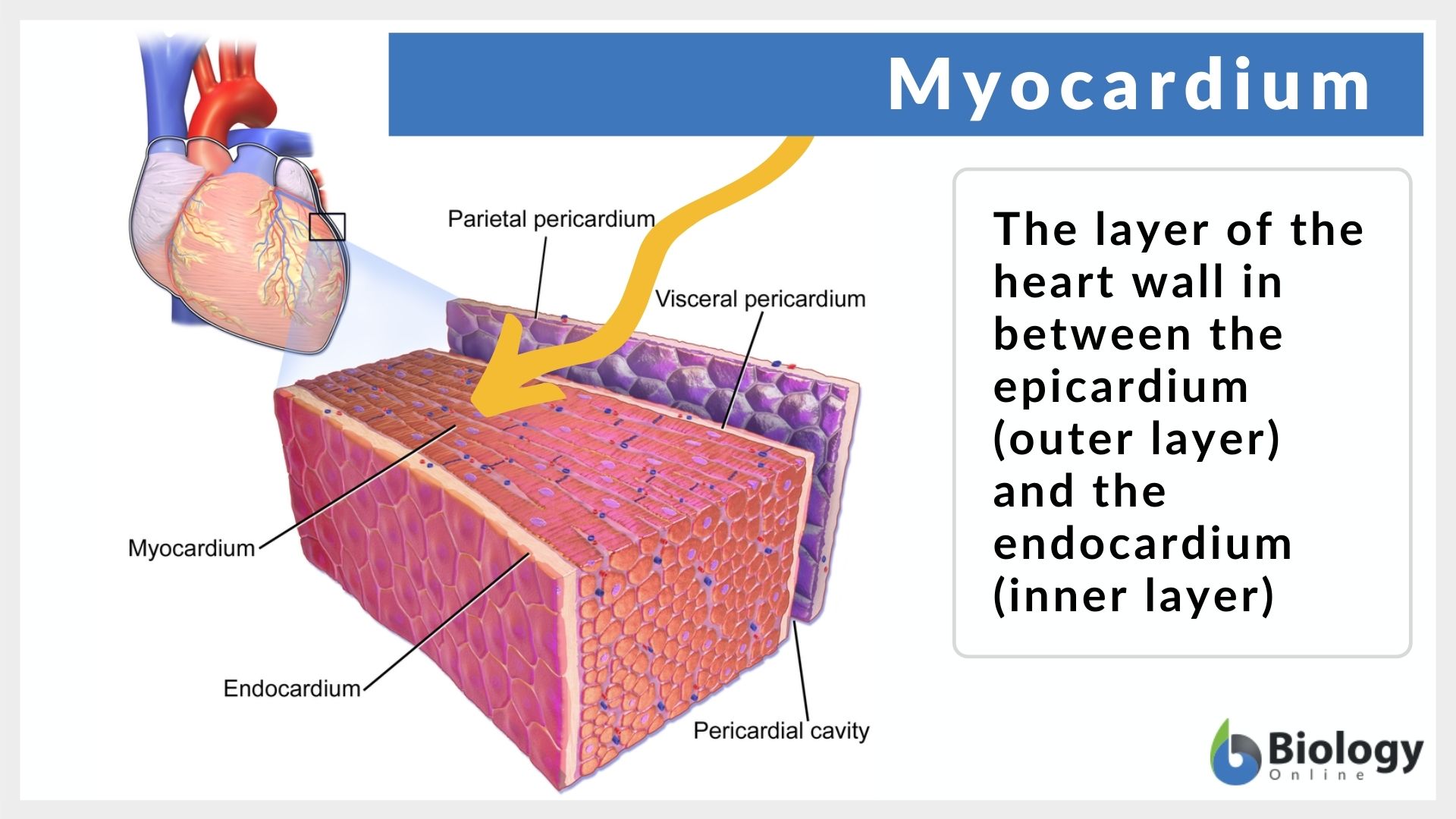
The heart wall: myocardium
in the middle, cardiac muscle
thickest layer, performs the work of the heart
thickness is proportional to the workload of the individual chamber
muscle is organized into bundles that spiral around the heart forming vortex of the heart
when ventricles contract they exhibit twisting or wringing motion the enhances blood ejection
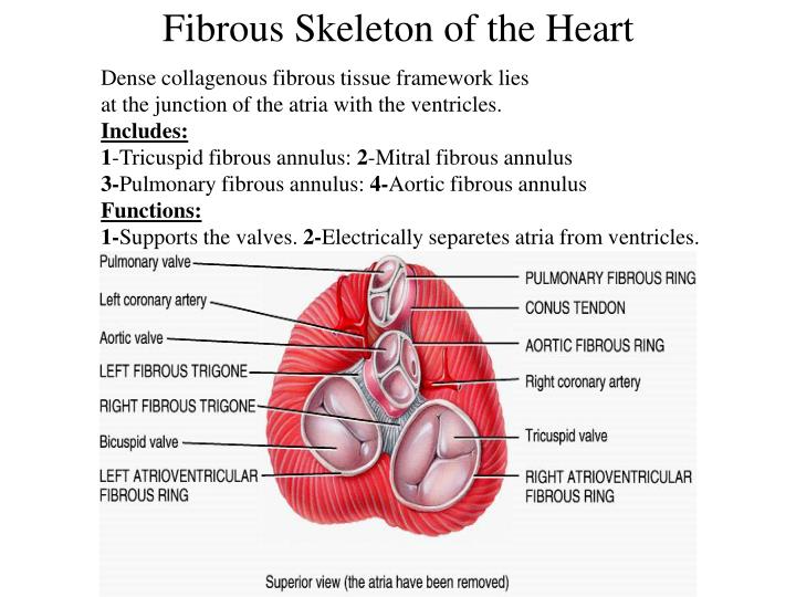
What is the fibrous skeleton?
fibrous skeleton is collagenous and elastic fibers, it is concentrated in the walls between heart chamber, fibrous rings around the valves and in sheets of tissue that interconnects the rings
it function to provide structural support for the heart, around valves and openings of the great vessels, holds these oriface open and prevents them from excessively stretching when blood comes in
anchors the cardiomyocytes and gives them something to pull against
nonconductor of electricity, serves as electrical insulation between the atria, and ventricles so that atria cannot stimulate ventricles directly the instulation is important to timing and coordination of electrical and contractile activity
some authorities think that elastic recoil of the fibrous skeleton may aid in refilling the hear with blood after each beat
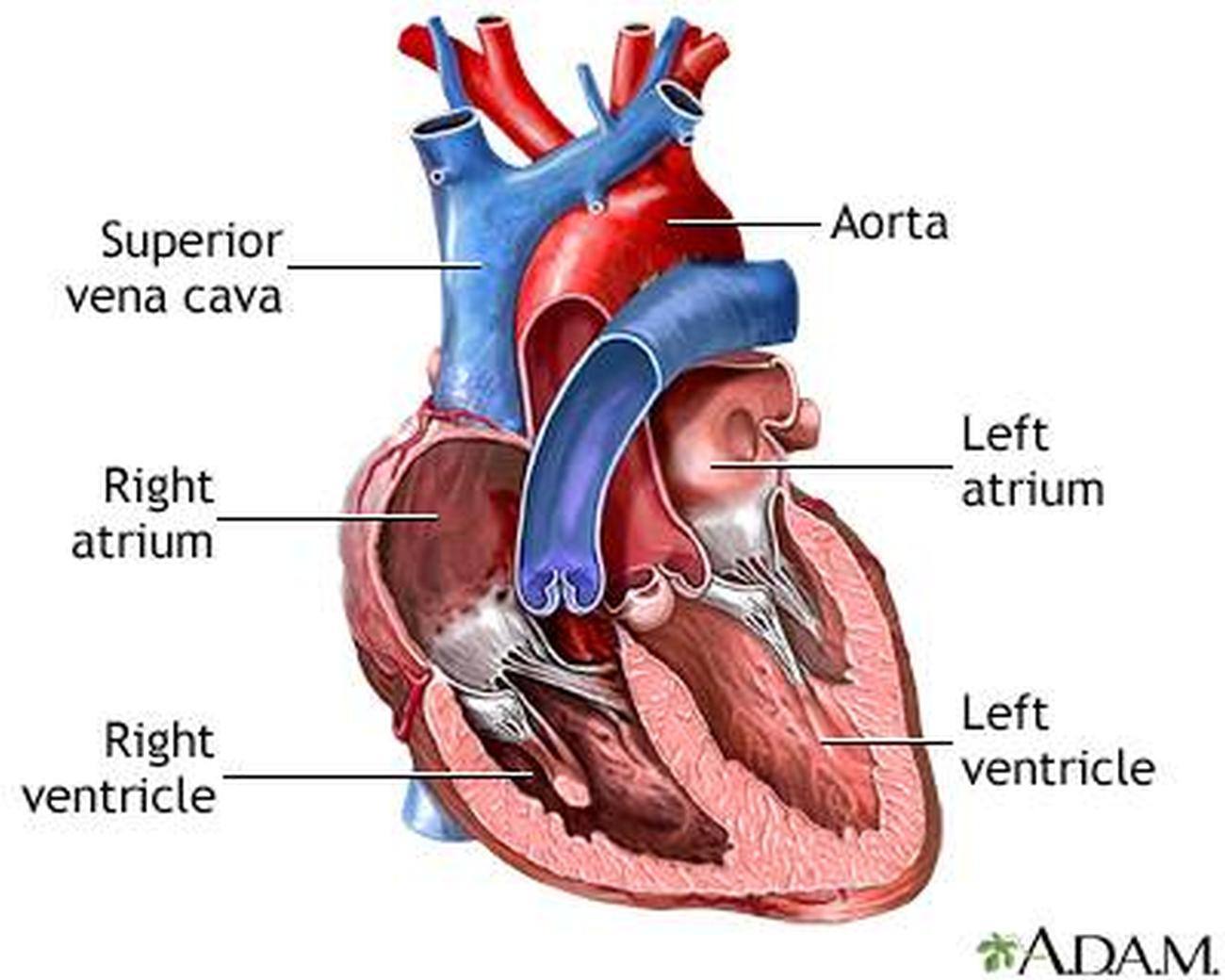
What are the chambers of the heart
4 chambers
Superior chambers: right and left atria
Inferior chambers: right and left ventricles
Right and Left Atrium (atria)
receiving chambers for blood returning to the heart by way of great veins
most of the mass is on the posterior side of the heart, only small amount is visible from the anterior view
Has earlike flap called auricle, slightly increase its volume
Thin flaccid walls corresponding to their light workload
Their job is to pump blood into ventricles below
Separated from each other by a wall called the interartrial septum
Both atrium, and both auricles have internal ridges of myocardium called pectinate mucles
Right and left ventricles
pumps that eject blood into the arteries and keep it flowing through the body
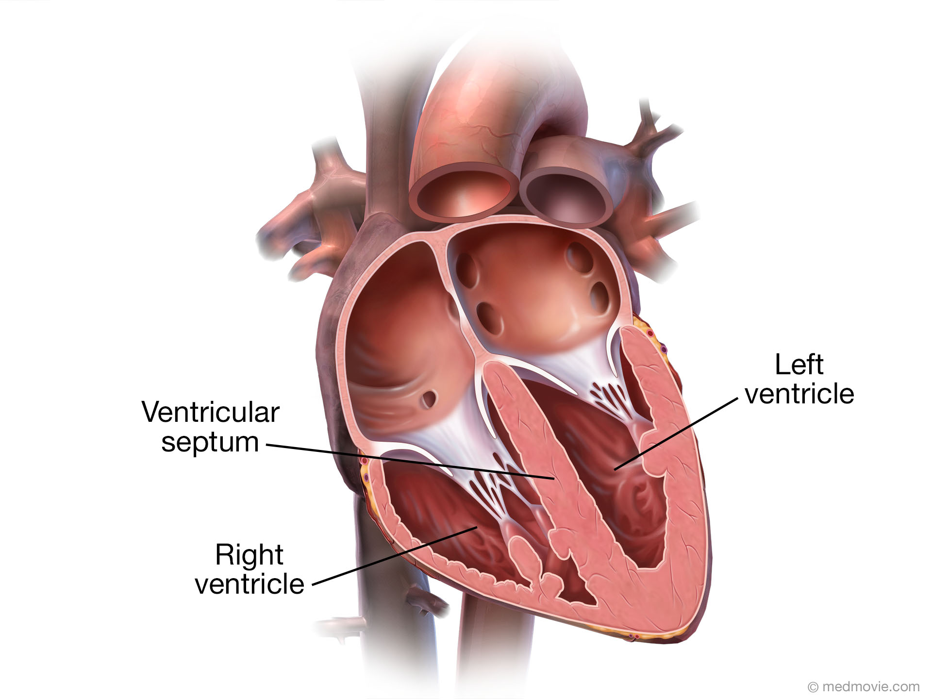
Right ventricle is most of the anterior of the heart
Left ventricle forms the apex and inferoposterior aspect
Ventricles are seperated by the interventricular septum
Right ventricle pumps blood only to the lungs and back to the left atrium, only moderately muscular
Left ventricle is 2 - 4 x as thick because it is the greatest workload, pumping blood through the entire body, roughly circular in cross sections
Right ventricle wraps around the left and is C shaped
both ventricles have internal ridges called trabeculae carneae, this is what keeps the ventricular walls from being like suction cups to one another, and allows them to refill
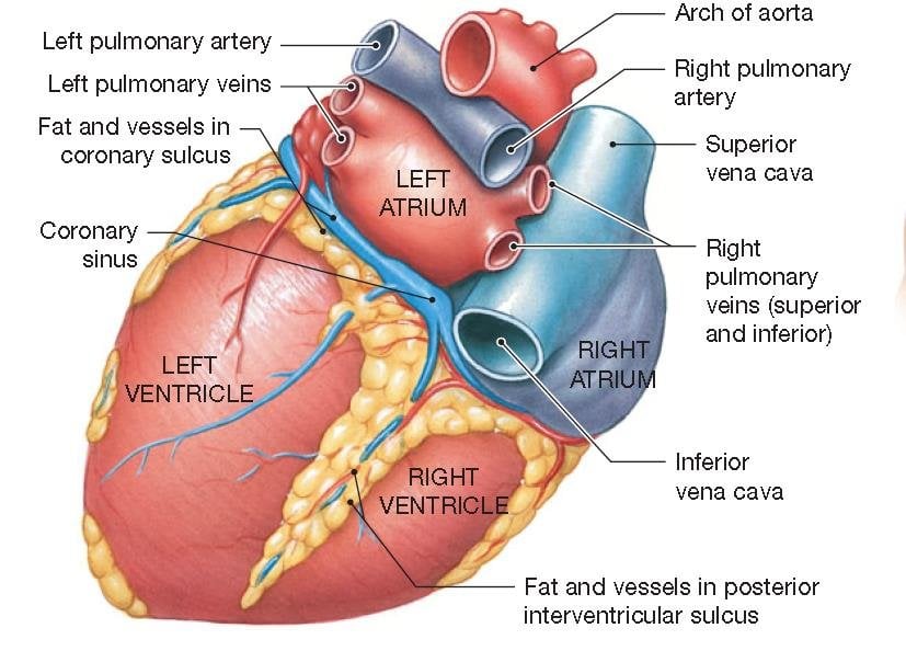
what are the three sulci?
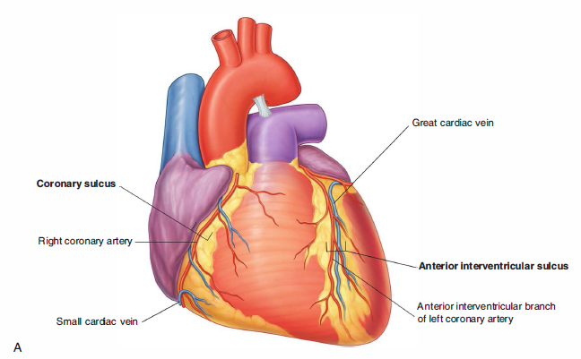
the boundaries of each part of the heart is marked by three sulci (grooves), filled with fat and coronary blood vessels
coronary sulcus encircles the heart near the base and separates the atria above from the ventricles below
the other two sulci extend obliquely down the heart from the coronary sulcus toward the apex
anterior interventricular sulcus - front of the heart
posterior interventricular sulcu - back of the heart
overlie the interventricular septum
hold the largest coronary vessels
What are the valves?
how the blood is able to be pumped effectively for one way flow
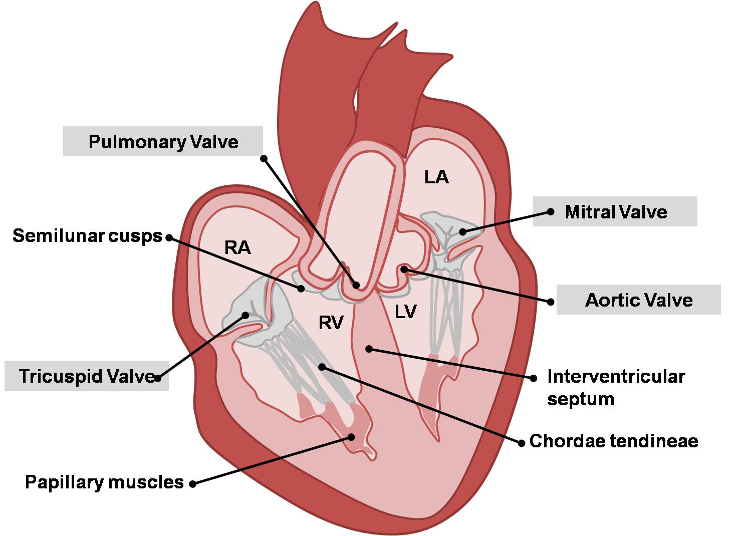
Valve between the atrium and its ventricle and another at the exit of each ventricle into the great artery
No valves that the great veins empty into atria
Each valve has two or three fibrous flaps called cusps or leaflets that are covered in endocardium
Antrioventricular valves
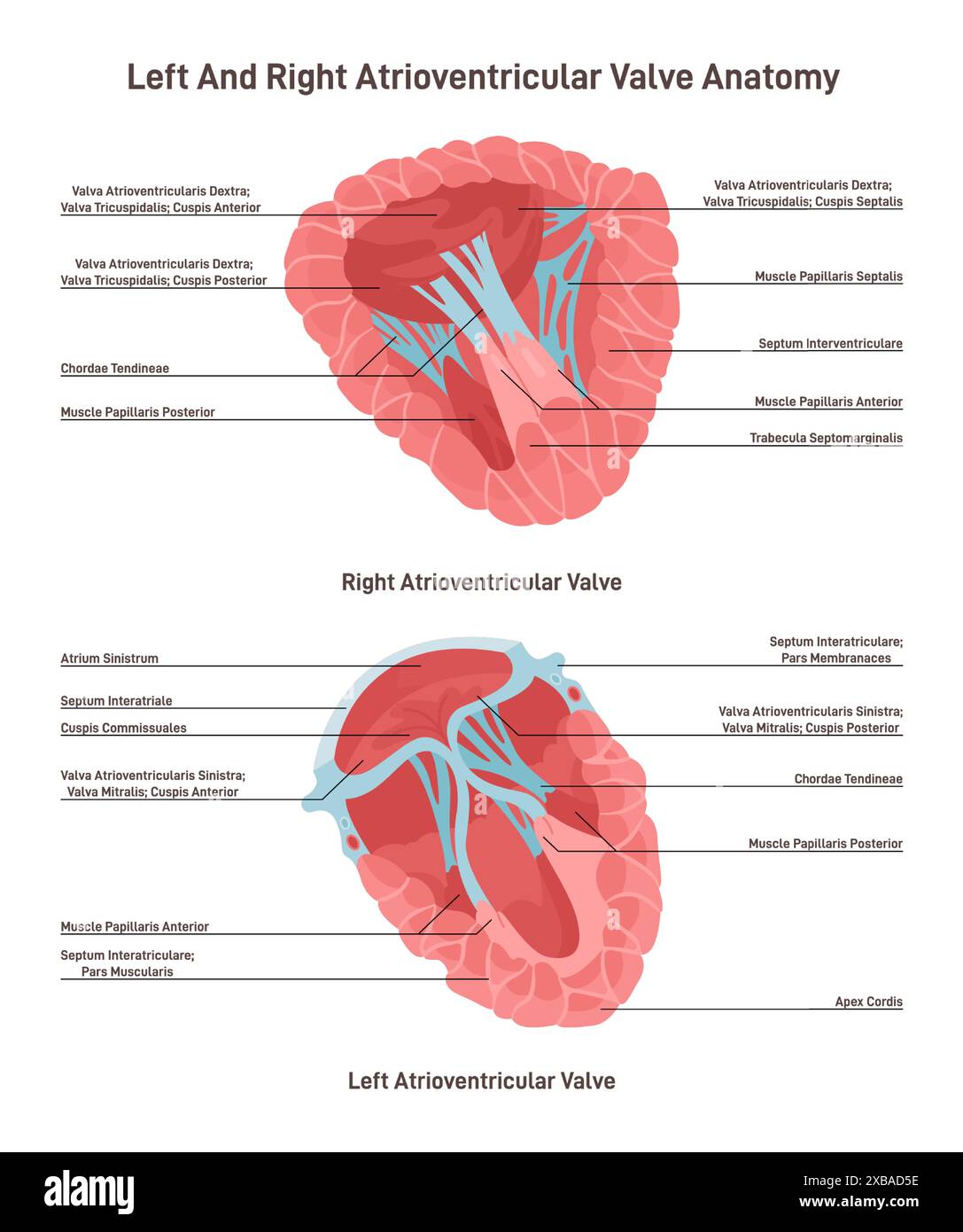
regular opening between atria and ventricles
the right AV (tricuspid) valve has 3 cusps
left AV valve has 2 cups, known as mitral valve
tendinous cords connect the valve cusps to the papillary muscles on the floor of the ventricle, preventing the AV valves from flipping inside out or bulging into the atria when ventricles contract
each papillary muscle has 2 or 3 basal attachments to the trabeculae of the heart wall
the attachment governs timing of electrical excitations of the papillary muscles and distribute mechanical stress
provide redundancy that protects an AV valve from complete mechanical failure if one attachment fails
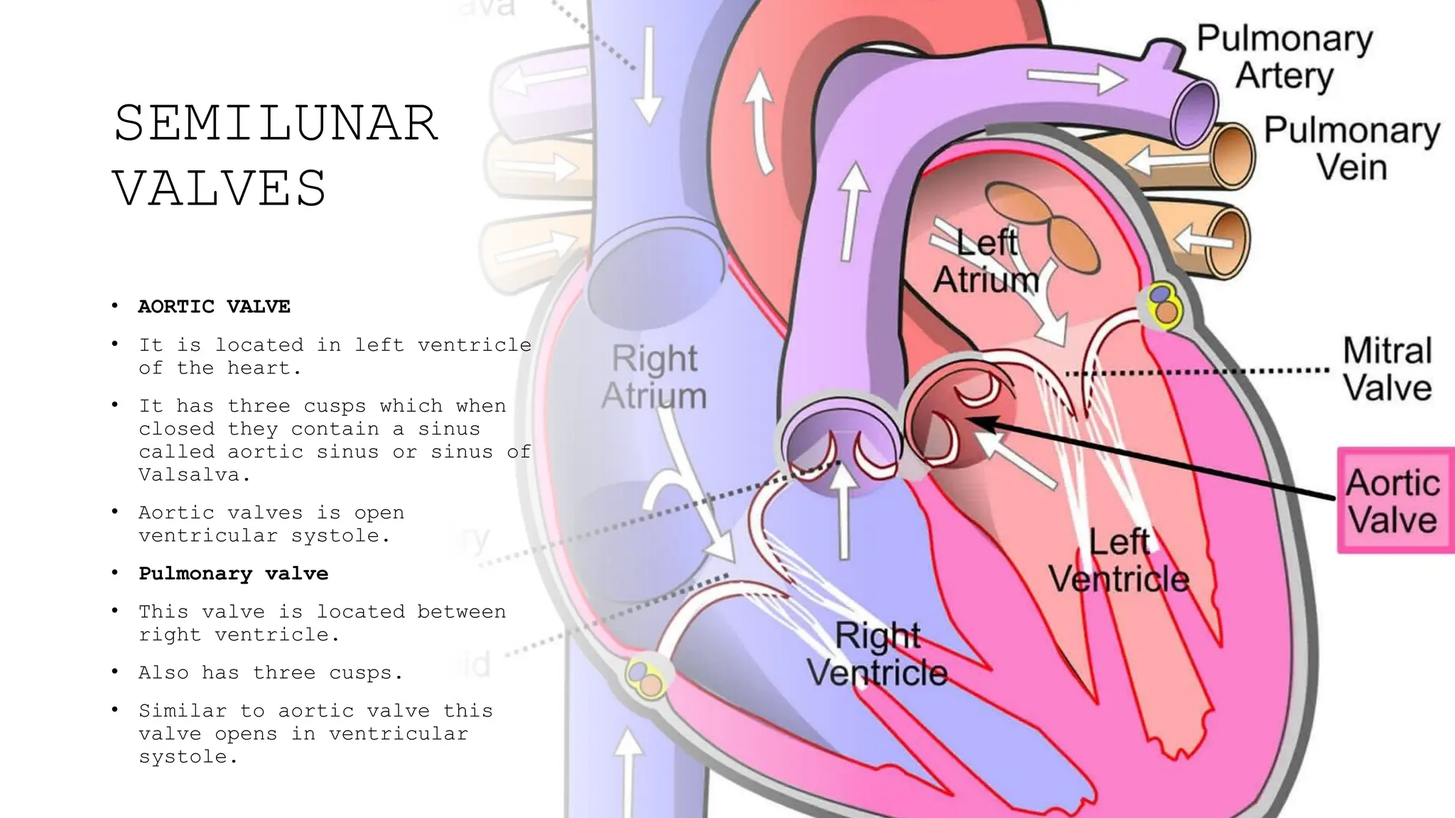
Semilunar Valves - pulmonary and aortic valves
regulate blood flow from ventricles into great arteries
Pulmonary valves: controls openings from the right ventricle into the pulmonary trunk
Aortic valve: controls opening from left ventricle into aorta
each has three cusps shaped like a shirt pocket
when blood is ejected from the ventricles, it pushes through the valves from below and preses cusps against arterial wall
when ventricles relax arterial blood flowes backward toward ventricles, but quickly fills the cusps
the inflated pocket meet at the center and quickly seal the opening, little blood flows back into the ventricles
because the valves are attached to the ventricle wall the way they are, they cannot prolapse and do not require teninous cords
the cusps are not muscular, they are pushed open and closed by changed in blood pressure that occurs when the heart chambers contract and relax
Blood flow
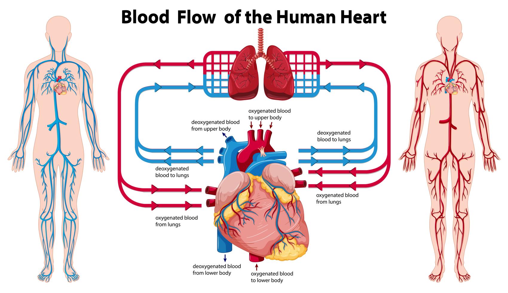
blood is kept separate on the left and right side of the heart
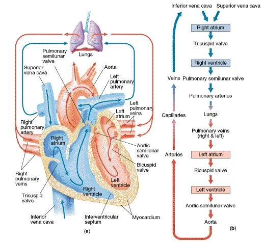
blood going through the systematic circuit returns through the superior and inferior venae cavae to the right atrium, flowing directly into the right atrium, through the right AV (tricupsid) valve into the right ventricle
when the right centricle contracts, it ejects blood thru pulmonary valve into pulmonary trunk on its way to the lungs to exchange carbon dioxide for oxygen
blood returns from lungs by two pulmonary veins on the left, and on two on the right, all four empty into the left atrium
blood flows through the left AV (mitral) valve into left ventricle
contraction of the left ventricle ejects the blood through the aortic valve into the ascending aorta, on its way through the systemic circuit again
Coronary circulation
blood vessels and flow of blood to supply the myocardium with oxygen
coronary blood vessels supply the myocardium with 250 mL blood/minute, 5% of circulating blood going to meet metabolic needs of the heart, it receives more than it “fair share” because of the work load
Arterial Supply
immediately after the aorta leaves the left ventricle, it goes to the right and and left coronary artery
orifices of these two arteries lie deep in the pockets formed by two of the aortic valve cusps
What does the Left Coronary Artery do?
Left Coronary Artery (LCA) travels through coronary sulcus under the left auricle and divides into two branches
1) the anterior interventricular branch: traveling down the anterior interventricular sulcus to the apex, rounds the bend, and travels a short distance up the posterior side of the heart. it joins the posterior interventricular branch, also called left anterior descending branch (LAD), this artery supplies blood to both ventricles and anterior 2/3s of the interventricular septum
2) circumflex branch: continues around the elft side of the heart in the coronary sulcus, gives off left marginal branch that passes down the left margin of the heart and finishes blood to the left ventricle. circumflex branch then ends on the posterior side of the heart. supplies blood to the left atrium and posterior wall of left ventricle
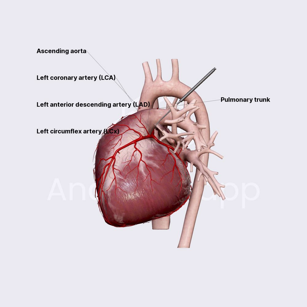
What does the right coronary artery (RCA) do?
supplies right atrium and sinuatrial node (pacemaker), continuing along the coronary sulcus under the right auricle and has two branches
1) right marginal branch, runs toward apex of the heart and supplies lateral aspect of the right atrium and ventricle
2) RCA continues around the right margin of the heart to the posterior side, sending a small branch to the atrioventricular node, gives off a large posterior interventricular branch. branch travels down corresponding sulcus and supplies posterior walls of both ventricles as well as posterior portion of the interventricular septum, ends by joining the anterior interventricular branch of the LCA
Why is blood supply so important to the heart?
The energy demand of the cardiac muscle is critical, an interruption of blood supply to any part of the myocardium can cause necrosis (tissue death) within minutes
Fatty deposit or blood clot in coronary artery can cause myocardial infraction (MI)/heart attack
Arterial Anastomses
Points where two arteries come together, allowing alternative routes of blood flow that can supply the heart tissue with blood if primary route is obstructed
When does the flow into the coronary arteries peak?
when the heart relaxes because
1) contraction of the myocardium squeezes the coronary arteries and obstructs blood flow
2) ventricles contract, aortic valve is forced open and is cusps cover opening to the coronary arteries, this blocks blood flowing back in
3) when they relax, blood in the aorta briefly surges back to the heart, filling aortic valve cusps and some flows back to the coronary arteries like water pouring into bucket and flowing through a hole in the bottom
blood flow increases during ventricular relaxation
What is venous drainage
route by which blood leaves an organ
after flowing through the capillaries of the heart wall, 5 - 10% of the coronary blood empties from multiple tiny vessels called small cardiac veins directly into heart chambers (especially right ventricle) , the rest returns to the right atrium following this route:
1) great cardiac vein: collects blood from the anterior aspect of the heart and travels alongside the anterior interventricular artery, carries blood from the apex toward the coronary sulcus, arcs around the left side of the heart and empties into the coronary sinus
2) posterior interventricular (middle cardiac) vein: found in posterior interventricular sulcus, collects blood from posterior aspect of the heart, carries blood from apex upward and drains into same sinus
3) left marginal vein: travels from a point near the apex up the left margin and also empties into the coronary sinus
4) coronary sinus: large transverse vein in the coronary sulcus on the posterior side of the heart, collects bloom from all three veins and some smaller ones, empties blood into right atrium
What is the heart beat?
contracts regular intervals, about 75 beats per minute (bpm) in resting adults
myogenic - the signal to beat is within itself
autorhythmic because it does not depend on the nervous system to beat
it has it’s own built in pace make and electrical system
What is angina pectoris?
Also known as myocardial infraction (heart attack)
Angina is sense of heaviness/pain in the chest from temporary/reversible ischemia (deficiency of blood flow to the cardiac muscle)
occurs when a partially blocked artery constricts
Oxygen deprived myocardium shifts to anerobic fermentation, producing pactate, stimulating pain receptors in the heart
Pain abates when artery relaxes and normal blood flow resumes
What is myocardial infraction (MI)?
Sudden death of myocardium, resulting in long term obstruction of the coronary circulation
Coronary arteries become obstructed by blood cot or fatty deposit (atheroma)
Cardiac muscle downstream from the obstruction dies, there is a sense of heavy pressure or squeezing pain in the chest “radiating” to the shoulder and left arm.
Some MI’s can be painless “silent” heart attacks - usually in elderly or diabetic people
Infractions weaken the heart wall, disrupt electrical conduction pathways and can lead to fibrillation and cardiac arrest
Heart failure is one of the leading causes of death in the US