3.2.2 - Cell cycle
1/23
Earn XP
Description and Tags
Name | Mastery | Learn | Test | Matching | Spaced |
|---|
No study sessions yet.
24 Terms
Mitosis
Parent cell divides to produce two genetically identical daughter cells
Needed for growth of multicellular organisms + repairing damaged tissues
Cell cycle
Contains interphase - cell growth + DNA replication
Interphase subdivided into 3 growth stages: G₁, S, G₂
Then mitosis
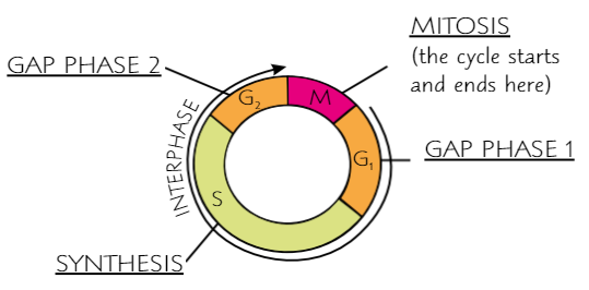
Gap phase 1
Cell grows; new organelles + proteins made
Synthesis
Cell replicates DNA, ready for mitosis
Gap phase 2
Cell keeps growing; proteins needed for cell division are made; DNA is checked for errors - cell dies if DNA incorrect
Interphase
(before mitosis in cell cycle)
Cell carries out normal functions + prepares to divide
Cell DNA unravelled + replicated
Organelles replicated
ATP content increased

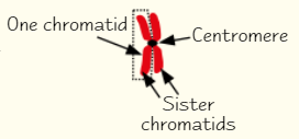
Important parts of chromosome in mitosis
As mitosis begins, chromosomes are made of two strands joined in middle by centromere
Separate strands called chromatids
There are 2 strands because each chromosome has made identical copy of itself during interphase
When mitosis ends, chromatids end up as one-strand chromosomes in daughter cells
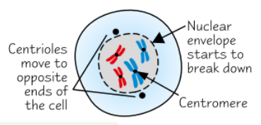
Prophase
Chromosomes condense - get shorter + fatter
Tiny protein bundles called centrioles start moving to opposite ends of cell
Forms network of protein fibres called spindle
Nuclear envelope breaks down → chromosomes lie free in cytoplasm
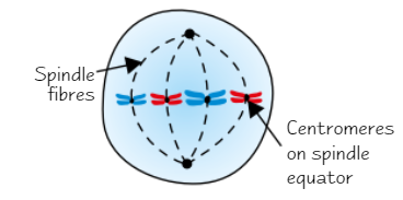
Metaphase
Chromosomes line up along middle of cell + become attached to spindle by centromere
Anaphase
Centromeres divide → sister chromatids separate
Spindles contract → pulls chromatids to opposite poles of spindle, centromere first
Makes chromatids appear v-shaped
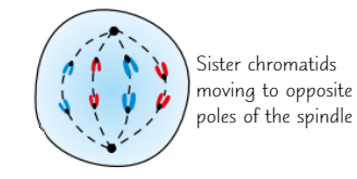
Telophase
Chromatids reach opposite poles on spindle
Chromatids uncoil → become long + thin
Now called chromosomes again
Nuclear envelope reforms around group of chromosomes → two nuclei
Cytokinesis finishes
Now two genetically identical daughter cells
Each cell starts interphase
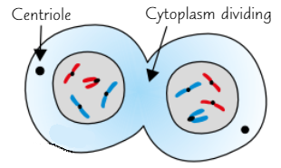
Cytokinesis
Division of cytoplasm, producing two new cells
Starts in anaphase, finishes in prophase
Cancer
Mitosis + cell cycle are controlled by genes
Normally, cells stop dividing after enough new cells are made
But if there’s mutation in gene that controls cell division, cells can grow out of control
Cells keep dividing, forming tumour
Cancer = tumour that invades surrounding tissue
General cancer treatment
Some treatments are designed to control rate of division in tumour cells by disrupting cell cycle → kills tumour cells
Treatments don’t distinguish tumour cells from normal cells (also kill normal body cells that are dividing) → tumour cells divide more frequently than normal ones, so treatments are more likely to kill tumour cells
Cancer treatment targeting G1
Some chemical drugs (chemotherapy) prevent synthesis of enzymes needed for DNA replication
If these aren’t produced, cell is unable to enter synthesis phase (S) → disrupts cell cycle → forces cell to kill itself
Cancer treatment targeting S phase
Radiation and some drugs damage DNA
DNA is checked for damage several times in cell cycle
If severe damage is detected, cell kills itself → preventing further tumour growth
Binary fission stage 1
Circular DNA + plasmid(s) replicate
Main DNA loop is replicated once, but plasmids can be replicated many times
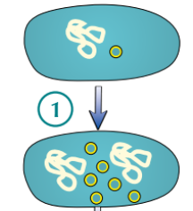
Binary fission stage 2
Cell gets bigger and DNA loops move to opposite ‘poles’ of cell
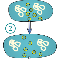
Binary fission stage 3
Cytoplasm begins to divide (and new cell walls begin to form)
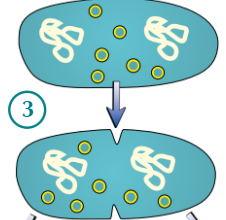
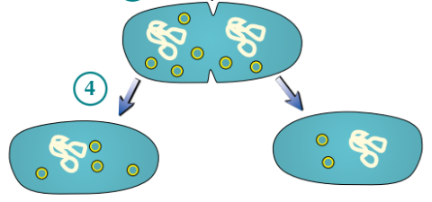
Binary fission stage 4
Cytoplasm divides → two daughter cells produced
Each daughter cell has one copy of circular DNA, but variable number of plasmid(s)
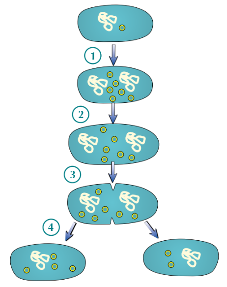
Binary fission
Circular DNA + plasmid(s) replicate
Main DNA loop is replicated once, but plasmids can be replicated many times
Cell gets bigger and DNA loops move to opposite ‘poles’ of cell
Cytoplasm begins to divide (and new cell walls begin to form)
Cytoplasm divides → two daughter cells produced
Each daughter cell has one copy of circular DNA, but variable number of plasmid(s)
Virus binding to host
Use attachment proteins to bind to complementary receptor proteins on surface of host cells
Diff viruses have diff attachment proteins, therefore require diff receptor proteins on host cells → some viruses can only infect one type of cell
Virus cell division
Because not alive, don’t undergo cell division
Virus replication
Inject DNA/RNA into host cell
Hijacked cell uses its ‘machinery’ to replicate viral particles