Retinal Vascular Diseases Part 2
 Call Kai
 Learn
Learn Practice Test
 Spaced Repetition
 Match
Match Flashcards
 Knowt Play
1/48

Earn XP
Description and Tags
Flashcards covering vocabulary and key diagnostic signs for Retinal Vein Occlusions, Retinal Artery Occlusions, Hypertensive Retinopathy, and Retinopathy of Prematurity.
Name | Mastery | Learn | Test | Matching | Spaced | Call with Kai | Chat |
|---|
No analytics yet
Send a link to your students to track their progress
49 Terms
pathogenesis of retinal vein occlusions
arteriosclerosis is an important causative factor for BRVO and CRVO.
in turn causes hypoxia of the retina drained by the obstructed vein
Arteriolosclerosis
Thickening and hardening of the arteriolar walls, which can compress the corresponding vein sharing a common adventitial sheath.
Common predisposing factors of RVO
Advancing age
hypertension
hyperlipidaemia
diabetes mellitus
raised intraocular pressure
oral contraceptive pills
Collaterals
Active remodeling of non-functional vascular network to bypass the site of obstruction and preserve the retinal tissue that is affected by ischaemia.
Branch Retinal Vein Occlusion (BRVO) Signs
VA is variable - dependent on extent of macular involvement
Dilatation and tortuosity of the venous segment further to the site of occlusion, attenuation closer to occlusion
flame-shaped, dot and blot haemorrhages, retinal oedema, and cotton wool spots
course of BRVO
acute features take 6-12 months to resolved and replaced with:
hard exudates, venous sheathing and sclerosis
collaterals which may be local / develop across the horizontal raphe
prognosis of BRVO
reasonably good
within 6 months 50% have 6/12 or better VA
management of BRVO
refer to ophthalmologist to monitor for sight-threatening conditions (e.g. macular oedema & neovascularisation)
Non-ischaemic Central Retinal Vein Occlusion (CRVO)
The most common type of CRVO presenting with sudden, unilateral blur vision (sudden painless loss of vision)
Signs of Non-ischaemic CRVO
VA: moderate to severe reduction
APD: absent / mild
tortuosity and dilation of all branches of CRV
dot and blot, flame-shaped haemorrhage, disc & macular oedema, variable cotton wool spots
Afferent Pupillary Defect (APD)
A condition where both pupils are equal in size, but when the affected eye is stimulated by light, neither pupil reacts; however, when the normal eye is stimulated, both react normally.
Prognosis of Non-ischaemic CRVO
reasonably good, 50% return to normal / near normal vision
conversion to ischaemic CRVO in 15% of cases in 4 months & 34% of cases in 3 years
Management of Non-ischaemic CRVO
refer to ophthalmologist to monitor for conversion to ischaemic CRVO
treatment of macular oedema with intravitreal steroid / anti-VEGF
Ischaemic Central Retinal Vein Occlusion (CRVO)
Characterised by rapid onset venous obstruction resulting in decreased retinal perfusion, capillary closure, and retinal hypoxia
Presentation of Ischaemic CRVO
sudden and severe visual impairment
Signs of Ischaemic CRVO
VA: usually counting finger worse
APD: marked
Severe tortuosity & engorgement of all branches of the CRV, extensive dot & blot and flame-shaped haemorrhages
cotton wool spots may be present
Complications of Ischaemic CRVO
may lead to profound vascular leakage, rubeosis iridis & neovascular glaucoma
fibrovascular membrane develops on the retina and contracts progressively, which may lead to tractional retinal detachment
Tractional Retinal Detachment
Separation of the neurosensory retina from the RPE (retinal pigment epithelium) by the contraction of a fibrovascular membrane in the absence of a retinal break.
Rubeosis iridis
The development of neovascularisation at the iris, occurring in 50% of eyes with ischaemic CRVO between 2 and 4 months.
Prognosis of Ischaemic CRVO
most acute signs resolve over 9-12 months
extremely poor due to macular ischaemia
rubeosis iridis develops in 50% of eyes between 2 and 4 months (causing neovascularisation glaucoma)
Management of CRVO
refer to ophthalmologist urgently for eyes with rubeosis iridis for laser pan-retinal photocoagulation (PRP)
Retinal Artery Occlusions
Atherosclerosis-related thrombosis at the level of the lamina cribosa (by far most common underlying cause if CRAO)
Risk factors of atherosclerosis
age
hypertension
diabetes
raised levels of LDL cholesterol
obesity
smoking
sedentary lifestyle
Carotid Embolism
The origin of the emboli is most often an atheromatous plaque at the carotid bifurcation
The emboli may be of the following types:
Cholesterol
Calcific
fibrin-platelet
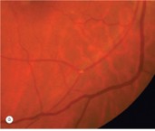
Hollenhorst plaques (Cholesterol)
Minute, bright, refractile, golden to yellow-orange cholesterol crystals often found at vessel bifurcations.
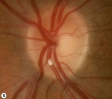
Calcific
single, white, non-scintillating particles
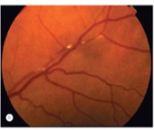
Fibrin-platelet
dull grey, elongated particles that may cause amaurosis fugax, occasionally complete obstruction
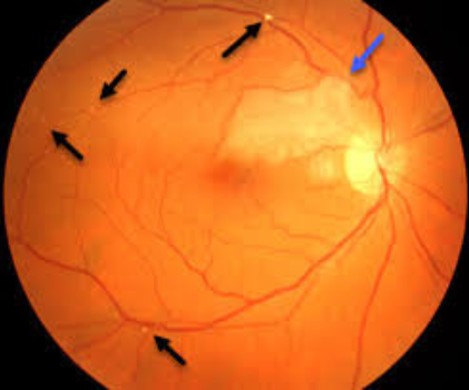
BRAO
CRAO
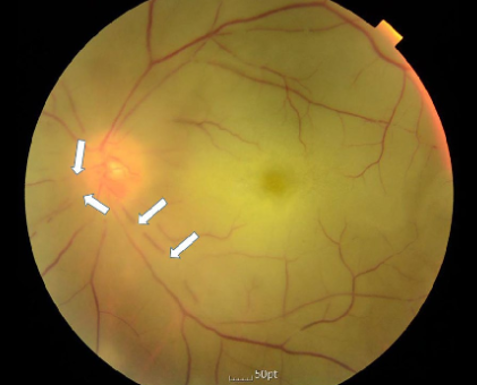
Amaurosis fugax
Painless transient monocular loss of vision, often caused by fibrin-platelet emboli.
Branch Retinal Artery Occlusion (BRAO) Presentation
Presents with sudden, profound altitudinal or sectoral VF loss
Signs of BRAO
narrowing of arteries & veins with sludging and segmentation of blood column
cloudy white retina that corresponds to the area of Ischaemia
emboli may be present
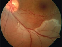
Embolic inferotemporal branch retinal artery occlusion
Prognosis of BRAO
poor unless obstruction can be relieved within a few hours
VF defect is permanent while the affected artery remains attenuated
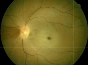
Segmentation
The sludging and breaking up of the blood column within a vessel, also known as 'cattle trucking' or 'boxcarring'.
Cherry-red spot
An appearance in CRAO where an orange reflex from the intact choroid stands out at the thin fovea in contrast to the surrounding pale, cloudy retina.
Cilioretinal artery
A vessel present in 20% of the population that arises from the posterior ciliary circulation to supply the macula and papillomacular bundle.
Central Retinal Artery Occlusion (CRAO) Management
Considered an emergency requiring immediate referral to the emergency department (less than 48 hours at presentation) to prevent permanent loss of vision.
Hypertensive Retinopathy (HR)
A spectrum of retinal vascular changes pathologically related to microvascular damage from elevated blood pressure, characterized by arteriolar narrowing and vascular leakage.
Macular star
A formation of hard exudates in the macula resulting from chronic retinal oedema in hypertensive retinopathy.
Arteriovenous (AV) nipping
A clinical sign of arteriolosclerosis involving thickening of the vessel wall at the points where arterioles and veins cross.
Copper-wiring
A Grade 3 arteriolosclerosis sign where there is obvious broadening of the arteriolar light reflex.
Silver-wiring
A Grade 4 arteriolosclerosis sign where the arterioles appear like white wires, associated with Grade 3 changes.
Salus sign
The deflection (a change of direction) of a vein at an arteriovenous (AV) crossing.
Bonnet sign
The banking (twisting) of a vein distal to an arteriovenous (AV) crossing.
Gunn sign
The tapering of a vein's width to a small point on both sides of an arteriovenous (AV) crossing.
Retinopathy of Prematurity (ROP)
Disorganised growth of blood vessels with fibrovascular proliferation affecting premature infants of very low birth weight (≤1500g) exposed to high oxygen concentrations.
Hyperoxia
High exposure to ambient oxygen concentrations which down-regulates VEGF in premature infants, potentially halting vessel migration and leading to ROP.
ROP Stage 1
The presence of a demarcation line in the retina.
ROP Stage 3
Extraretinal fibrovascular proliferation extending from a ridge into the vitreous.