091 Module 2 - Ultrasound Transducers and the Soundbeam
1/62
There's no tags or description
Looks like no tags are added yet.
Name | Mastery | Learn | Test | Matching | Spaced |
|---|
No study sessions yet.
63 Terms
Name the three main transducer groups.
Linear array
Curved linear array
Phased sector array
List the different names for a phased sector array.
sector probe
cardiac probe
Explain the meaning of a 1D phased array. Draw an example of a 1D vs a 2D.
1D: single row of crystals
Phased array: crystals are stimulated electronically

Describe the display format, frequency range, and clinical usage of sector transducers.
pulses originate from the same starting point → very small nearfield
small footprint for rib access
pie shaped displays
prioritize temporal resolution
BW = 1-5 MHz (adult) and 6-12 MHz (pediatric)
cardiac application
Describe the display format, frequency range, and clinical usage of linear transducers.
pulses originate from different starting points and move in the same direction (scan lines are parallel)
various sizes of footprints (larger than sector)
rectangular image format (nearfield size = farfield size)
BW = 12-3 MHz, 12-5 MHz, 18-7 MHz
superficial structure application
breast
tendons and muscle
intraoperative
detail resolution more important than temporal resolution
Nearfield is also called the ______ zone.
Fresnel
Farfield is also called the ______ zone.
Fraunhofer
Describe the display format, frequency range, and clinical usage of curved (convex) linear transducers.
pulses originate from different starting points and travel in different directions
wide near and farfield
larger footprint of various sizes
BW = 5-1 MHz, 6-2 MHz, 9-4 MHz
deep structure applications
abdominal
neonatal
endocavity
detail resolution more important than temporal
List some abilities of 3D matrix transducers.
uniform slice thickness
no focal point in elevational plane
volumetric imaging (3D imaging)
scan in multiple planes at the same time
Name the most simple transducer and its applications.
Pedof transducer
continuous wave
single crystal design
Doppler applications only
used for cardiac and peripheral vasculature
only graph, no image
Define and explain Huygen’s principle.
Each individual crystal creates its own wavelet which interact to become a wavefront.
Define wavelet.
Sound produced from 1 transducer crystal
Define wavefront.
Multiple wavelets interacting.
Define sound beam.
Multiple wavefronts interacting.
Name the dimensions of the sound beam. Which dimensions create the imaging plane?
axial - length
lateral - width
elevational - depth
Axial and lateral create the imaging plane.
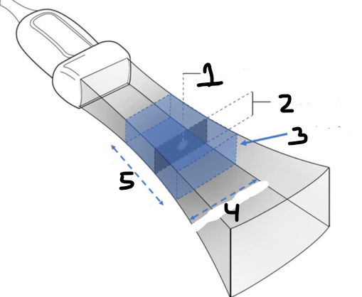
Label this diagram.
focal point
elevational plane
imaging plane
lateral resolution
axial resolution
Axial resolution, lateral resolution, and elevational resolution can be grouped together as…
Detail resolution
SPL is proportional to _____, which is inversely proportional to _____.
wavelength, frequency
What determines the area of best elevational resolution?
The focal point of the elevational plane is determined by the type of the transducer.
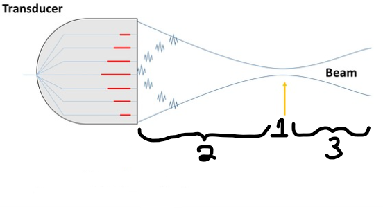
Label the diagram. Provide 3 different terms for #2 and 2 different terms for #3.
focal zone
Fresnel zone / near zone length (NZL) / nearfield
Fraunhofer zone / farfield
What separates the nearfield and farfield?
Focal point
Where along the sound beam should the structure of interest be?
In the nearfield or focal zone.
Write the equation for NZL.
NZL = [D2 (mm) x f (MHz)] / 6
When aperture increases, the focal point moves _____ to the transducer.
closer
Where along the sound beam is the best lateral resolution? Why?
Focal point, ↓area → ↑intensity
Structures within the focal zone have increased ______ and _______ resolution.
lateral, contrast
Define resolution.
Processing or distinguishing somethin into its consituent parts.
List the three types of ultrasound imaging resolution.
detail resolution
contrast resolution
temporal resolution
Give another term for detail resolution.
Spatial resolution
List some factors determining temporal resolution.
FR
FT
PRP/PRF
depth
FOV
#scan lines
scan line density
Improving the ______ will improve contrast resolution.
SNR
Define noise in terms of contrast resolution. Where is it mostly seen?
Low level echoes returning from artifact interactions.
Mostly seen in anechoic spaces
Give an example of why noise in anechoic spaces are detrimental to diagnoses.
Noise artifact can present as pathology.
i.e. noise and thrombus look very similar in anechoic spaces
What are the units of measurement for temporal resolution?
Hz = frames/sec
When an ultrasound image has choppy sequences, the ______ resolution is low.
temporal
Calculate FR if PRP=3ms and #lines=300. Is this scan diagnostic?
PRP = 3ms
#lines = 300
FT = PRP x #lines
FT = 3ms x 300
FT = 900ms
FR = 1/FT
FR = 1/900ms
FR = 0.0011kHz
FR = 1.1Hz
This is very low temporal resolution as there is only 1.1 frames per second. FR<15Hz so this scan is not diagnostic.
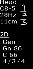
Label the settings listed in this image.
transducer BW in MHz
FR
depth
List some frame time variables that can be changed by the sonographer.
Maximum depth
# of scan lines
transducer type
FOV size
scan line density
multi-focus
THI
CD/PD
At the same depth, which transducer has the highest frame rate? Why?
Sector probe - fewest # scan lines and smallest FOV
Decreasing from 10cm depth to 5cm depth, what happens to FR?
↑FR
If scan line density is increased, what will happen to the FR? What kind of resolution(s) does this change?
↓FR → ↓temporal resolution
↑ lateral resolution
What is a reasonable FR before the human eye is capable of detecting “flicker” or “motion blur” or slow motion effect?
15Hz
<15Hz is not diagnostic because the temporal resolution is too slow.
Define scanned modes of imaging and give some examples.
2D B-mode
3D B-mode
Colour Doppler
Power Doppler
Imaging mode that requires multiple scan lines across a FOV.
Define non-scanned modes of imaging and give some examples.
M-mode
PW and CW Spectral Doppler
Imaging mode that uses one scan line to produce a graph.
Describe the purpose of nuchal translucency measures. What kind of resolution is required to be high?
Measure of the fetus to predict genetic abnormalities. Requires high detail/spatial resolution to keep the measurement accurate.
Axial resolution =
SPL/2
Axial resolution distinguishes to structures on the ____ axis as the sound beam.
same
If SPL=1mm, what is the required distance between structures to be distinguished.
Axial resolution = SPL/2
AR = 1mm/2
AR = 0.5mm
Any structures equal or greater than 0.55mm is distinguished.
The SPL is 3mm, are reflectors 1mm apart distinguished? What about 2mm?
AR = SPL/2
AR = 3mm/2
AR = 1.5mm
1mm<1.5mm → 1mm apart is distinguished
2mm>1.5mm → 2mm apart is not distinguished
Name 2 influences on axial resolution.
transducer damping
change the number of cycles in the pulse → change PD → change SPL
frequency
change wavelength → SPL
Lateral resolution distinguishes to structures on the ____ axis as the sound beam
perpendicular
Lateral resolution =
lateral beam width
Lateral resolution is also called…
transverse resolution
Azimuthal resolution
Sound beams are not uniform in beam width due to _____, which happens in the nearfield, and _____, which happens in the farfield.
convergence, divergence
Lateral resolution is affected by amount of penetration. True/False.
False - axial resolution is impacted by degree of penetration through frequency.
Where is the worst lateral resolution. Why?
Farfield
Sound beams diverge → ↑area → ↓intensity and ↑beam width
What is the smallest distance between laterally adjacent structures that are distinguished?
2mm
Slice thickness refers to which plane?
Elevational plane.
How is the elevational focal point determined in a 1D array vs 2D array?
1D array - focal point is fixed and determined by lenses or curved crystals.
2D array - focal point is not fixed and can be changed to reduce artifact
Reflectors within the elevational plane can cause ______ ______ ______ in the imaging plane.
partial volume artifact
Where is PVA mostly seen in the image?
Anechoic spaces
What is another name for partial volume artifact?
Slice thickness artifact
Explain why a PVA may appear when a vessel is in long axis but not in short axis.
In long axis, the curved radius of the vessel is in the elevational plane, causing the echo to reflect at an oblique angle that enters the imaging plane. The vessel should be anechoic, hence PVA is seen in the lumen.
In short axis, there is no curved reflector in the elevational plane, hence echoes in the elevational plane reflect at a perpendicular angle and do not enter the imaging plane.