15. Parasitic & tumour diseases of digestive system in respective pig categories
1/62
Earn XP
Description and Tags
(Protozoa – Giardiosis, Eimeriosis & isosporosis of piglets, cryptosporidiosis, Balantiosis; Helminths – Strongylosis, Ascaridosis, Hyostrongylosis, Trichurosis, Oesophagostomosis, Macracantorynchosis, papillomatosis, Intestinal adenomatosis).
Name | Mastery | Learn | Test | Matching | Spaced | Call with Kai |
|---|
No analytics yet
Send a link to your students to track their progress
63 Terms
What are examples of protozoa causing digestive disease in swine?
Cystoisospora suis
Giardia duodenalis
Eimeriosis spp.
Balantidium coli
Cryptosporidium spp.
What is the only species of Cystoisospora that infects pigs?
Cystoisospora suis
What is the LC of Cystoisospora suis?
Direct, shed unsporulated oocyst, sporulated oocyst have 2 sporocysts with 4 sporozoites each → piglets ingest sporulated oocyst → located intracellularly in enterocytes of jejunum and ileum
What age of piglets is most susceptible to Cystoisospora suis infection?
5-15 days of age
What are the clinical signs of Cystoisospora suis infection in piglets?
Pasty to watery diarrhoea, yellowish colour, foul and milky smell, decreased body weight gain, unlevelled litter
What diagnostic method is used for Cystoisospora suis infection?
Flotation and coproscopic detection of oocysts, necropsy findings
What is the treatment for Cystoisospora suis?
Toltrazuril 20 mg/kg. Usually unsuccessful.
What hygiene measures should be taken to prevent Cystoisospora suis?
Cleaning and disinfecting farrowing pens
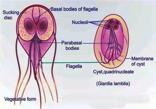
What is the spp of Giardia that infects pigs?
Giardia duodenalis assemblages A-H.
What is the size of Giardia duodenalis?
8-12 µm
What stages of life cycle does Giardia duodenalis have?
Direct life cycle
Trophozoite (replicative) colonises the mucosal surface of the duodenum and jejunum
Cyst (infective) stages
What clinical signs are associated with Giardia duodenalis infection?
Soft to watery faeces with mucus and fat, flatulence, and anorexia
How is Giardia duodenalis diagnosed?
Using the flotation method for cysts (4 nuclei) with Faust's solution for three consecutive days (intermittent shedding) at 400x magnification
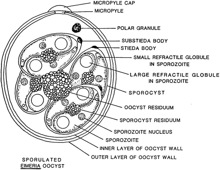
Which Eimeria species infect pigs and where?
Eimeria debliechi (SI, jejunum)
E. polita (SI)
E. scabra (SI, ceacum)
E. spinon.
What is a common clinical sign of Eimeria infection?
Poor growth, sloppy diarrhoea (varies in colour, can be bloody), dehydration, and wasting. None in adults (carriers)
How is Eimeria infection diagnosed?
Flotation (4 sporocysts each with 2 sporozoites)
What is the treatment for Eimeria infection?
Toltrazuril, potentiated sulphonamides
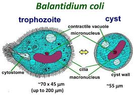
What spp causes Balantiosis in pigs?
Balantidium coli
What is the life cycle of Balantidium coli in pigs?
Cysts are ingested, excystation occurs in the small intestine, and trophozoites colonise the large intestine. Usually asymptomatic
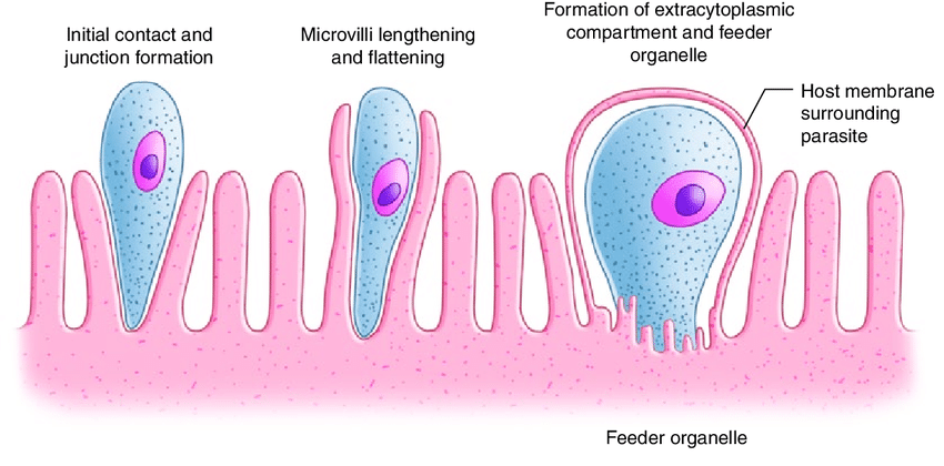
Where does Cryptosporidium predilect in pigs?
Intestine, located intracellularly, but with extracytoplasmic location
Which Cryptosporidium species are significant in pigs?
C. suis, C. parvum, C. scrofarum
What diagnostic method is used for Cryptosporidium in pigs?
Faecal smears stained with Ziehl-Neelsen or Kinyoun stain (better for pigs). PCR, ELISA (coproantigens)
What is the treatment for Cryptosporidium infections in pigs?
No specific anti-cryptosporidial drugs; treatment may include antibiotics like spiramycin and halofunginone
What is a common characteristic of cryptosporidiosis in immunocompetent hosts?
It is usually self-limiting
What can severe cryptosporidiosis lead to in very young, old, or immunocompromised pigs?
Long-lasting, severe diarrhoea
What are the helminths affecting the digestive system?
2 - 2 - 1 - 1 rule (SI - LI - Stomach - Lungs)
SI: Ascaris suum, Strongyloides ransomi
LI: Trichuris, Oesophagostomum
Stomach: Hyostrongylus
(Lungs: Metastrongylus)
What is the most common species of Strongyloides in pigs?
Strongyloides ransomi
How are Strongyloides transmitted to pigs?
Percutaneously (L3), perorally via faeces (no migration), and lactogenic
What are the routes of migration/transmission of Strongyloides?
Tracheal
Somatic → lactogenic transmission
Autoinfection
What is characteristic of the parasitic Strongyloides and where does it develop?
Parasitic Strongyloides are female-only and develop in the small intestine
What happens during lactogenic infection of Strongyloides in pigs?
Hypobiotic larvae in the mammary glands are transmitted to piglets through milk, with no migration in the host (sow)
What is autoinfection in Strongyloides?
L1 larvae hatch from eggs in the intestine and cause reinfection without leaving the host
What are the clinical signs of heavy Strongyloides infections in pigs?
Blood-streaked, mucoid diarrhoea, retarded growth, unsightly skin, dehydration, damage to lungs (haemorrhages & inflammation), and death
What diagnostic methods are used for Strongyloides in pigs?
Egg or larval detection in faeces using flotation, larvoscopic methods (Baermann), Koga agar plate, serology, and PCR
What treatments are available for Strongyloides in pigs?
Ivermectin, doramectin, levamisole, thiabendazole
What is the most common pig parasite?
Ascaris suum
Where do adult Ascaris suum parasites live in pigs?
In the lumen of the small intestine, predominantly the jejunum
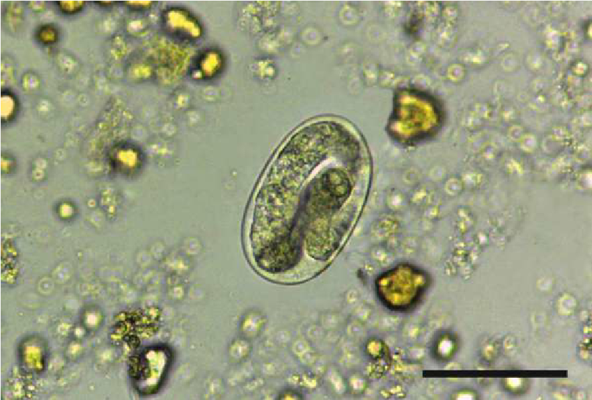
What is the life cycle of Ascaris suum?
Eggs with infectious L3 enter the host orally, undergo entero-hepatic-pulmonary migration, and mature in the small intestine
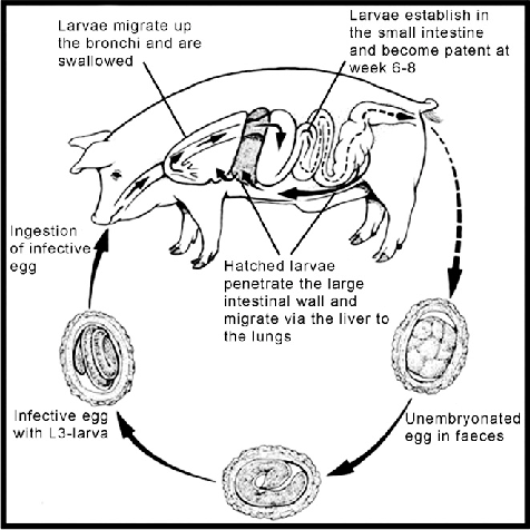
What are the pathogenesis effects of Ascaris suum in pigs?
Liver (milk spots), lungs (verminous pneumonia and haemorrhages), and intestine (thickening and atrophy)
What are the clinical signs of Ascaris suum infection in pigs?
Usually asymptomatic, but may include osmotic diarrhoea, reduced food intake, respiratory signs, and poor weight gain
How is Ascaris suum controlled in pigs?
Deworming after weaning (week 3), regrouping (week 8), week 58, and three times a year for sows/boars using benzimidazoles and macrocyclic lactones
What is the spp of Oesophagostomum in pigs and where does it predilect?
Oesophagostomum dentatum and is found in the colon and caecum
What is characteristic of Oesophagostomum dentatum?
Forms nodules in colon and caecum
What are the clinical signs of Oesophagostomum dentatum infection in pigs?
Frequently asymptomatic, but heavy infections cause inappetence, weight loss, diarrhoea, reduced weight gain, reduced feed efficiency, reduced litter sizes, and reduced milk production
What diagnostic method is used for Oesophagostomum dentatum?
Egg detection in faeces using flotation and coproculture for L3 differentiation
What treatments are available for Oesophagostomum dentatum?
Benzimidazoles (fenbendazoles), levamisole, macrocyclic lactones (ivermectin), and targeted treatment with nitroxinil or pyrantel due to resistance
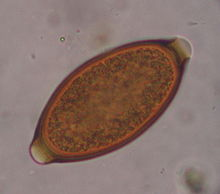
Name the Trichuris spp and where it infect pigs?
Trichuris suis (whipworm), infects the large intestine of pigs
What is the life cycle of Trichuris suis?
L1 hatches from the egg in the small intestine, cecum, or colon, invades the mucosa, moults to L2, and establishes under the epithelial layer to become adults
What are the clinical signs of heavy Trichuris suis infections in pigs?
Mucous diarrhoea, weight loss, colitis, hypoalbuminaemia, and retarded development
How is Trichuris diagnosed?
Flotation. Detection of lemon-shaped egg
What is the main treatment for Trichuris suis in pigs?
Benzimidazoles (several doses to target adults) and macrocyclic lactones
What Hyostrongylus spp and where does it infect pigs?
Hyostrongylus rubidus (red stomach worm), found in stomach
Which group of pigs are more commonly affected by Hyostrongylus?
Living outside
Intensive rearing
How is Hyostrongylus rubidus transmitted?
Ingestion of eggs
What are the clinical signs of Hyostrongylus rubidus infection in pigs?
Haemorrhagic gastritis with diarrhoea and anaemia.
Larval stage irritate glands (hypertrophic stomatitis), adult on mucosal surface (haemorrhages)
What spp is the giant thorny-headed worm and how does it infect pigs?
Macracanthorhynchus hirudinaceus, attaches to the intestinal mucosa and penetrates the intestinal wall, causing inflammation
What intermediate host carries the eggs of Macracanthorhynchus hirudinaceus?
Dung beetle
What are the clinical signs of Macracanthorhynchus hirudinaceus infection in pigs?
Low-level infections are often asymptomatic, but heavy infections may cause inappetence and weight loss
What treatments are available for Macracanthorhynchus hirudinaceus in pigs?
Levamisole and ivermectin
How can Macracanthorhynchus hirudinaceus be differentiated from Ascaris suum?
The anterior end has a retractable proboscis with recurved hooks. (Can be differentiated from Ascaris by pushing on the proboscis)
What are examples of tumours affecting the digestive system in swine?
Papillomatosis
Intestinal adenomatosis
What is papillomatosis in pigs?
A rare condition characterised by solitary or multiple nodules on the face and genitals caused by the papilloma virus
What is intestinal adenomatosis in pigs?
A common diarrhoeal disease in growing-finishing and young breeding pigs characterised by hyperplasia and inflammation of the ileum and colon caused by Lawsonia intracellularis