AP1 CH15: Special Senses
1/100
There's no tags or description
Looks like no tags are added yet.
Name | Mastery | Learn | Test | Matching | Spaced | Call with Kai |
|---|
No analytics yet
Send a link to your students to track their progress
101 Terms
Gustation
- Taste
- Molecules have to be dissolved in water
- Flavor in food involves smell, texture, and appearance
- Cranial nerves VII, IX, X send taste sensations to the gustatory nucleus of the medulla oblongata
Tongue and taste bud diagram
Sweet, salty, bitter, sour, and savory (umami)

Ageusia
Absence of taste
Hypogeusia
Decrease in taste sensation, is not strong
Causes of ageusia and hypogeusia
- Upper respiratory tract infection
- Drugs: valium, amphetamines, tranquilizers, etc.
- Injury to unmyelinated nerves or to cranial nerves leading from oral mucosa to brainstem
- Oral infection/inflammation
- Mucositis damages microvilli
- Nutritional deficiencies: zinc, copper, nickel
- Inherited (Riley-Day Syndrome)
Treatments for ageusia and hypogeusia
-Treat the pathology/infection
- Address vitamin deficiency or hormonal imbalance
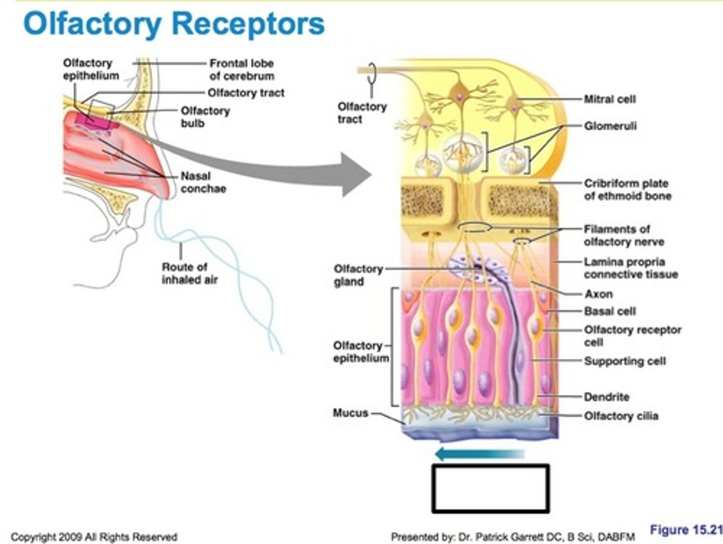
Olfaction
- Smell
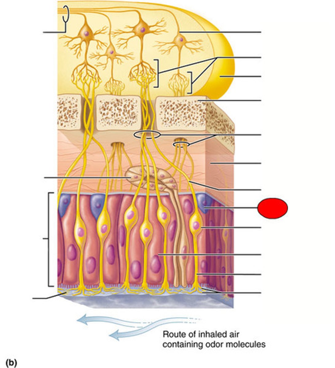
Anatomy of olfaction
Olfactory mucosa on the roof of the nasal cavity contains 10-20 million olfactory neurons, each has 10-20 cilia (olfactory hairs)
Chemicals must be volatile and water-soluble
Physiology of Olfaction
Some odors stimulate nociceptors that trigger the trigeminal nerve
Quick to adapt
Olfaction mechanism
Odorant binds to receptor
Receptor activates G protein
G protein activated adrenylate cyclase
Adrenylate cyclase converts ATP to cAMP
cAMP opens a cation channel, allowing Na+ and Ca2+ influx and causing depolarization

Smell diagram
Anosmia
- Caused by head injuries; tear the olfactory nerves, nasal cavity inflammation, or aging
- Obstruction by polyps; treatment includes topical or systemic corticosteroid or surgical removal of polyps
- Damage to sensory epithelium after severe cold/allergy/smoking
- Zinc deficiency
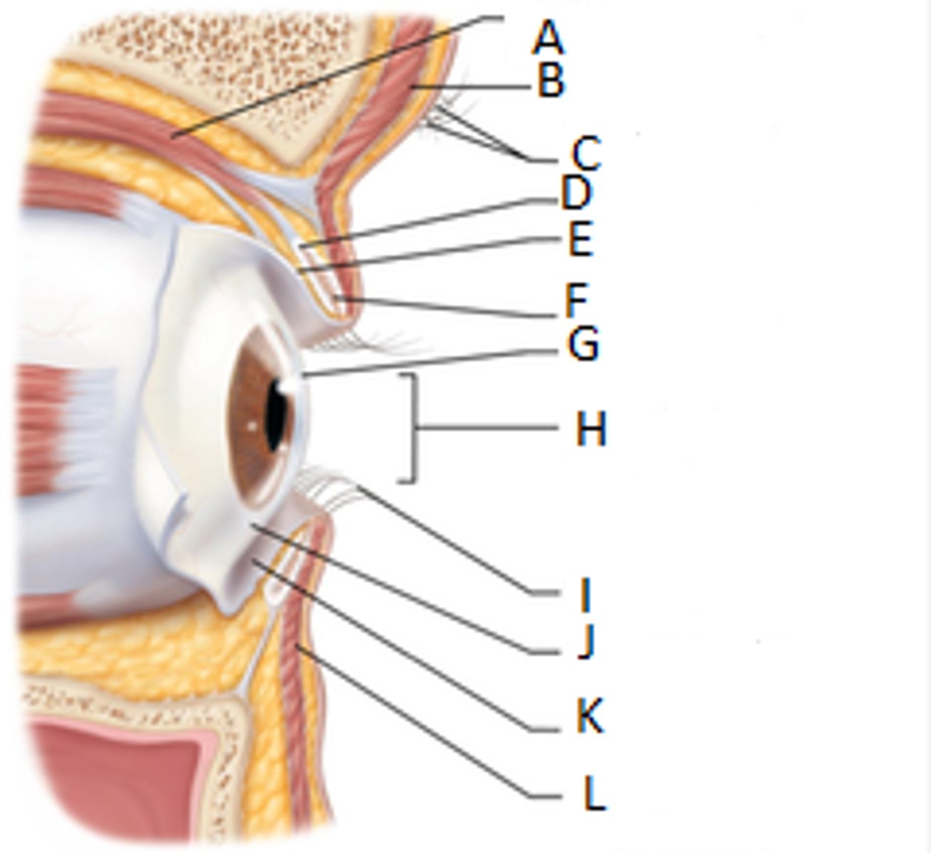
Lateral view of eye
- conjunctiva: lines eyelids as palpebral, lines the eyeball as ocular; lubricates and protects
- lacrimal apparatus: lacrimal gland and associated ducts, secretes tears
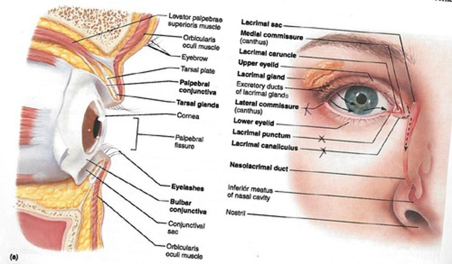
Eyebrow
To reduce glare
Eyelids (palpebrae) and Conjunctiva
Protection
Lacrimal Apparatus
Tear ducts and the glands associated with making tears
To flush the eyes out, keep moist
Contain mucus/antibodies/lysozyme, enter the eye through superolateral excretory ducts, exit the eye medially through lacrimal punctum, and drain into nasolacrimal duct
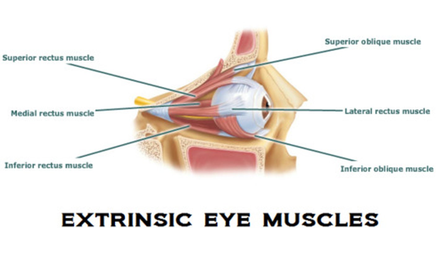
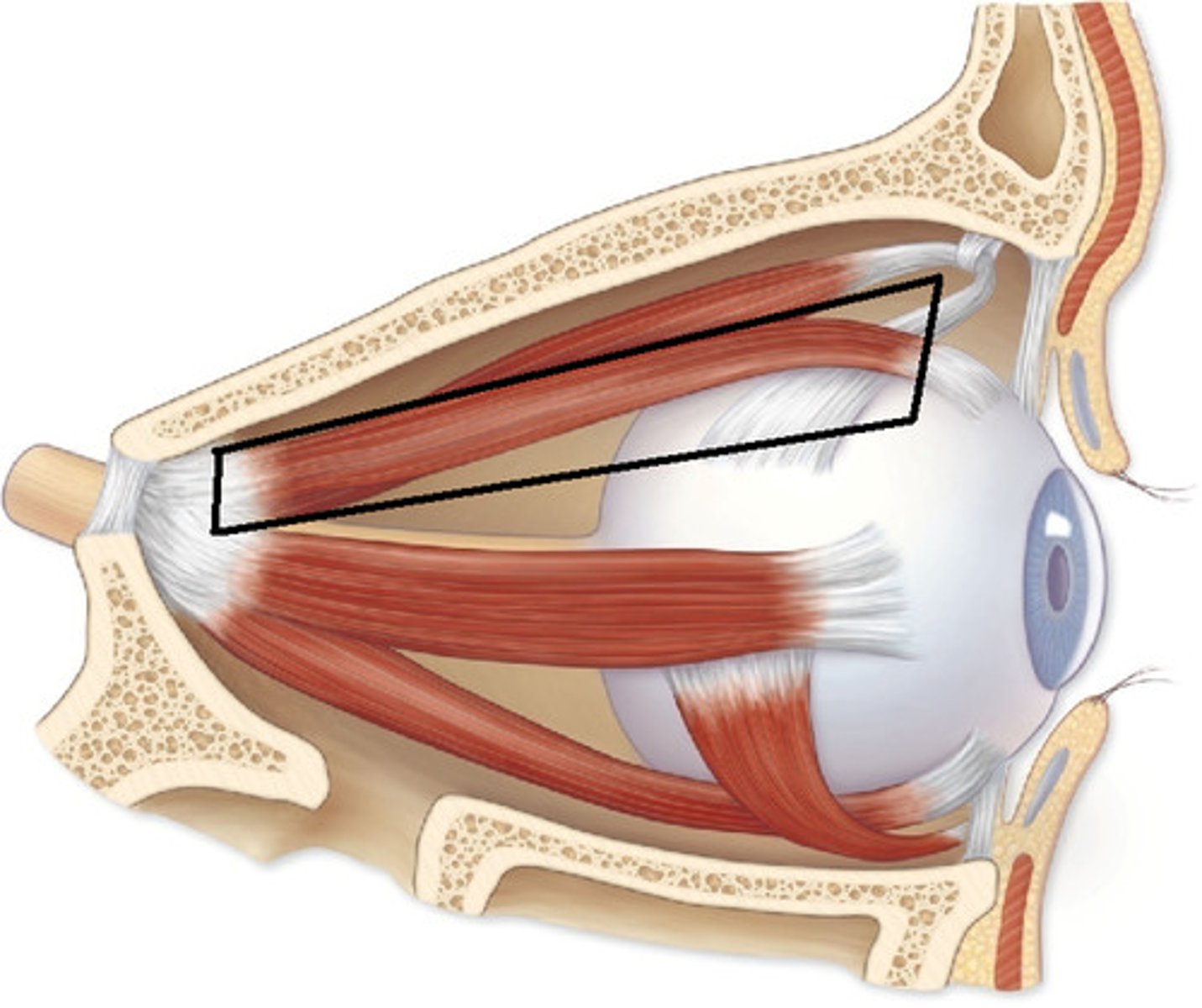
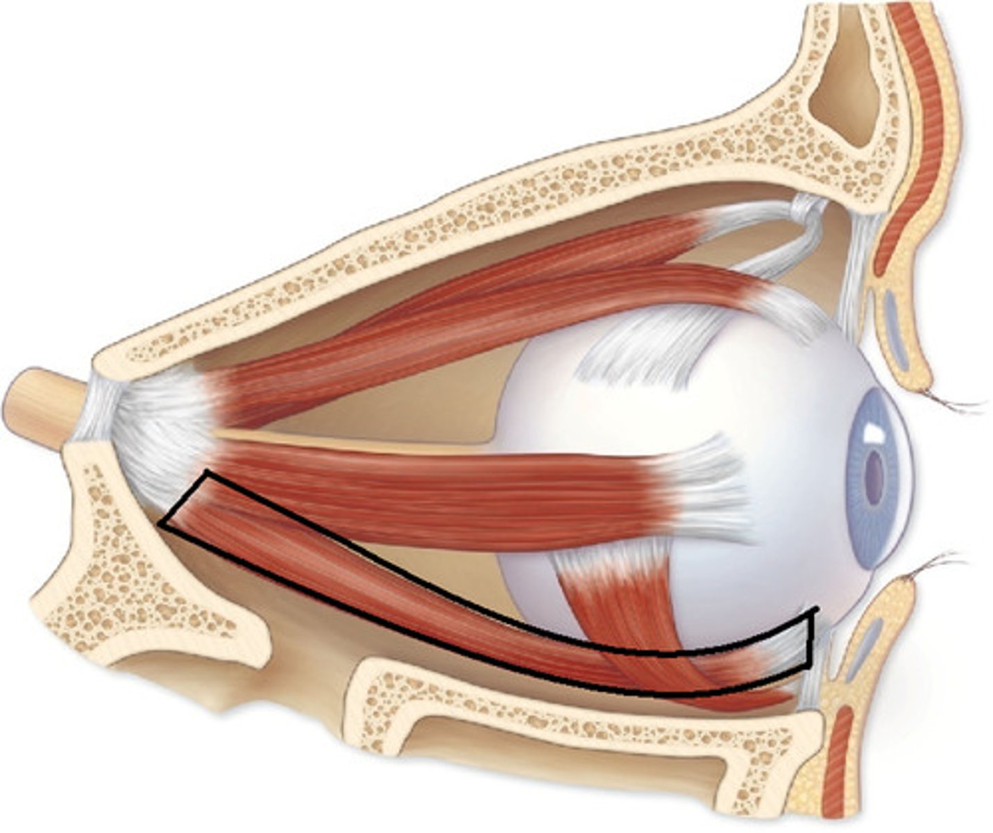
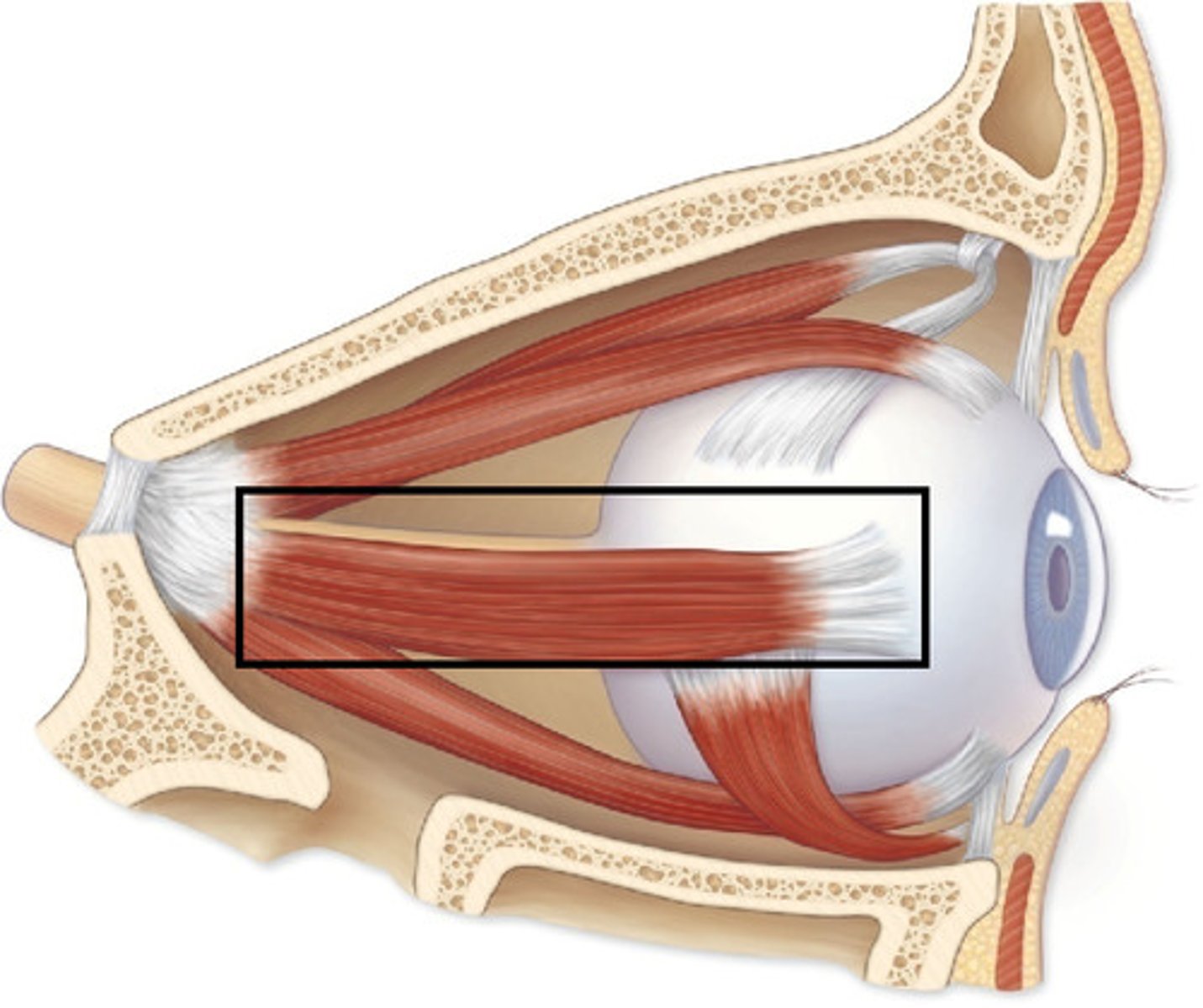
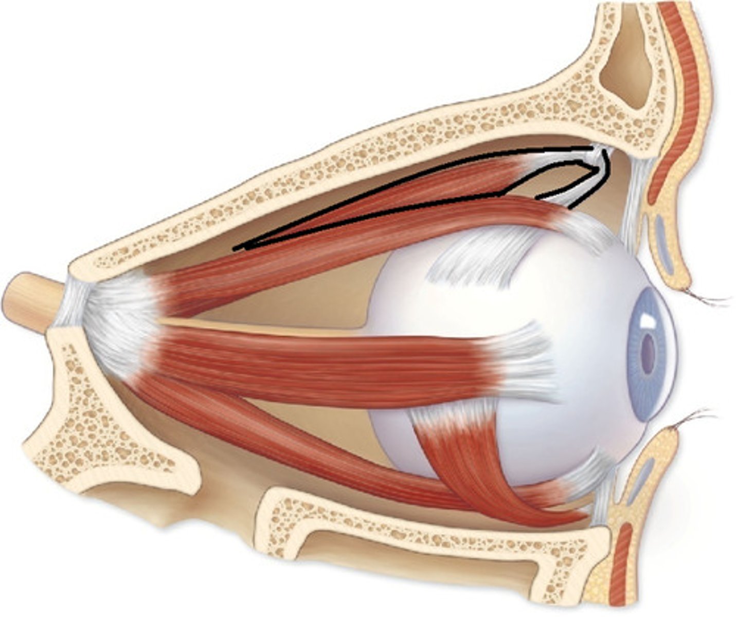
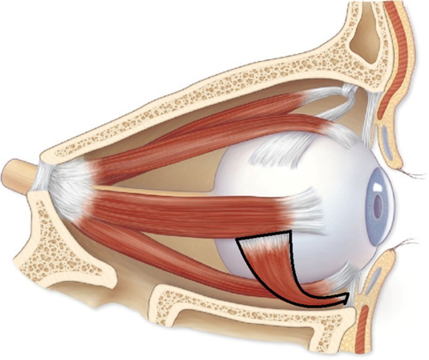
Extrinsic eye muscles
- Two basic types of eye muscles
Saccades movements
Scanning movements: tracking moving objects
Maintains shape of eyeball
Saccades
Small, jerky movements
Superior rectus
Elevates eye and turns it medially
Inferior rectus
Depresses eye and turns it medially
Medial rectus
Moves eye medially

Lateral rectus
Moves eye laterally
Superior oblique
Depresses eye and turns it laterally
Inferior oblique
Elevates eye and turns it laterally
Conjunctivitis
Pink eye
Contagious, can be caused by infection
Treatment: eye drops, medicine
Strabismus
- Congenital weakness of the external eye muscles
- Affected eye rotates medially/laterally
- Treatments: eye exercises, surgery
Olfactory cilia
Receptors for smell

Olfactory sensory neuron
A bipolar (double-ended) nerve cell that captures odorant molecules and initiates the neural signals for smell.
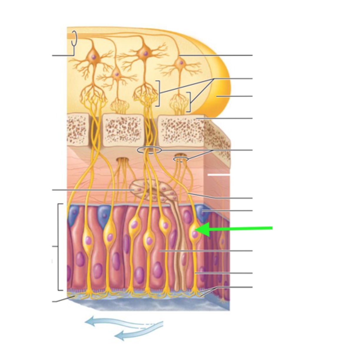
Olfactory stem cell
Replace old sensory neurons every few months
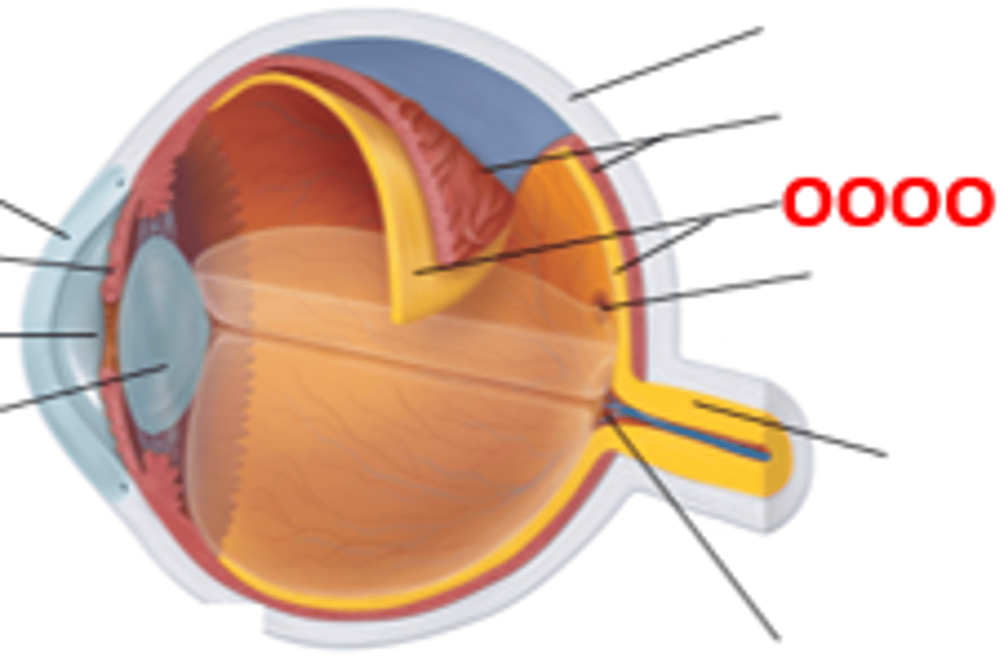
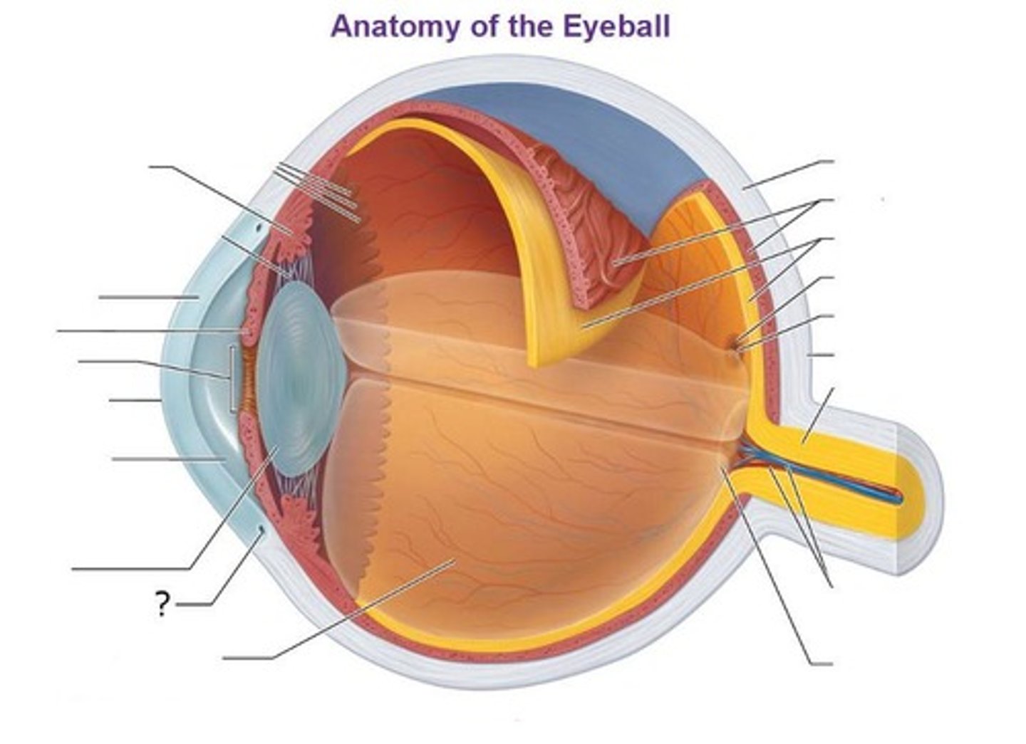
Anatomy of the eye
- Three tunics (layers)
- Tunica fibrosa
- Tunica vasculosa
- Tunica interna
~ Ciliary body
~ Iris controls the diameter of the pupil, contains chromatophores with varying quantities of pigment
Tunica fibrosa
Outer layer that includes sclera and transparent cornea
Tunica vasculosa
Middle layer that has a layer to keep the inner eye dark, has a choroid, supplies eye tissue with oxygen and nutrients
Tunica internosa
Inner layer that is the retina
Ciliary body
Forms a muscular ring around the lens, changing the shape of the lens to focus
B

Suspensory ligament
Connects the lens to the ciliary body
Cornea
G, the transparent layer forming the front of the eye
The clear tissue that covers the front of the eye
Iris
E, a ring of muscle tissue that forms the colored portion of the eye around the pupil and controls the size of the pupil opening
- iris is two muscles; sphincter pupillae and dilator pupillae

Pupil
- Sphincter pupillae muscle contracts with parasympathetic activation (pupil constricts)
- Dilator pupillae muscle contracts with sympathetic activation (pupil dilates)
F

Lens
H, the transparent structure behind the pupil that changes shape to help focus images on the retina

Aqueous humor
Anterior segment of eye
G

Vitreous humor
Posterior segment of eye
J
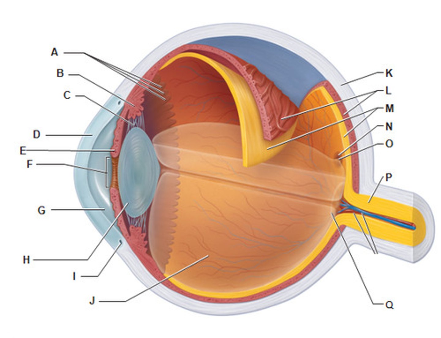
Sclera
K, white of the eye

Retina
- Most posterior layer of the retina is pigmented epithelium
- Neural apparatus has three principal cell layers
- Contains sensory receptors that process visual information and sends it to the brain
retinal cells
- rods: contain rhodopsin (pigment) for colorless vision in dim light
- cones: bright light, sharp color vision
- bipolar cells (middle layer): synapse with photoreceptors and ganglion cells after receiving an image
- ganglion cells (outer layer): receive input from bipolar neurons

Macula lutea
N, a yellowish central area of the retina that is rich in cones and that mediates clear detailed vision
- Directly posterior to the lens
- Contains the fovea centralis

Fovea centralis
O, area consisting of a small depression in the retina containing cones and where vision is most acute

Optic nerve
The nerve that carries neural impulses from the eye to the brain

Optic disk
A hole in the retina where the optic nerve fibers exit the eye, no vision here (blind spot)
Q

Choroid
L; Middle, vascular layer of the eye, between the retina and the sclera

Central artery and vein of the retina
19
Supply retina with nutrients, only area in body where blood vessels can be directly observed
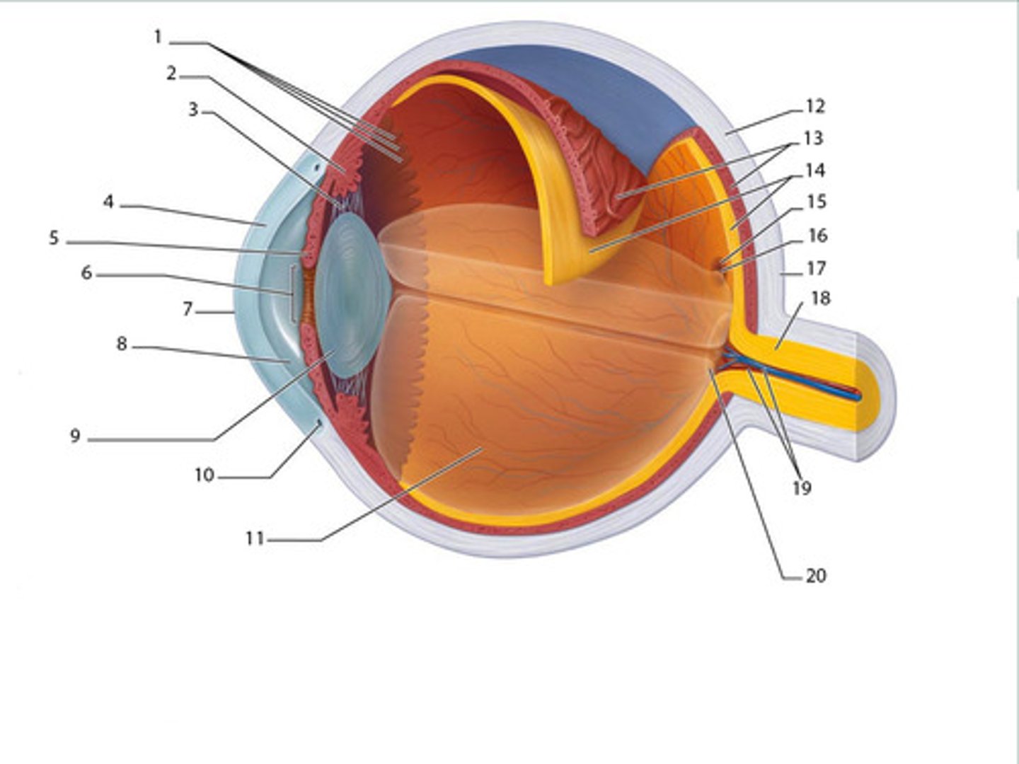
Ora serrata
A, the serrated boundary between the ciliary muscle and the retina

Suspensory ligament
C, attaches the lens to the ciliary body

Scleral venous sinus
Drains the aqueous humor from the eye
Optical apparatus
Admits and refracts light rays, then focuses them on the retina
Parasympathetic stimulation
Constricts the eye with the sphincter pupillae muscle
Sympathetic stimulation
Dilates the eye with the dilator pupillae
Neural apparatus
Has three principal layer
Rods
Contain the pigment rhodopsin
Colorless vision in dim light
Cones
Bright light and are responsible for both sharp and color vision
Bipolar cells
The middle layer, are those that rods and cones synapse with after receiving an image
Ganglion cells
Receive input from bipolar neurons
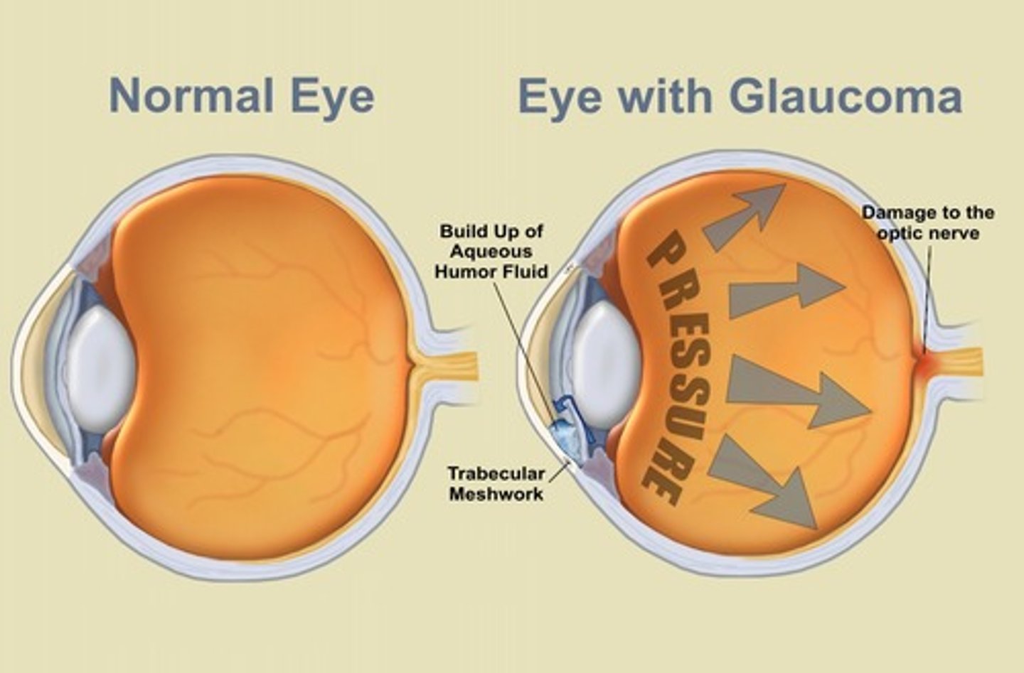
Glaucoma
- Drainage of aqueous humor is blocked, pressure increases, retina and optic nerve compressed, looks like tunnel vision
- Treatment: eye drops (miotics) increase rate of aqueous humor drainage, and possible surgery
Cataract
- "Waterfall"
- Lens thickens and hardens: inadequate nutrients to deep lens fibers, clumping of crystallin proteins
- Treatment: surgically remove defective lens, replace with artificial one
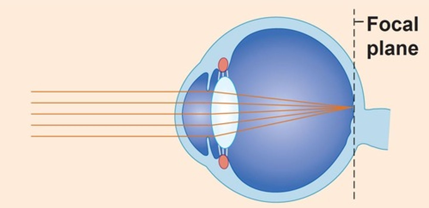
Emmetropic eye
Normal, focal point on retina
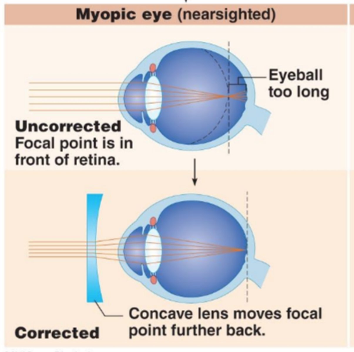
Myopic eye (myopia)
Eyeball too long, causes myopia (nearsightedness), focal point is in front of the retina; concave lens
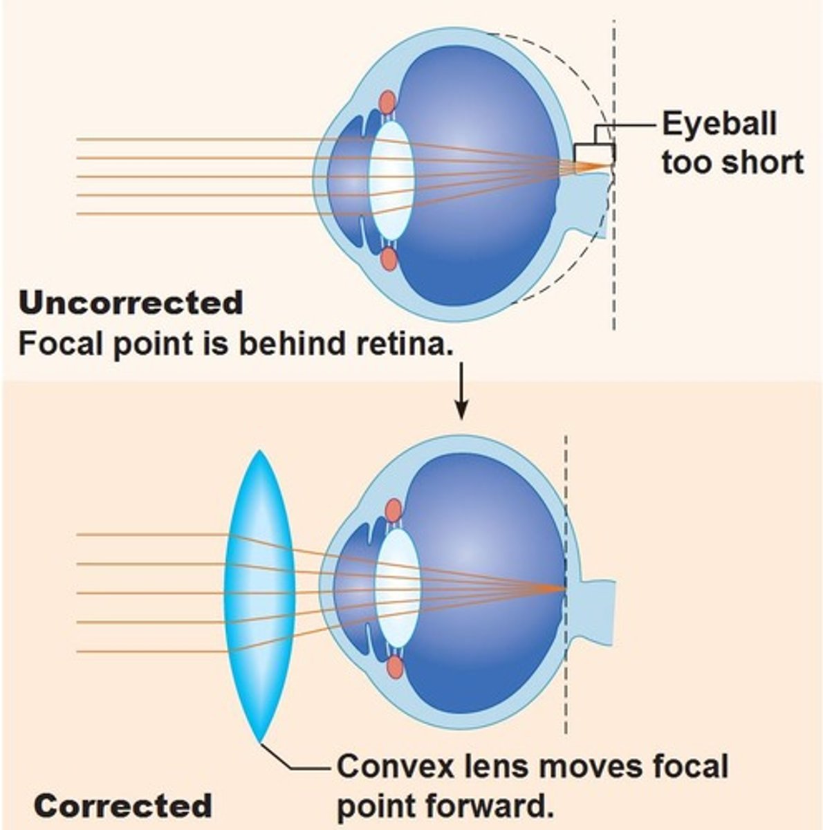
Hyperopic eye (hyperopia)
Eyeball too short, causes hyperopia (farsightedness), focal point is behind the retina; convex lens
Nyctalopia
- Night blindness
- Rod function is defective
- Most common cause is prolonged vitamin A deficiency, rod degeneration
Image formation
Pupillary constrictor narrows the pupil to admit less light to the eye
Pupillary dilator widens the pupl to admit more light
Photopupillary reflex
Photo pupillary reflex
Reflex; the constriction of pupils when they are exposed to bright light, consensual
Refraction
- As light enters the eye, it's refracted by the cornea
- The aqueous humor doesn't reflect light well
- Most of the bending of light occurs in the lens
Near response
- Lens must change shape to focus light on the retina (flattens for distance, bulges for close) by means of ciliary muscle
- Convergence of the eyes orients visual axis to focus on the fovea centralis of each eye
- Lens accommodates; constriction of the pupil adjusts the amount of light, reduces spherical aberration
- Elasticity decreases with age
Near point of vision
The closest an object can be and still come into focus
Color vision
- Made possible by three sets of cones named for the absorption peaks of their visual pigments
- Blue: 420nm
- Green: 531nm
- Red: 558nm

Color blindness
- Lack of one or more cone types
- Most common in males (8-10%)
- Red-green is most common
Stereoscopic vision
Combination of two retinal images to give a 3-D perceptual experience; depth perception

Visual projection pathway
- The optic nerves converge to form optic chiasma. Then, fibers continue as optic tracts
- Hemidecussation
- Optic tracts pass around the hypothalamus to the lateral geniculate body of the thalamus
- Second-order neurons arise here to form optic radiation of fibers in the cerebrum
- These projects to the primary visual cortex of the occipital lobe (conscious perception of image)

Hemidecussation
Occurs within the chiasma, is an X
Sound
Any audible vibration of molecules
Measured in pitch and loudness
External ear
- 3 parts
- Outer auricle
- Auditory canal
- Tympanic membrane (eardrum) is very sensitive to pain
Pitch
- Determined by the frequency of vibration
- Human ears hear from 20-20,000 HZ (hertz)
Loudness
- Perception of amplitude (intensity) of frequency
- Loudness is expressed in decibels, with 120-140 causing pain in most people
Hearing loss
Prolonged exposure to sounds over 90 dB
Auricle
The outer projection, funnels vibrations toward the auditory canal and eardrum
External auditory canal
Leads to eardrum, lined with protective ceruminous glands and hairs
Middle ear
- Malleus (hammer)
- Incus (anvil)
- Stapes (stirrups)
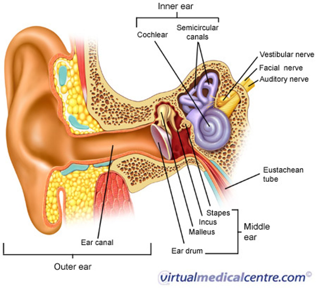
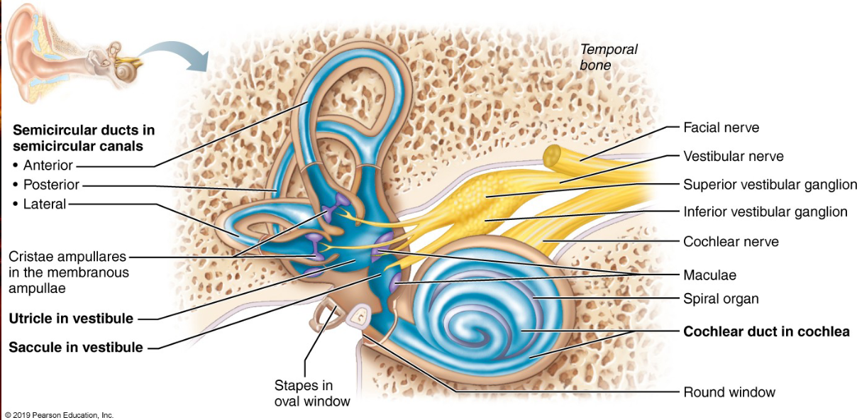
Inner ear
- Housed within temporal bone
- Between bone and membrane is a fluid called perilymph
- Endolymph fills the chamber with the membranous labyrinth
Perilymph
Fluid between bone and membrane of inner ear
Endolymph
Fills chamber with the membranous lymph
Organ of Corti
- Basilar membrane supports the organ of Corti containing hair cells, each with stereocilia
- The tips of the stereocilia shear against an overlying tectorial membrane
- IHCs and OHCs
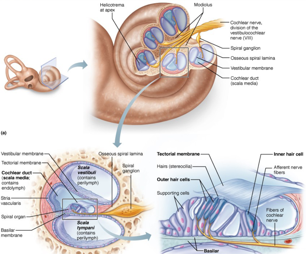
Inner hair cells (IHCs)
Contains stereocilia, send actual hearing impulses
Outer hair cells (OHCs)
Contains stereocilia, adjust the response of the cochlea to different frequencies
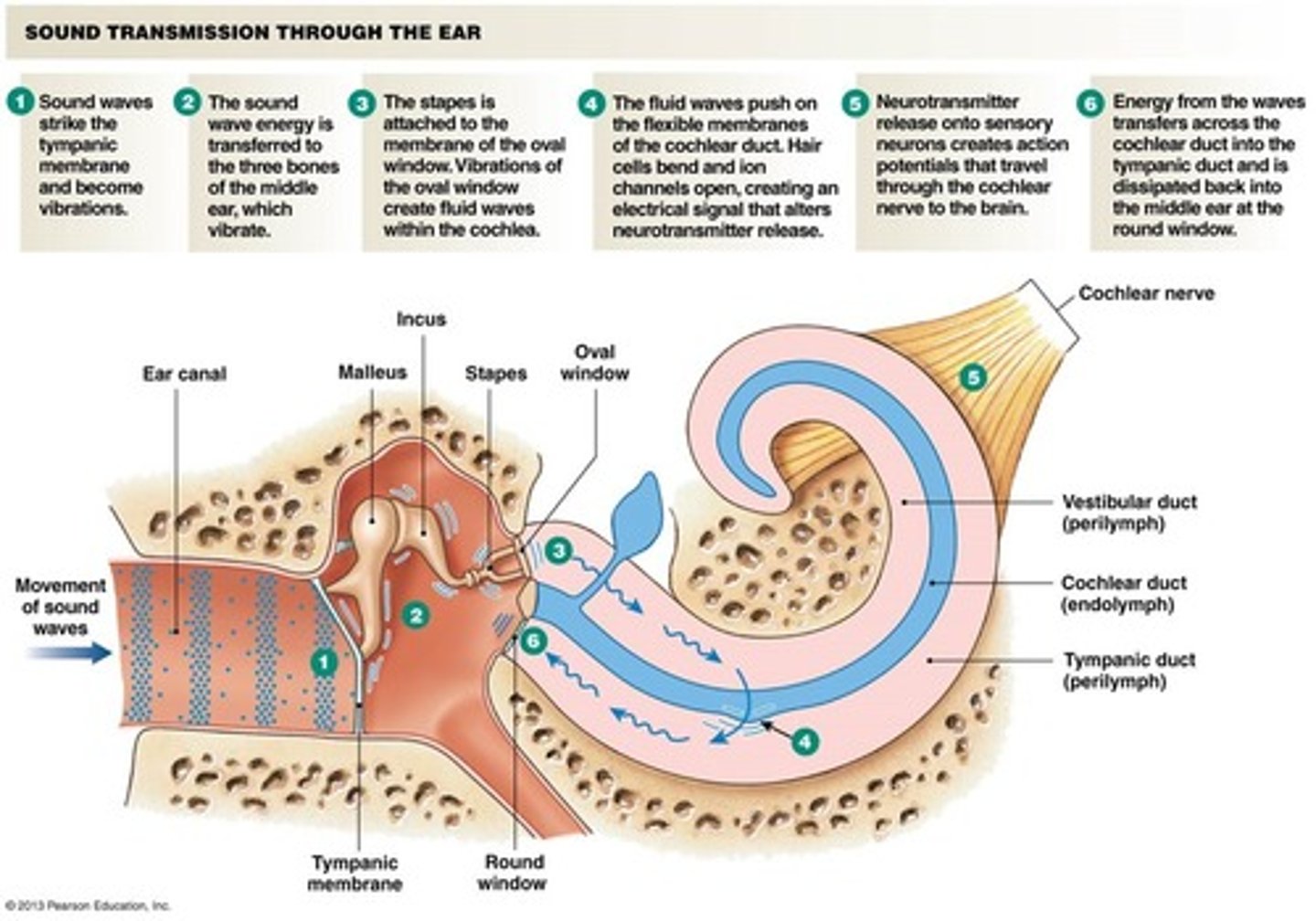
Physiology of hearing
- Auditory ossicles function: to concentrate the energy from the eardrum to a smaller oval window:
The ossicles and eardrum are protected by the tympanic reflex in response to loud noises (not effective for sudden loud noises)
The middle-ear muscles tighten up before you speak to protect your ears from the volume of your own voice
Stimulation of Cochlear Hair Cells
- Auditory ossicles vibrate against the oval window (sets up vibrations within the fluid-filled inner ear)
- Endolymph of cochlear duct vibrates, causing hair cell stereocilia to move against tectorial membrane
- Bending of stereocilia causes depolarization of hair cell; bending in the opposite direction closes the potassium ion channel while the cell hyperpolarizes
- Hair cell releases neurotransmitter during depolarization, generating an AP to cochlear nerve
Sound transduction
- Loud sounds produce vigorous vibrations of Organ of Corti, exciting more cells over larger area
- Higher AP frequency = loud sound
- A sound causes a standing wave in the basilar membrane
- Lower frequency sounds cause a peak amplitude at distal end of the organ of Corti
- Higher frequency sounds are detected closer to the proximal end
Conduction deafness
Hinderance to sound conduction (earwax buildup, perforated eardrum)
Earaches, otitis media and otosclerosis of ossicles
Surgical treatment, replace ossicles or excess tissue
Sensorineurial deafness
Direct damage to neural structurews
Presbycusis, the inability to hear high-pitched sounds
Common because of age and exposure to loud noises
Cochlear implants
Equilibrium
Static and Dynamic Equilibrium
Maculae and utricle (horizontal) and saccule (vertical) respond to changes in gravity and linear acceleration
Static equilibrium
When you’re sitting or standing, still
Dynamic equilibrium
During movement
Meniere’s syndrome
Labyrinth disorder affecting both semicircular canals and the cochlea
required attacks of vertigo (dizziness), nausea, vomiting, difficulty standing, tinnitus (ringing or clicking in ears)
Treatment: mild = attention drugs, moderate = low-salt diet/diuretics to decrease endolymph volume, severe = surgically drain excess endolymph