Retinal Physiology
1/85
Earn XP
Description and Tags
These flashcards cover key concepts from the lecture on Retinal Physiology, focusing on eye anatomy, functions, and related conditions.
Name | Mastery | Learn | Test | Matching | Spaced |
|---|
No study sessions yet.
86 Terms
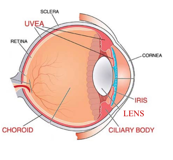
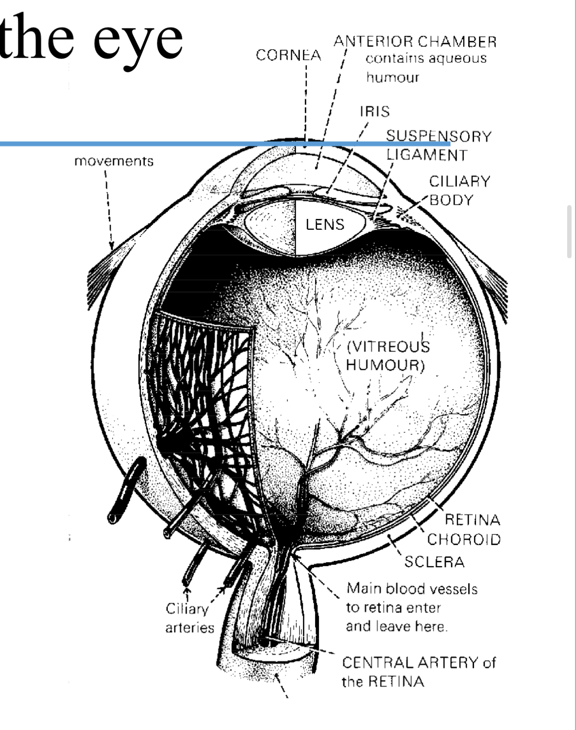
What are the three layers of tissue that enclose the eye?
The outer layer (cornea and sclera), which fuses with dura around the optic nerve and meet at the limbus
the middle layer (uvea) which is pigmented
the inner layer (retina).
What is the primary refractive surface of the eye?
What does light go through before touching the retina
The cornea.
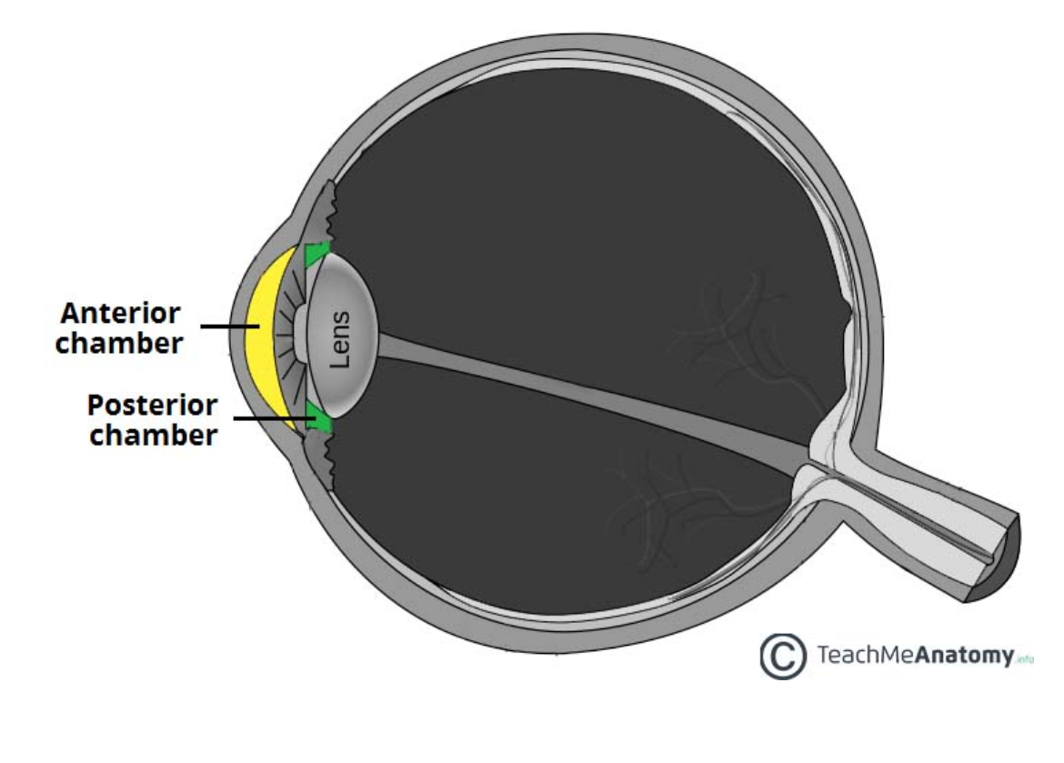
Through the cornea, into the anterior chamber with aqueous humour,
then into posterior chamber through lens
Then into vitreous humour
Then retina

What fluid fills the anterior chamber of the eye? What’s it made and secreted by ?
Where is the AC and PC
Aqueous humour - made and secreted by the ciliary body.
AC is between iris and corneas innermost surface
PC is a small space anterior to lens but behind iris with aqueous humour too
What composes the middle layer of the eye?
The uvea, which includes the iris, the ciliary body, and the choroid.
What is the role of the vitreous humour?
A watery gel and it holds the shape of the eye fixed and rigid to maintain focusing accuracy.
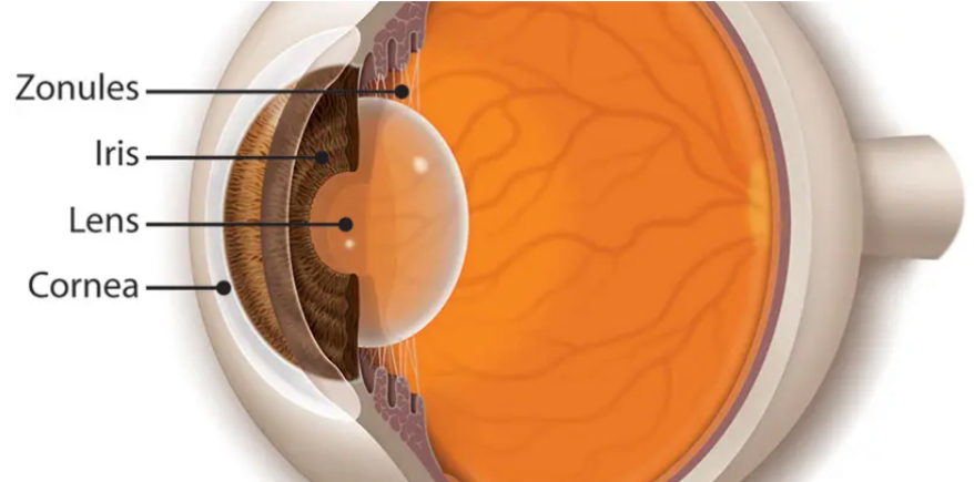
What is the cornea and how is the shape maintained ?
The main refractive / focusing surface of the eye , since medium changes from air to transparent tissue
Surface is kept spherical by intraocular pressure between 10 and 21 mm HG
Where is the choroid and what does it contain
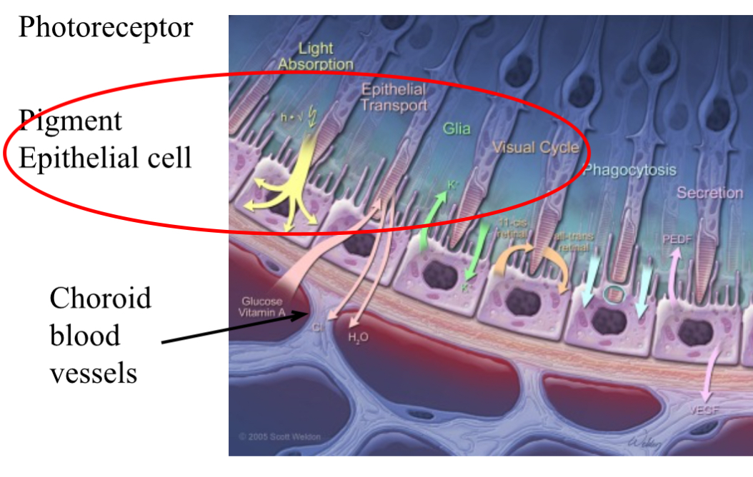
It sits between the sclera and the retina. and it contains pigment epithelial cells which prevent light scattering and reflection
It also contains blood vessels to supply the retina
How does the ciliary body keep the shape of the lens
Has smooth muscle bundles that shape the lens . It also produces and secretes aqueous humour into the posterior chamber.
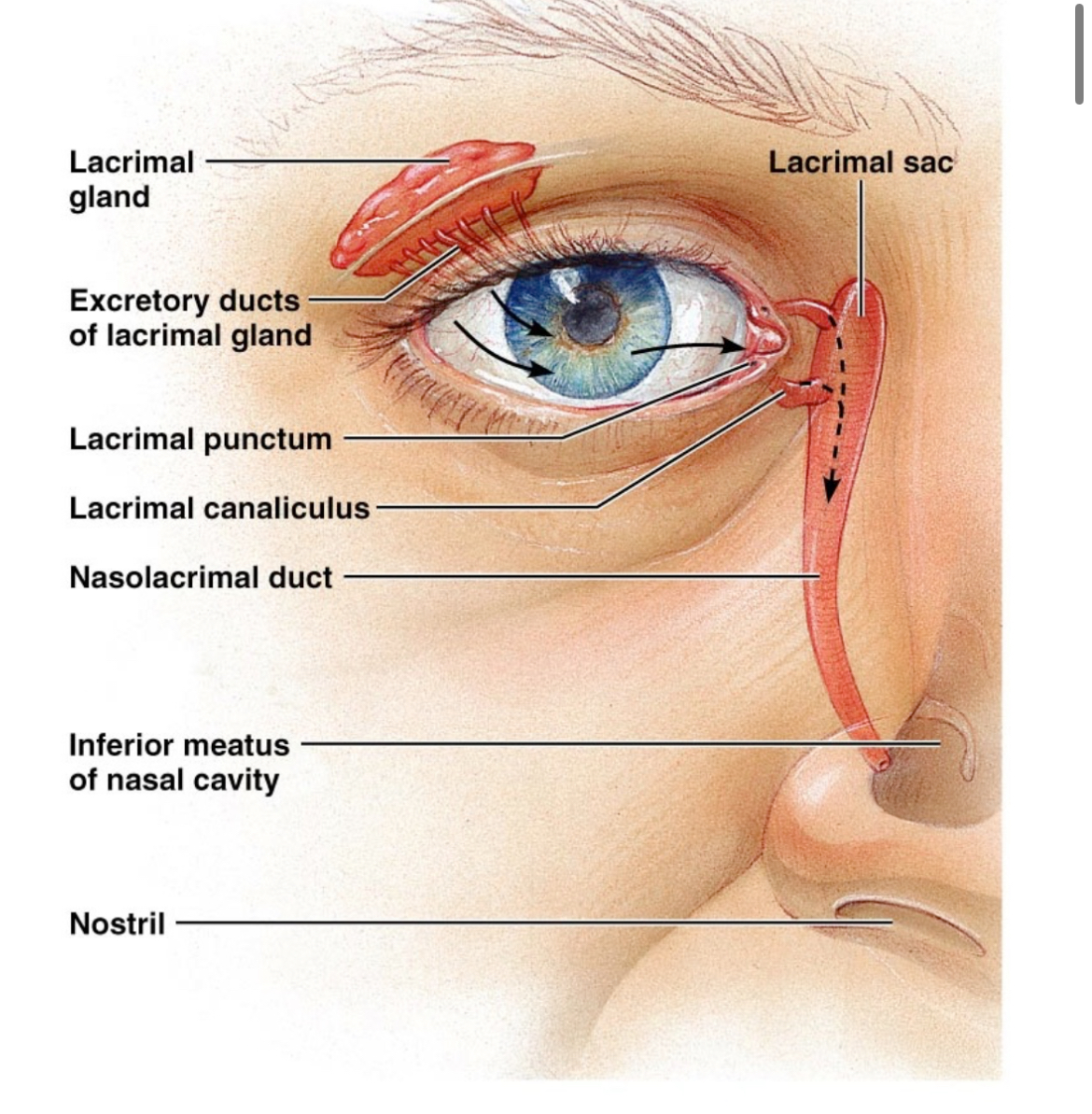
What glands secrete tears?
The lacrimal glands.
How does the pupil function in relation to light?
The iris constricts or dilates the pupil, controlling the amount of light that passes through.
What is the function of goblet cells in the conjunctiva?
They produce mucin, which is part of the tear film.
What type of cells are in the conjunctiva
Stratified columnar epithelium, goblet cells and capillaries
What does the lens do
What is it held in place by
How does the shape change
Allows eye to focus on objects at various distances
Zonular fibrils
Changes due to ciliary muscles - flattens for distance and bulges for close
What is the distance range of pupil diameter?
3-7 mm.
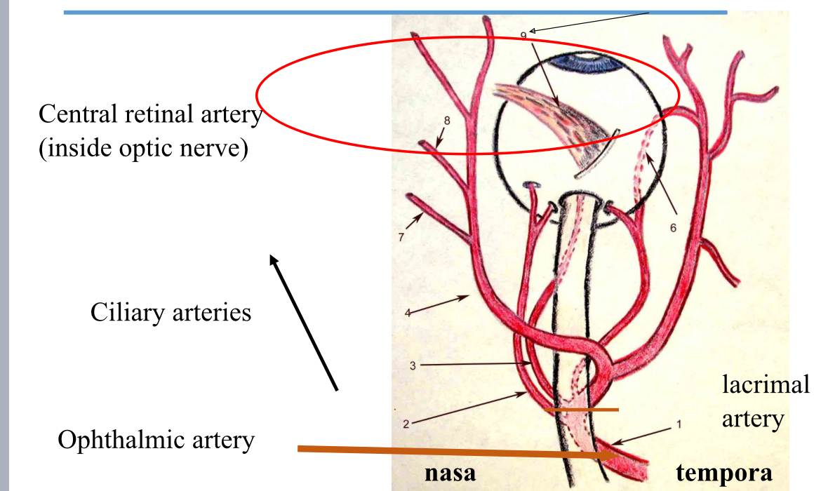
What supplies the inner retina with blood?
The central retinal artery.
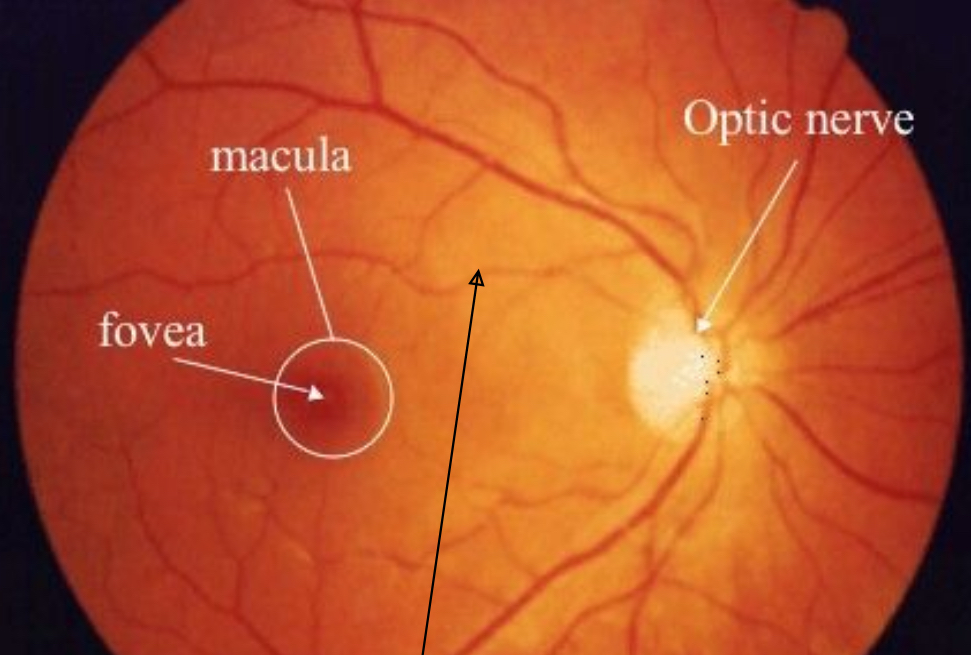
What is the nerve fibre layer made of in the retina
Ganglion cell layer axons which meet at the optic disc to form the optic nerve
What are the two main types of photoreceptors in the retina?
Rods and cones.
What part of the retina has the highest visual acuity?
What is it made up of and for what ?
The fovea.
Cones for normal visual activity.
Damage is more serious than to peripheral retinal damage.
What happens to rods in bright light?
They are suppressed in daylight and do not differentiate color.
What is the normal range for intraocular pressure?
10 - 21 mmHg.
What is glaucoma?
Degeneration of the optic nerve due to raised intraocular pressure.
What are the main risk factors for glaucoma?
Age over 40, family history, ethnicity (African, Hispanic, Asian), and eye trauma.
What is the process by which tears drain from the eye?
Tears exit the eye medially via the lacrimal punctum and drain into the nasolacrimal duct.
Where is aqueous humour produced?
The ciliary body.
What are the three layers of the tear film?
Mucin gel layer, aqueous layer, and oil layer.
What is the function of the choroid?
To provide blood supply to the retina and prevent light scattering.
What is cataract formation?
Opacification of the lens which affects vision.
Which artery supplies the eye?
The ophthalmic artery.
What is the role of the ciliary body?
To shape the lens and produce aqueous humour.
What is the difference between rods and cones in terms of light sensitivity?
Rods are very sensitive to light, while cones mediate the ability to see detail and are active in daylight.
What is the blind spot?
The area where visual axons leave the eye to form the optic nerve and where there are no photoreceptors.
Also where vessels enter and exit eye.
What could happen if the drainage of aqueous humour is blocked?
It could lead to increased intraocular pressure and potentially glaucoma.
What happens to the lens during vision focusing for distance?
The lens flattens.
What happens to the lens during vision focusing for near objects?
The lens bulges.
What is the role of pigment epithelial cells in the retina?
To prevent light scattering and supply the photoreceptors.
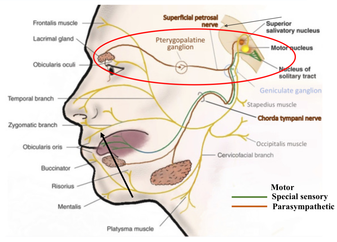
What are lacrimal glands ?
How is secretion stimulated ?
Lacrimal glands secrete tears which provide an optically smooth refracting surface.
Stimulation is stimulated by parasympathetic innervation by the facial nerve CN7.
How do tears enter the eye
Via excretory ducts which exit eye medially into the lacrimal puncture and drain into the nasolacrimal duct.
Where do parasympathetic efferents to the lacrimal gland synapse ?
In the pterygopalatin ganglion.
So parasympathetic nervous system is for rest and digest and efferents carry signals away from the brain to the target organ ( lacrimal glands).
The prepaying ganglion is a cluster of nerve cells located in deep face behind the maxilla. Acts as a relay station to connect to another nerve cell.
Basically nerve signals from the brain tell lacrimal gland to make tears travel through a parasympathetic pathway.
What are the 3 layers of tear film
Mucin gel layer - by conjunctival goblet cells
Aqueous layer - by lacrimal gland
Oil layer - on surface, by meibonian glands on eyelids.
Function of tear films -4
Optically smooth refracting surface
Removes debris from ocular surface
Has antibodies and lysozymes to prevent bacterial growth
Allows transmission of oxygen on the vascular cornea
what adaptation is there for the eye ( photoreceptors) to cope with high oxygen demands ?
the retina has a DUAL blood supply
What is a dual blood supply
the inner retina, ganglion and bipolar cells are supplied by central rental artery.
Photoreceptors supplied by choroid
Both the central retinal arteries and ciliary arteries branch off the ophthalmic artery - which is a branch off the internal carotid artery.
What is the Choroid
Network of capillaries supplied by the ciliary arteries which penetrate the sclera at the back of the eye.
What is the function of the aqueous humour?
To provide nutrition to the avascular cornea and help maintain intraocular pressure.
What is the blood supply to the photoreceptors and how
Oxygen diffuses into photoreceptors from the choroid capillaries. Through layer of cells known as pigment epithelial cells
What is the primary component of the outer segment of photoreceptors?
Disks of membrane containing photopigments (rhodopsin in rods and opsins in cones).
What happens to the worn out cells of the photoreceptors
Epithelial cells phagocytose the worn out ends of the photoreceptors and transfer the debris into the capillaries of the choroid .
What happens in phototransduction when light hits the photoreceptors?
It leads to hyperpolarization of the cell and a decrease in glutamate release.
What cells are photoreceptors?
What cells are in fovea
Rods and cones
Cones
What are rods
There is only one type and they are sensitive to light.
They are active in the dark and have no colour differentiation. Night vision.
Low resolution becUse many rods synapse to one bipolar cell
What do cones do ?
When are they active ?
help to see detail
Active in the day light and there are three types - red and green and blue to different wavelengths
High resolution , one cone is one bipolar cell.
3 iodopsin pigments
What does the stacking arrangement of discs do for photo-reception on rod and cone cells ?
Allows light to pass though in sequence, so higher chance of photon interacting with molecule of pigment.

Why do cones have a shorter outer segment ?
As there is enough light in the day to guarantee a photon will react with a photo pigment
In the dark how does depolarisation of photoreceptors occcur
There is a constant inward leak of sodium in the outermost part of the receptor. It keeps the cell depolarised and tonically releasing glutamate from the synaptic ending.
But light hyper polarises the cell and stops the release of glutamate
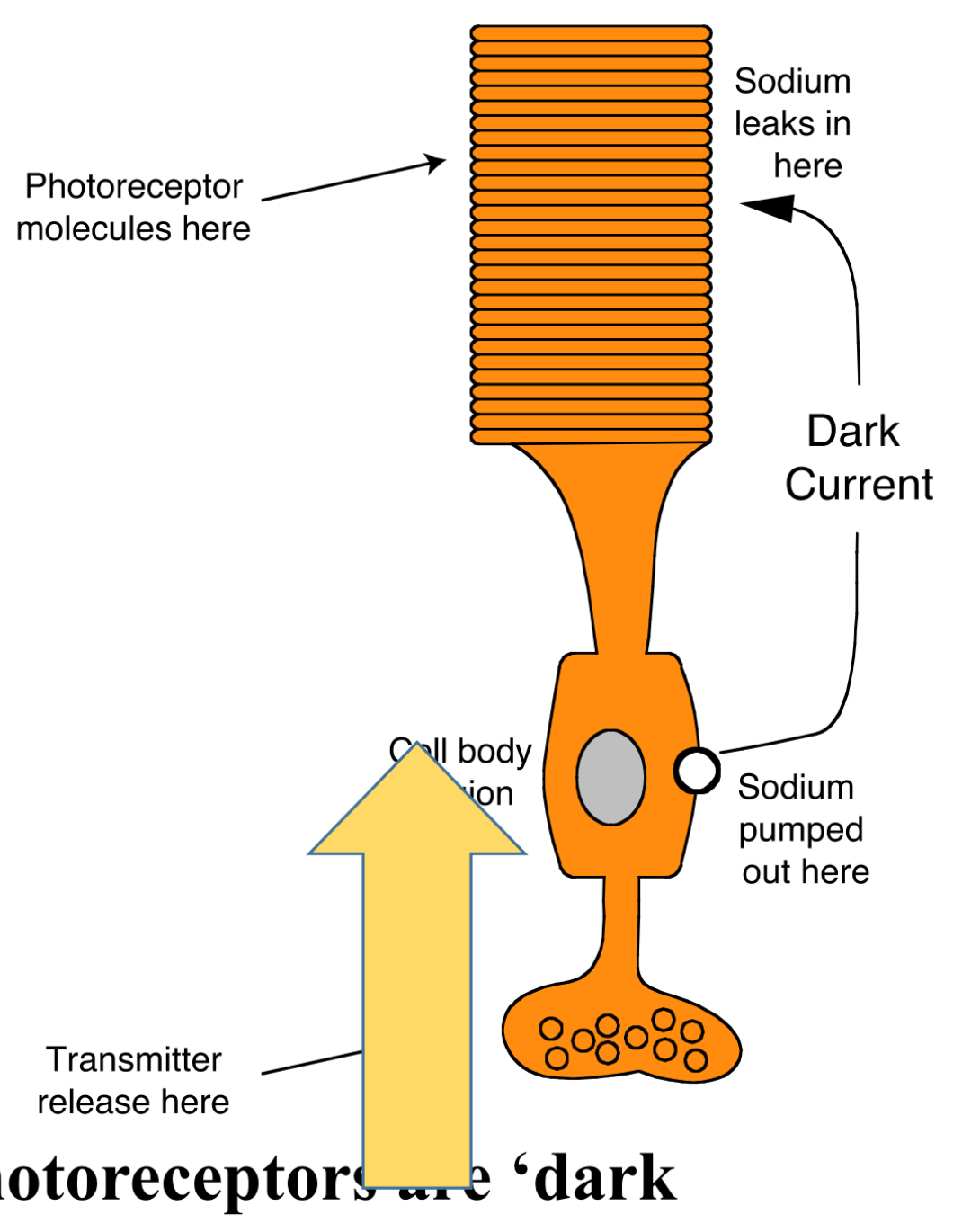
What is the main neurotransmitter released by photoreceptors in the dark?
Glutamate.
What does absorption of light do to rhodopsins shape ? IN RODS
it changes the shape
then acts via a G protein called transduction to reduce the levels of cyclic GMP in the rod.
THIS reduction closes Na channels and allows cell to hyper polarised and stop releasing glutamate.
Inhibits
What does light do cones
It has opsins to absorb light
Explain signal transduction
Basically glutamate is continuously released from synaptic endings at the end of rod and cone cells in the dark. Then bipolar cells depolarise and release neurotransmitters into the ganglion cells.
the ganglion cells project their axons into the optic nerve and therefore signal this light to the brain.
What is signal transduction
how a signal or nerve impluse is passed from one pathway to another.
What’s the difference between off and on bipolar cells
OFF bipolar cells depolarise in the dark and are inactive when light is present.
ON bipolar cells depolarise in response to light
Difference between ganglion and retinal cells structure
Ganglion cells have axon that project into the optic nerve.
Retinal cells are inter neurons

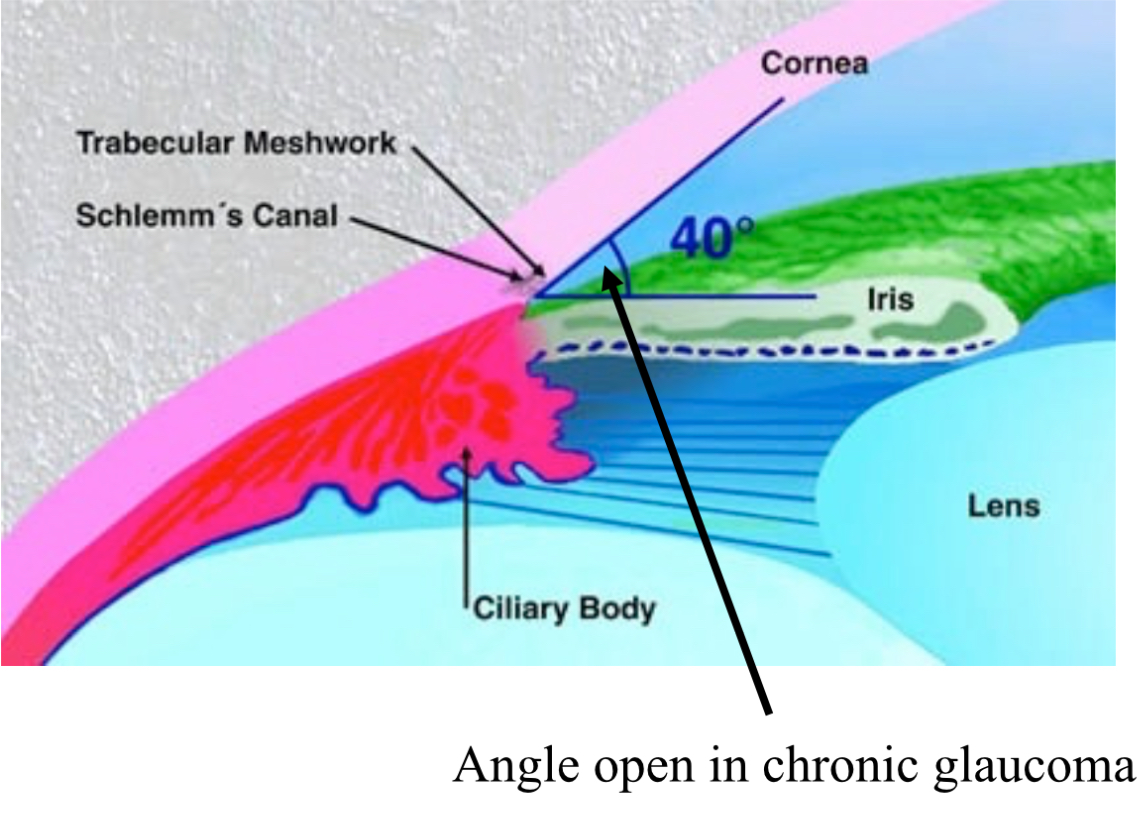
Which two structures are involved in the drainage of aqueous humour?
Trabecular meshwork and canal of schlemm
What is intraocular pressure ?
The difference between formation and drainage of aqueous humour. Normally at 10-21 mug
What happens when drainage of fluid is blocked ?
Intraocular pressure rises and this leads to a glaucoma
What is a glaucoma
Degeneration of optic nerve due to raised intraocular pressure.
What are the primary types of glaucoma?
Primary open-angle glaucoma and primary angle-closure glaucoma.
Difference between primary open angle glaucoma and primary closed angle glaucoma ?
primary open angle is slow and progressive. When the trabecular mesh work is gradually blocked. The angle between iris and cornea remain the same.
Primary closed angle is when the agile between the cornea and iris is reduced. So fluid cannot pass through canal of schlemm . More painful and vision loss.
What is the role of myelinated nerve fibers in signal transduction from photoreceptors?
They carry signals to the brain about light sensation.
What types of medications are used to treat glaucoma?
Prostaglandin analogs, beta-adrenergic receptor antagonists, alpha2-adrenergic agonists, and carbonic anhydrase inhibitors.
What do glaucoma meds do
Reduce pressure by reducing the formation of aqueous humour or increasing drainage - normally in the form of eyedrops
Although a laser trabeculoplasty can be done to unblock the trabecular mesh work.
What is the consequence of prolonged exposure to ultraviolet light on the eye?
It can catalyze free radical formation which may damage the lens, leading to cataracts.
Also lower levels of antioxidants can cause them
Risk factors of cataracts
Uv light , lower antioxidant levels. Diabetes and hypertension.
Vit c and E reduce risk because it increases antioxidants . Alkaline nature of Aq humour activates vitamin c
What are some signs and symptoms of cataracts?
Clouded vision, night vision difficulties, sensitivity to light, and seeing halos around lights.
What surgical procedure is performed for cataracts?
Phacoemulsification - which is the removal of the natural lens and replaced with an artificial lens
What does the term 'visual acuity' refer to?
The clarity or sharpness of vision, primarily determined by the fovea.
What type of vision are cones responsible for?
Color vision and high-resolution vision in bright light.
Which component of the eye is avascular?
Cornea.
What is the role of the lens in focusing light?
To fine-tune the focus of light onto the retina.
What is the typical thickness of the central cornea?
540 micrometers.
What symptom may indicate a problem with the vitreous humour?
Flashes or floaters in the vision.
What type of epithelium lines the conjunctiva?
Stratified columnar epithelium.
In the context of ocular anatomy, what does 'myopia' refer to?
Nearsightedness, where distant objects appear blurred.
What role does the ciliary muscle play during accommodation?
It contracts to change the shape of the lens for focusing.
What is one effect of diabetes on eye health?
Increased risk of cataracts and glaucoma.
What are glycosaminoglycans (GAGs) in the context of tears?
Molecules in the mucin gel layer that help stabilize the tear film.