cr - image review
 Call Kai
 Learn
Learn Practice Test
 Spaced Repetition
 Match
Match Flashcards
 Knowt Play
1/17

There's no tags or description
Looks like no tags are added yet.
Name | Mastery | Learn | Test | Matching | Spaced | Call with Kai | Chat |
|---|
No analytics yet
Send a link to your students to track their progress
18 Terms
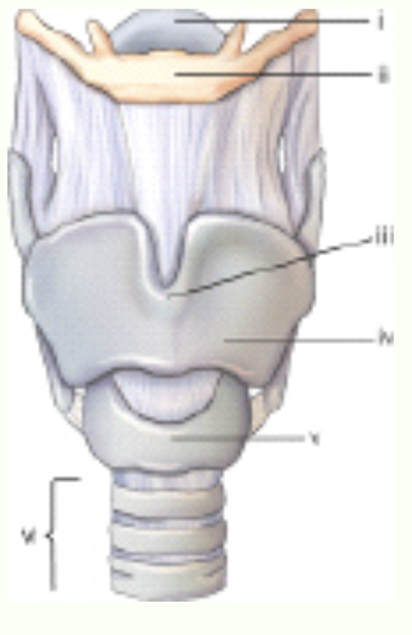
Label this image.
i - epiglottis
ii - hyoid bone
iii -
iv -
v -

Label this image.
1 - fundus
2 -
3 -
4 - cystic duct
5 -
6 -
7 -
8 -
9 - pancreatic duct
10 -
11 -
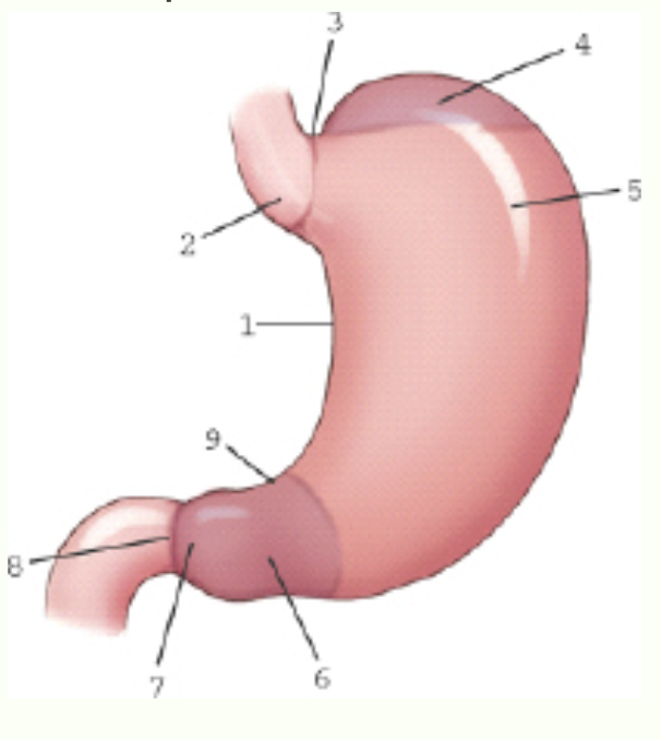
Label this image.
1 -
2 - incisura cardiaca
3 - cardiac notch
4 -
5 -
6 - pyloric portion
7 -
8 -
9 -
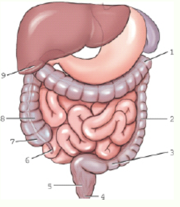
Label this image.
1 - left colic flexure
2 -
3 -
4 -
5 -
6 - vermiform appendix
7 -
8 -
9 -
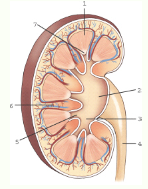
Label this image.
1 - medulla
2 -
3 - major calyx
4 -
5 -
6 -
7 -
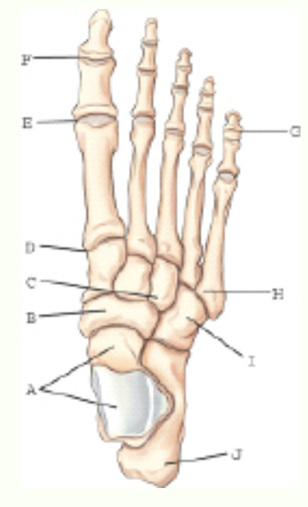
Label this image.
A - talus
B - navicular bone
C - third/lateral cuneiform
D -
E -
F -
G -
H -
I -
J -
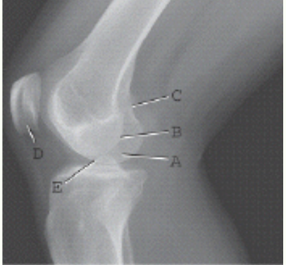
Label this image.
A - medial condyle of the femur
B -
C - adductor tubercle
D -
E -
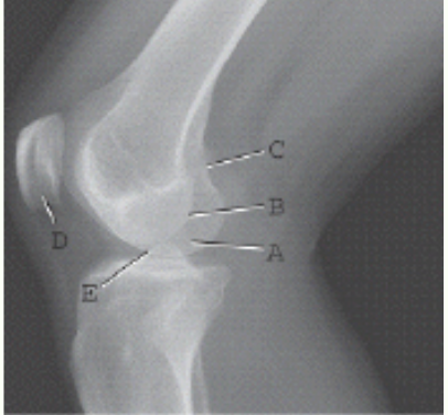
This radiograph represents which of the following positions?
Mediolateral projection of the knee but underrotated toward the IR

Label this image.
A -
B -
C -
D -
E -
F -
G - ischial tuberosity
H -
I -
J - anterior iliac spine (ASIS)
K -
L -
M - ischium
N -
O -
P - ischial spine
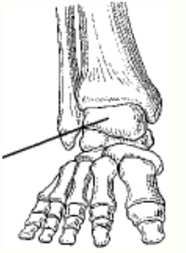
The bone identified in the figure above is the:
talus

Which projection is demonstrated in the figure above?
axial calcaneus (plantodorsal)
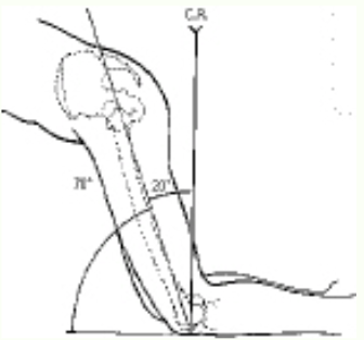
The patient position and central ray shown in the figure above will demonstrate the:
intercondylar fossa

The patient position and central-ray method demonstrated in the figure above is the:
Camp-Coventry (intercondylar fossa)
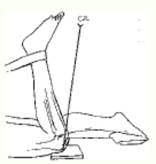
The projection of the patella demonstrated in the figure above is the:
Settegast

Which part of the knee is identified on the figure above?
medial meniscus
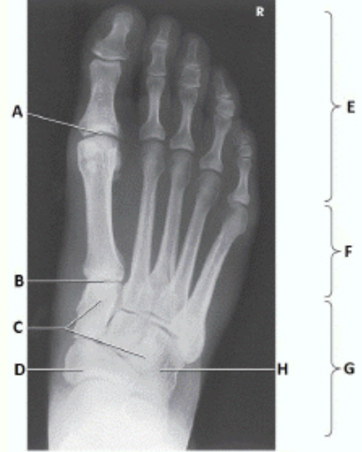
Label this image.
A -
B -
C -
D -
E -
F -
G - tarsals
H -
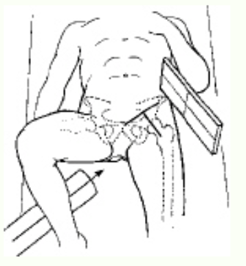
Which projection of the hip is shown in the figure above?
axiolateral

This projection is correctly referred to as:
carpal canal, inferosuperior projection.
carpal tunnel, inferosuperior projection.
Gaynor-Hart method.
all of the above.
all of the above.