Biochemistry II Module of the MCAT Self Prep eCourse: Lesson 7: Membrane Receptors (Pro)
1/39
There's no tags or description
Looks like no tags are added yet.
Name | Mastery | Learn | Test | Matching | Spaced | Call with Kai |
|---|
No analytics yet
Send a link to your students to track their progress
40 Terms
Lesson 7: Membrane Receptors
Lesson 7: Membrane Receptors
Membrane receptors are _________________ that communicate with the outside environment of a cell.
(A) Phospholipid Bilayers
(B) Steroid Hormones
(C) Glycosidic Linkages
(D) Integral Proteins
(D) Integral Proteins
Membrane receptors are Integral Proteins that communicate with the outside environment of a cell.
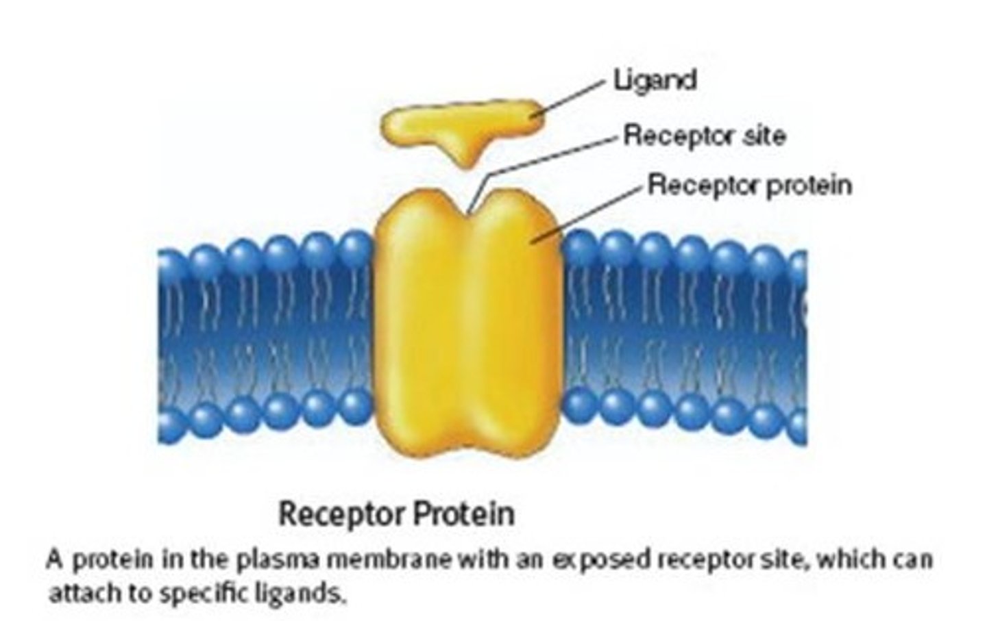
CRB Why could effective Membrane Receptors not be Embedded Proteins?
If Membrane Receptors were Embedded Proteins, then they either would not be able to bind the extracellular signal or would not be able to have any interactions with the inside of the cell.
In other words, an effective Membrane Receptor must both be able to interact with both the extra- and intra-cellular environments!
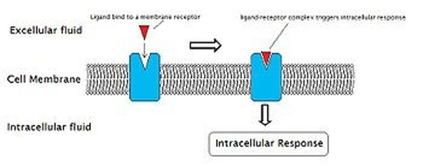
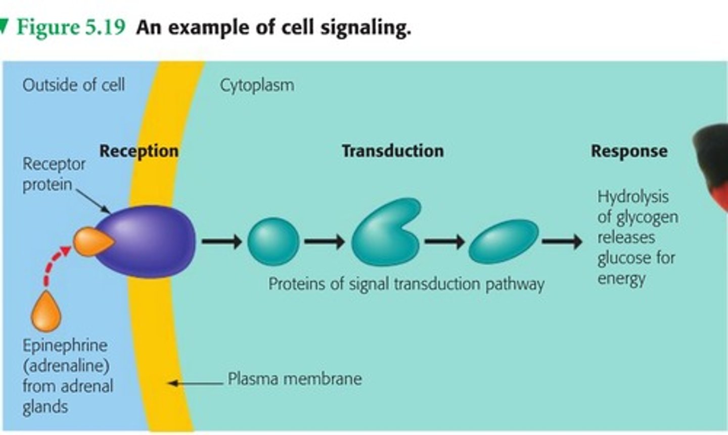
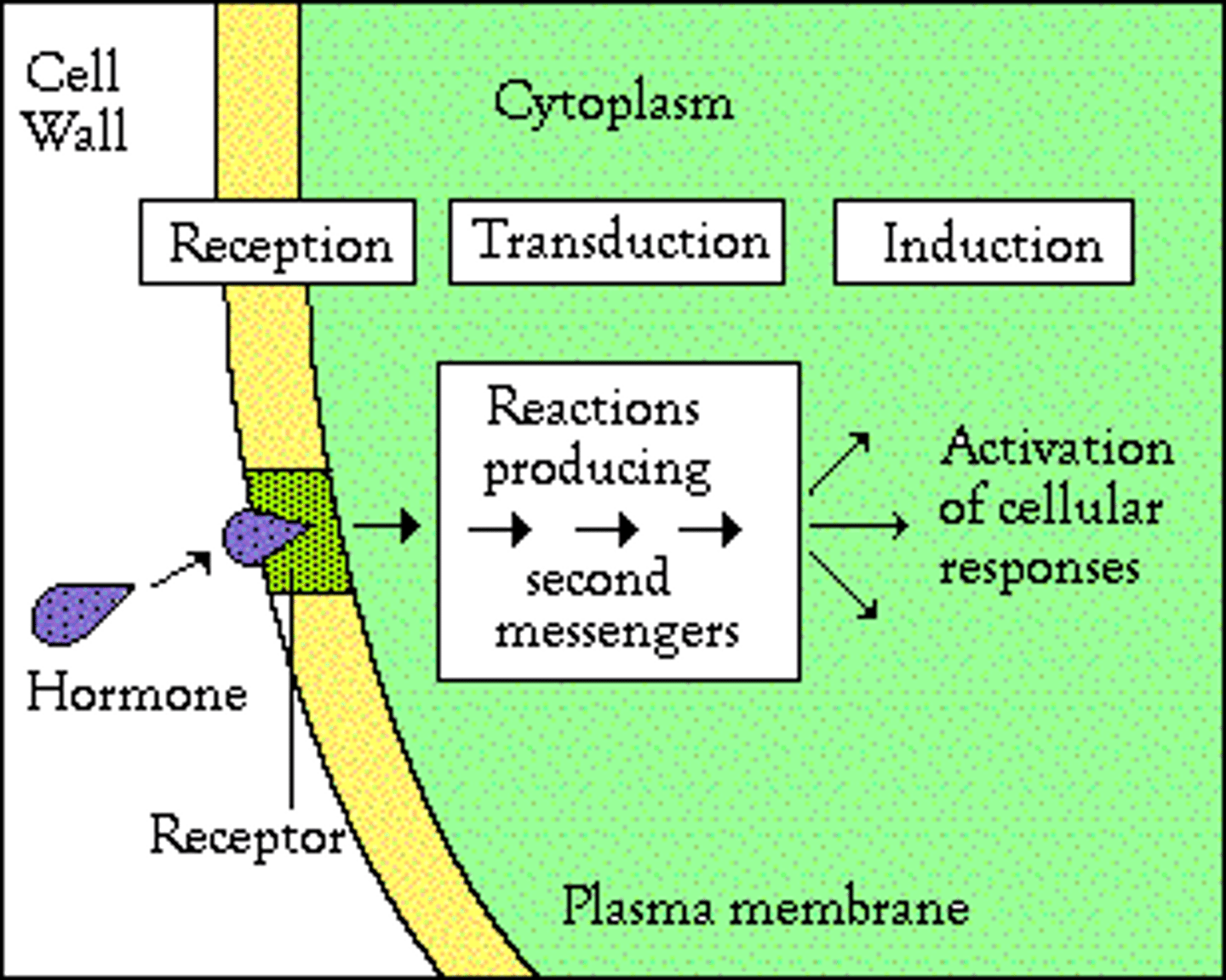
Describe the process of Signal Transduction.
Signal Transduction is the binding of an extracellular ligand to a membrane receptor, creating a ligand-receptor complex. This then results in an intracellular response.
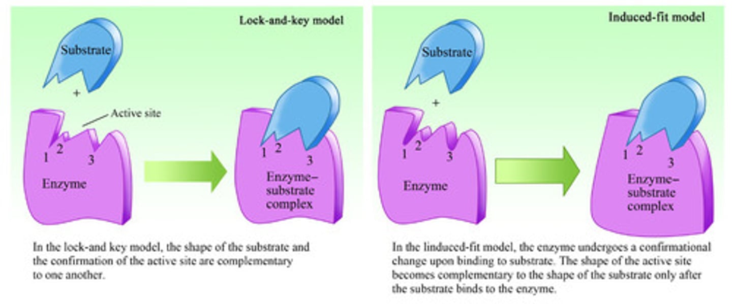
Compare the "Lock and Key" with the "Induced Fit" model.
The "Lock and Key" model is the idea that ligands are perfectly shaped to only fit into certain enzymes (in this case membrane receptors).
The "Induced Fit" model is the idea that ligands are not perfectly shaped but that when they bind to enzymes, the conformation of both the ligand and enzyme will change to fit tightly. This is the "induced fit."
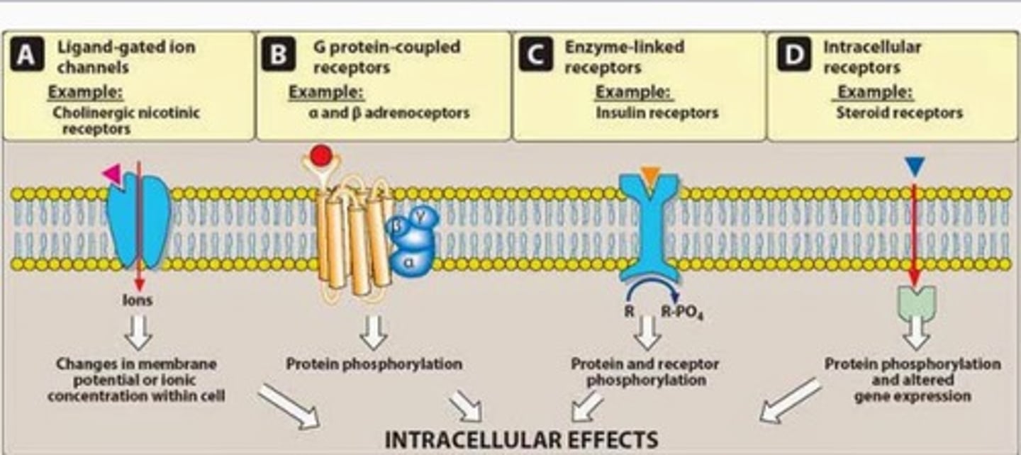
Which of the following are examples of Membrane Receptors?
I. Ligand-gated Ion Channels
II. G-protein Coupled Receptors
III. Enzyme Linked Receptors
(A) I and II Only
(B) II and III Only
(C) I and III Only
(D) I, II, and III
(D) I, II, and III
The following are examples of Membrane Receptors:
I. Ligand-gated Ion Channels
II. G-protein Coupled Receptors
III. Enzyme Linked Receptors

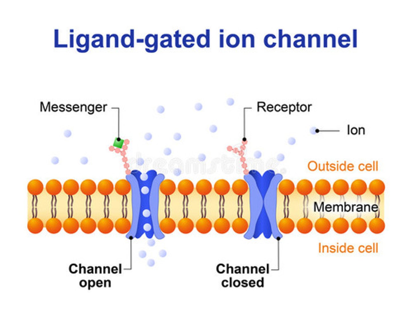
Ion Channel Linked Receptors are also known as:
(A) Ligand-gated Ion Channels
(B) G-protein Coupled Receptors
(C) Enzyme Linked Receptors
(D) Membrane Receptors
(A) Ligand-gated Ion Channels
Ion Channel Linked Receptors are also known as Ligand-gated Ion Channels.
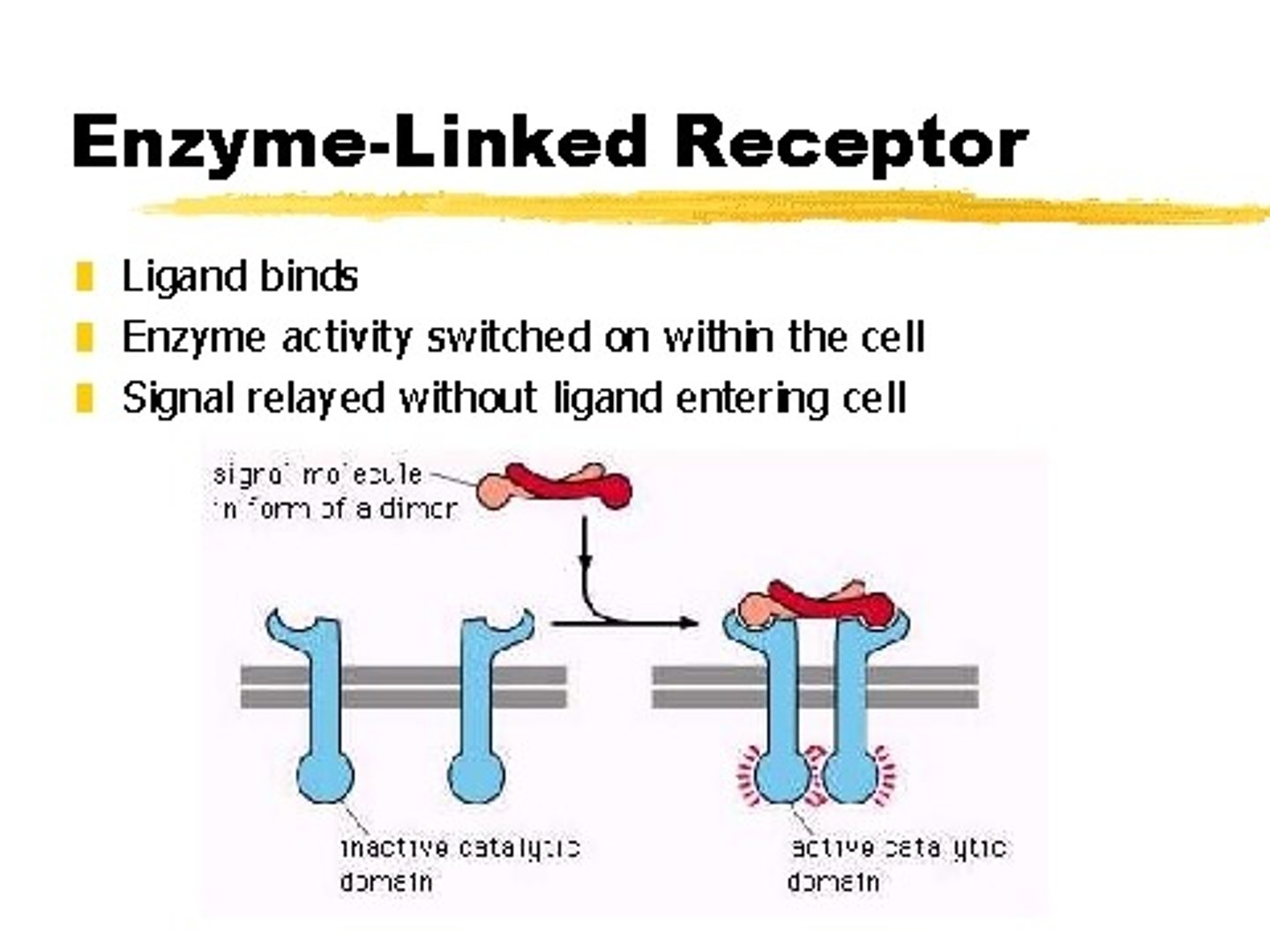
Catalytic Receptors are also known as:
(A) Ligand-gated Ion Channels
(B) G-protein Coupled Receptors
(C) Enzyme Linked Receptors
(D) Membrane Receptors
(C) Enzyme Linked Receptors
Catalytic Receptors are also known as Enzyme Linked Receptors.
What is the key difference between each type of Membrane Receptor:
(1) Ligand-gated Ion Channels
(2) G-protein Coupled Receptors
(3) Enzyme Linked Receptors
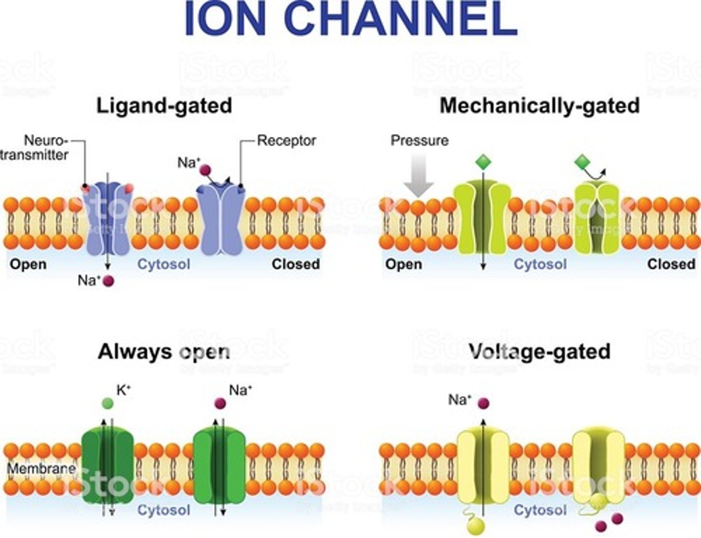
Ligand-gated Ion Channels are transmembrane ion channels that open and close in response to the binding of a ligand, allowing ions to flow in or out of a cell.
G-protein Coupled Receptors are a unique receptor type that includes the activation of a G-protein.
Enzyme Linked Receptors act as both a membrane receptor and as an enzyme. Binding of the ligand activates the catalytic activity of the receptor.
Compare Ligand-gated Ion Channels to Voltage-gated Ion Channels and Stretch-activated Ion Channels.
Ligand-gated Ion Channels are opened and closed due to the binding of a ligand.
Voltage-gated Ion Channels are opened and closed due to a change in the electrical environment (or the membrane potential).
Stretch-activated (or Mechanically-gated) Ion Channels are opened and closed due to stretching or deformation of the cell membrane.
CRB The Nernst Equation can determine the Membrane Potential for a specific molecule based upon its charge, the environmental conditions [usually simplified to a constant], and the concentration of the molecule intra- and extra-cellularly. Write out the Nernst Equation.
E = (61.5/z)log([ion]out/[ion]in)
E = Membrane Potential (mV)
z = Charge of the Ion
[ion]out = Ion Concentration Outside Membrane (mol/L)
[ion]in = Ion Concentration Inside Membrane (mol/L)
Note that 61.5 is the constant accounting for enviornmental conditions.
G Protein Coupled Receptors are found in:
I. Archaea
II. Prokaryotes
III. Eukaryotes
(A) I Only
(B) III Only
(C) I and II Only
(D) II and III Only
(B) III Only
G Protein Coupled Receptors are found in Eukaryotes.
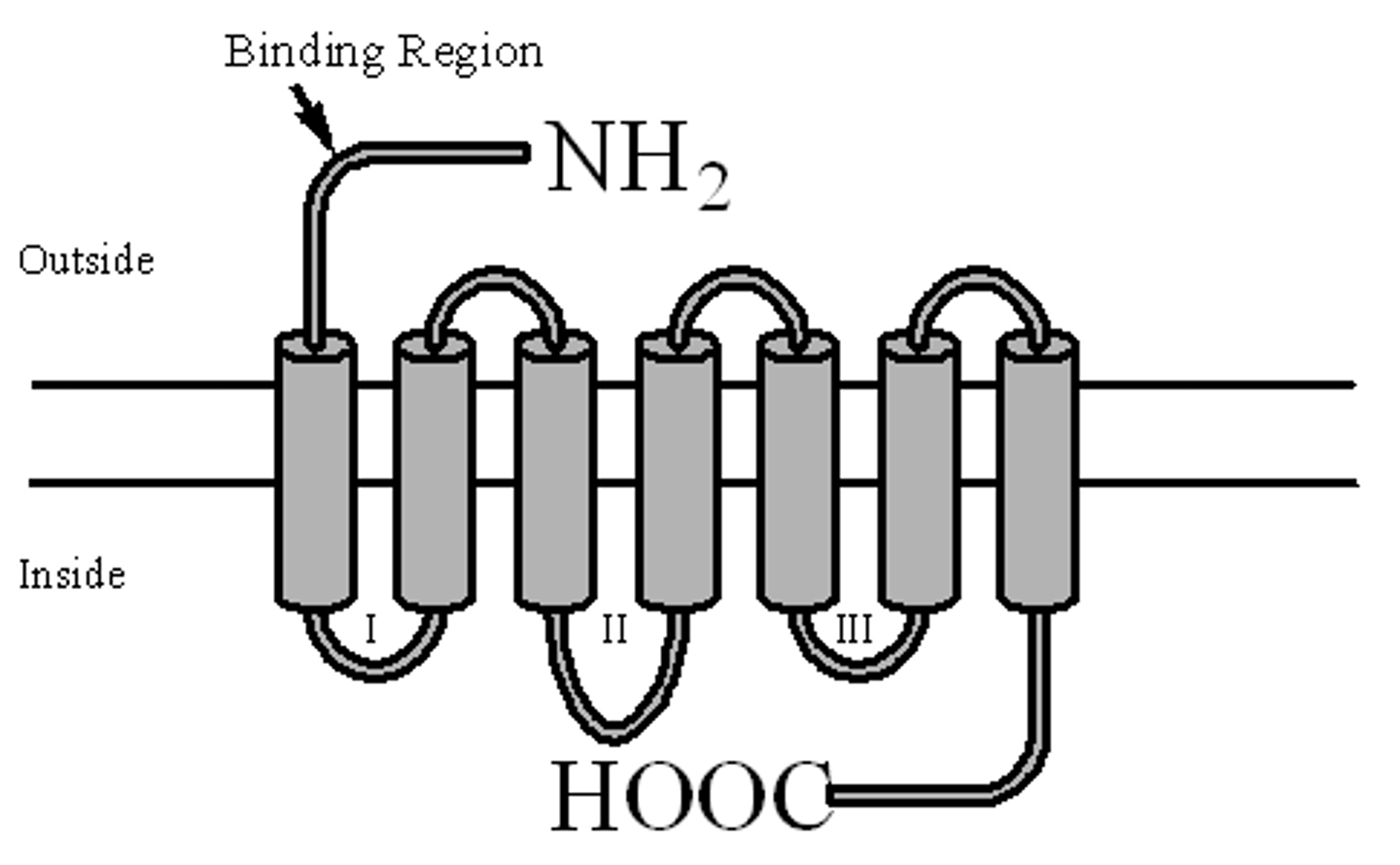
What does it mean that GPCRs are "7 Transmembrane Receptors"?
It means that their protein spans the membrane 7 times!
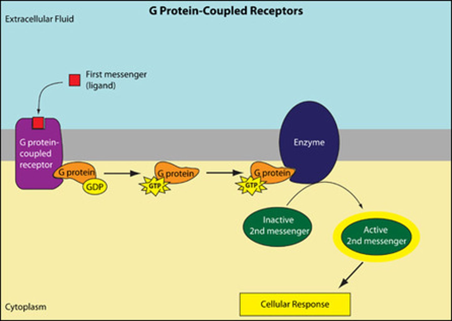
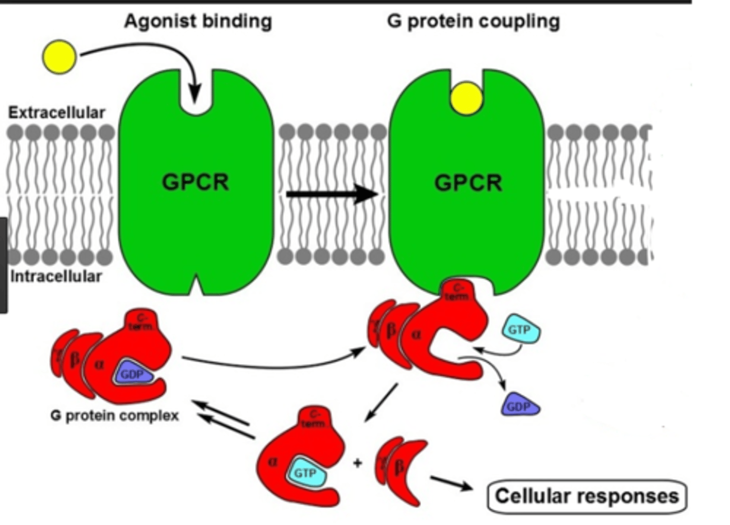
The GPCR mechanism is extremely important to know. Please draw out this mechanism, including the α, β, and γ subunits of the G Protein.
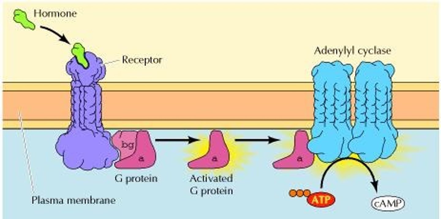
Epinephrine is a classic example of a molecule that binds to a GPCR. In this case, the activated α-subunit binds to Adenylate Cyclase. What does Adenylate Cyclase then do?
Adenylate Cyclase converts ATP into cAMP (Cyclic AMP), which will then activate and alter other proteins within the cell, ultimately resulting in increased heart rate, dilated blood vessels, glycogenolysis, etc.
What is/are the secondary messenger(s) in this instance?
I. α-subunit
II. cAMP
III. GPCR
(A) I Only
(B) II Only
(C) III Only
(D) I and II Only
(D) I and II Only
Secondary Messengers are any molecule that transmits a signal from the cell membrane. GPCR would not be included since it constitutes the protein that receives the signal whereas the G Protein and cAMP transmit signals from the cell membrane to the intracellular environment.
Enzyme Linked Receptors have which of the following domains?
I. Ligand Binding Domain
II. Enzymatic Domain
III. DNA Binding Domain
(A) I and II Only
(B) II and III Only
(C) I and III Only
(D) I, II, and III
(A) I and II Only
Enzyme Linked Receptors have Ligand Binding Domains and Enzymatic Domains but not DNA Binding Domains.
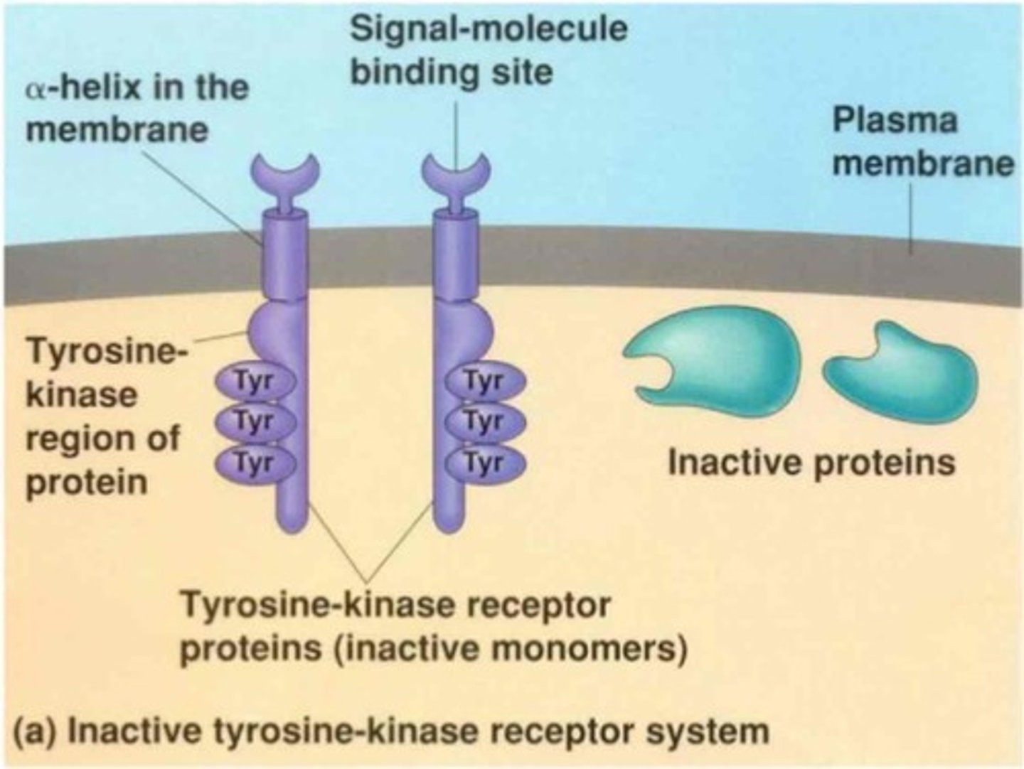
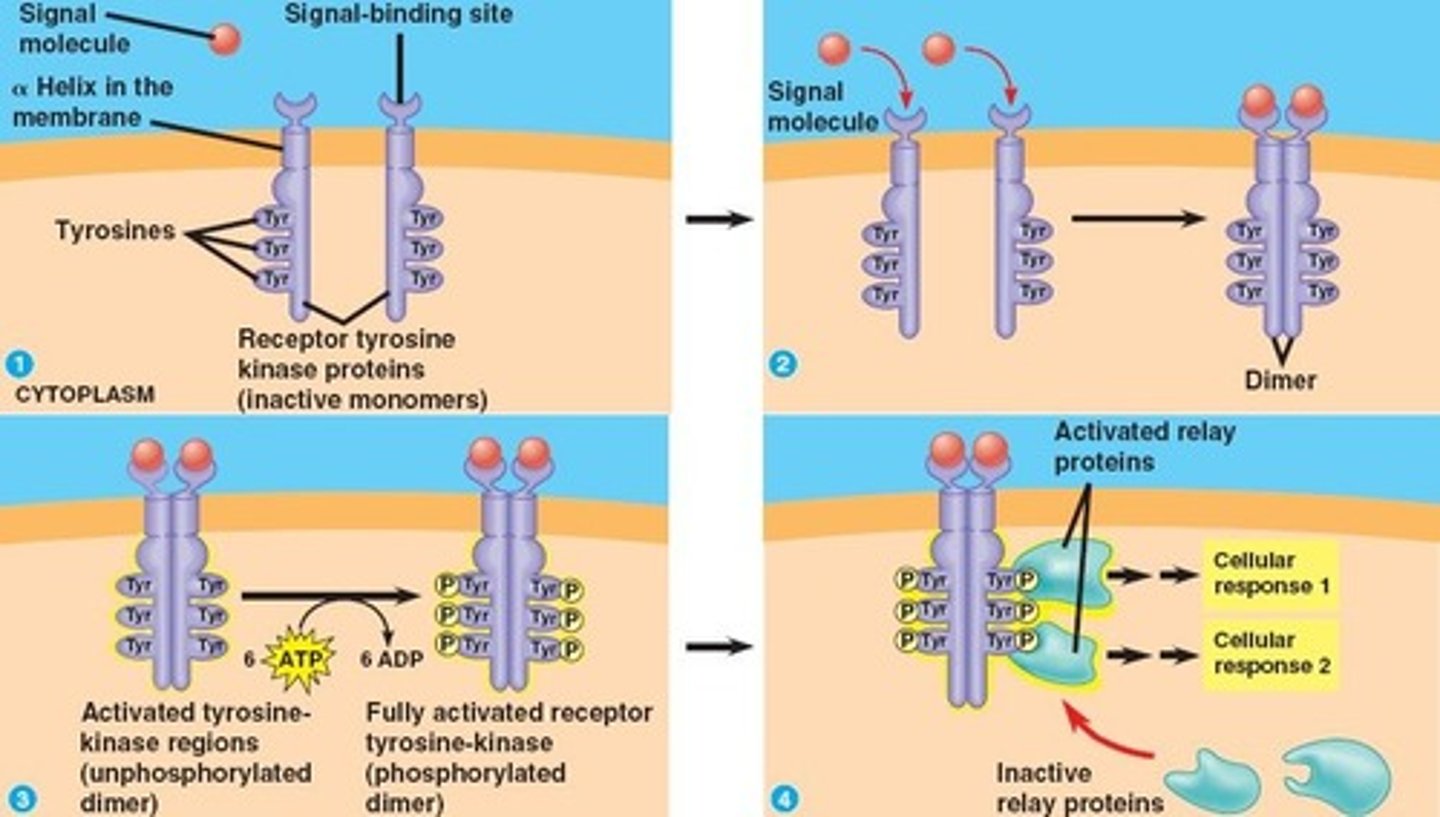
Receptor Tyrosine Kinases (RTKs) are classic examples of Enzyme linked receptors. After a ligand binds to the Ligand Binding Domain, what cascade of events occurs in order to activate intracellular proteins?
Upon activation, neighboring RTKs will move close together and form cross-linked dimers. Next the Tyrosine amino acids in the Enzymatic Domain will phosphorylate each other. Now, these Phosphorylated Tyrosine amino acids can bind to intracellular proteins and activate them.
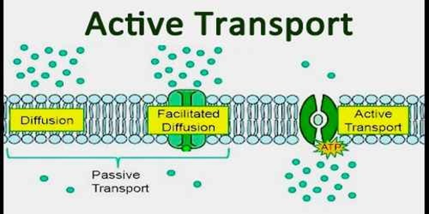
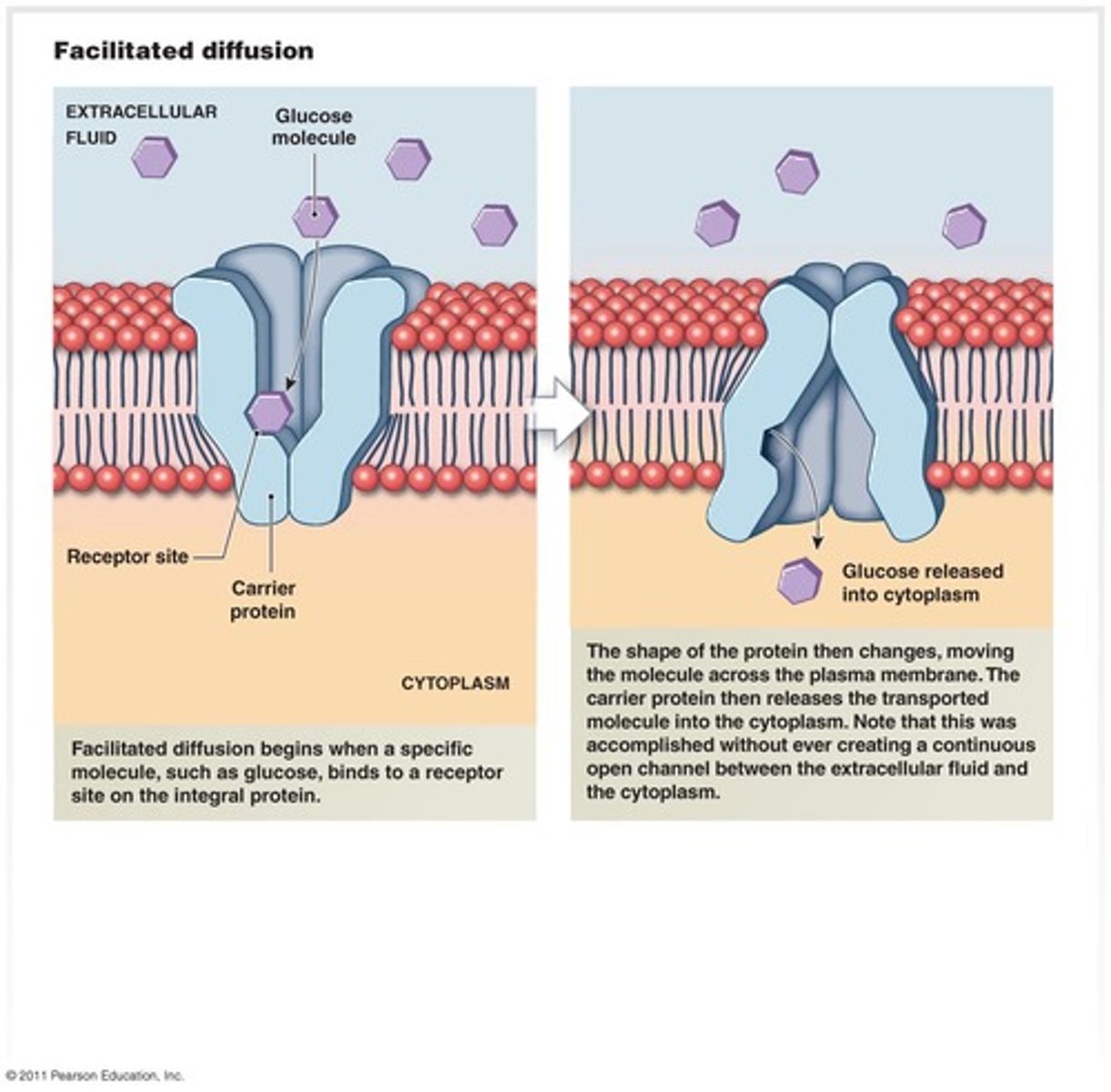
True or False? Facilitated Diffusion is an example of Active Transport because it utilizes a protein.
False. Active Transport is defined as transmembrane transport that utilized energy. Facilitated Diffusion uses a transport protein, but it does not require energy. For this reason, Facilitated Diffusion is NOT an example of Active Transport.
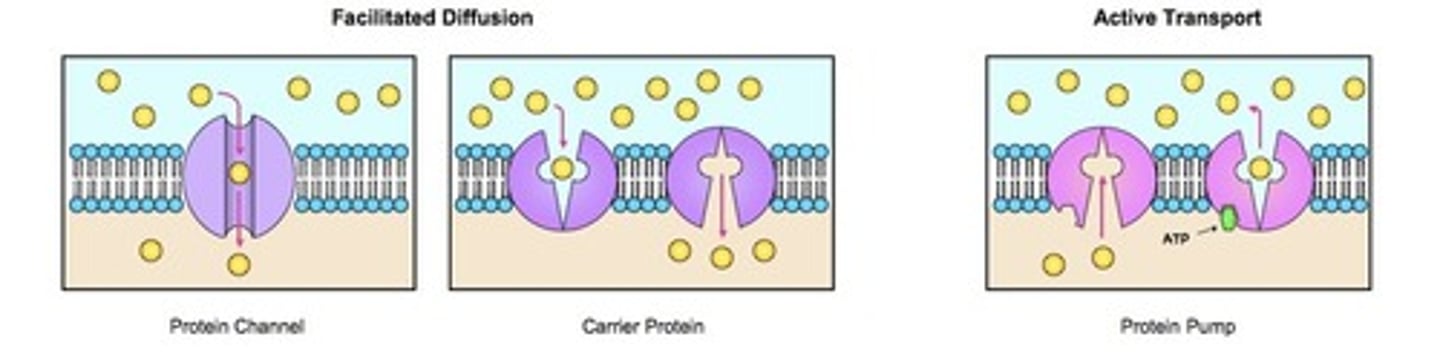
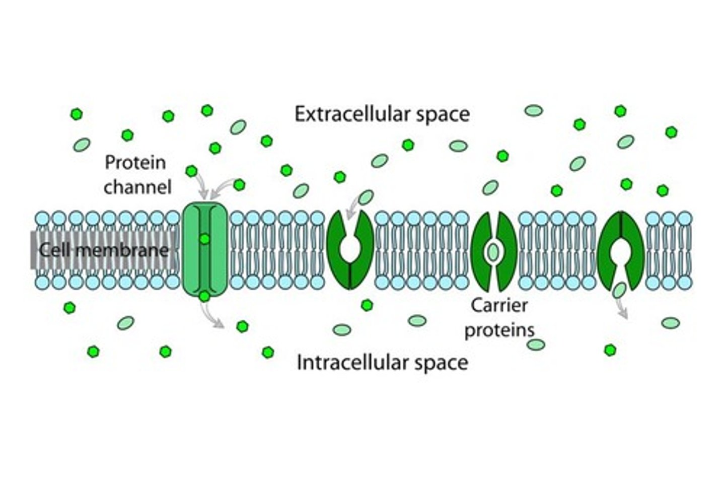
CRB What is the purpose of having Carriers and Channel Proteins for Facilitated Diffusion?
These molecules that use Carriers or Channel Proteins for facilitated diffusion are not able to diffuse across the phospholipid bilayer, but have the concentration gradient required to want to diffuse across the membrane.
In other words, Channel Proteins and Carriers enable the diffusion that cannot occur directly across the Cell Membrane.
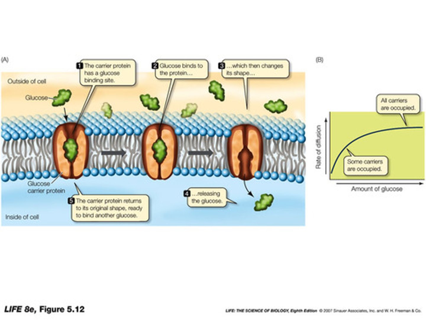
CRB Which of the following statements about Carriers are true?
I. Carrier proteins will never be open to both sides of the cell membrane simultaneously.
II. Carrier Proteins, when alternating which side is open, take on a closed state called the Occluded State.
III. Carrier Proteins, for the above reasons, are often considered similar to revolving doors.
(A) I only
(B) I and II only
(C) II and III only
(D) I, II and III
(D) I, II and III
Each of the following statements about Carriers are true:
I. Carrier proteins will never be open to both sides of the cell membrane simultaneously.
II. Carrier Proteins, when alternating which side is open, take on a closed state called the Occluded State.
III. Carrier Proteins, for the above reasons, are often considered similar to revolving doors.
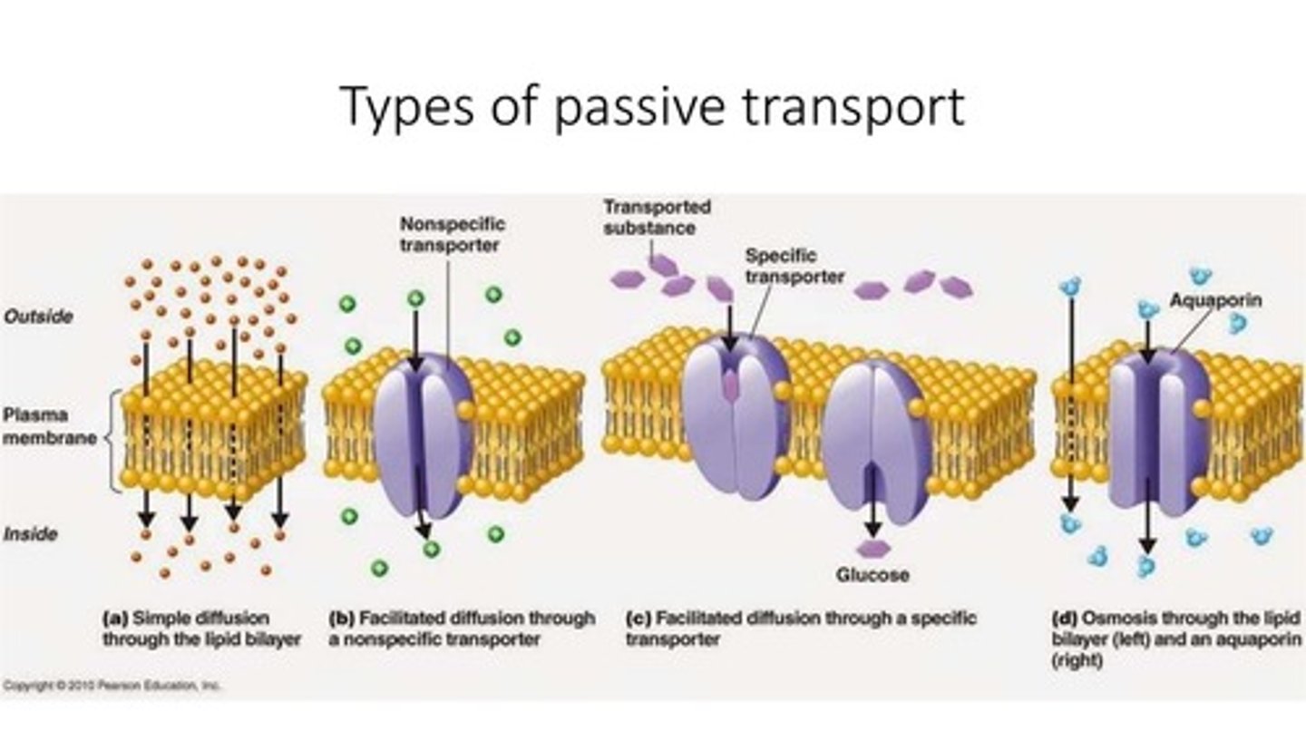
Compare the four major types of Passive Transport:
(1) Diffusion
(2) Osmosis
(3) Filtration
(4) Facilitated Diffusion
Diffusion is when a substance simply diffuses/transports directly through the cell membrane without the assistance of a channel/protein.
Osmosis is the movement of water across a cell membrane.
Filtration is special type of Diffusion in which some substances are allowed to diffuse while others are not.
Facilitated Diffusion is diffusion that occurs with the assistance of a protein without the utilization of energy.
CRB Which of the following types of transport have a >0 ΔG?
I. Passive Transport
II. Primary Active Transport
III. Secondary Active Transport
(A) I only
(B) II only
(C) I and III only
(D) II and III only
(D) II and III only
Primary and Secondary Active Transport both have a positive ΔG, meaning they require energy.
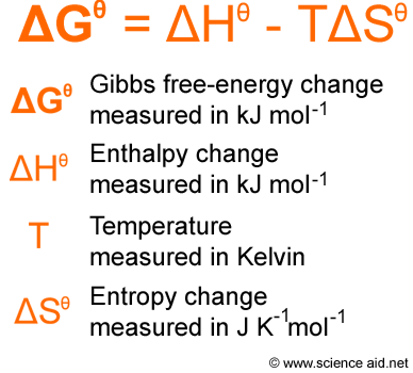
CRB Recall the equation for Gibbs Free Energy. What must be increasing to make Passive Transport have a negative ΔG?
It is illogical to think that this Passive Transport will significantly increase temperature, so this Passive Transport must increase Entropy.
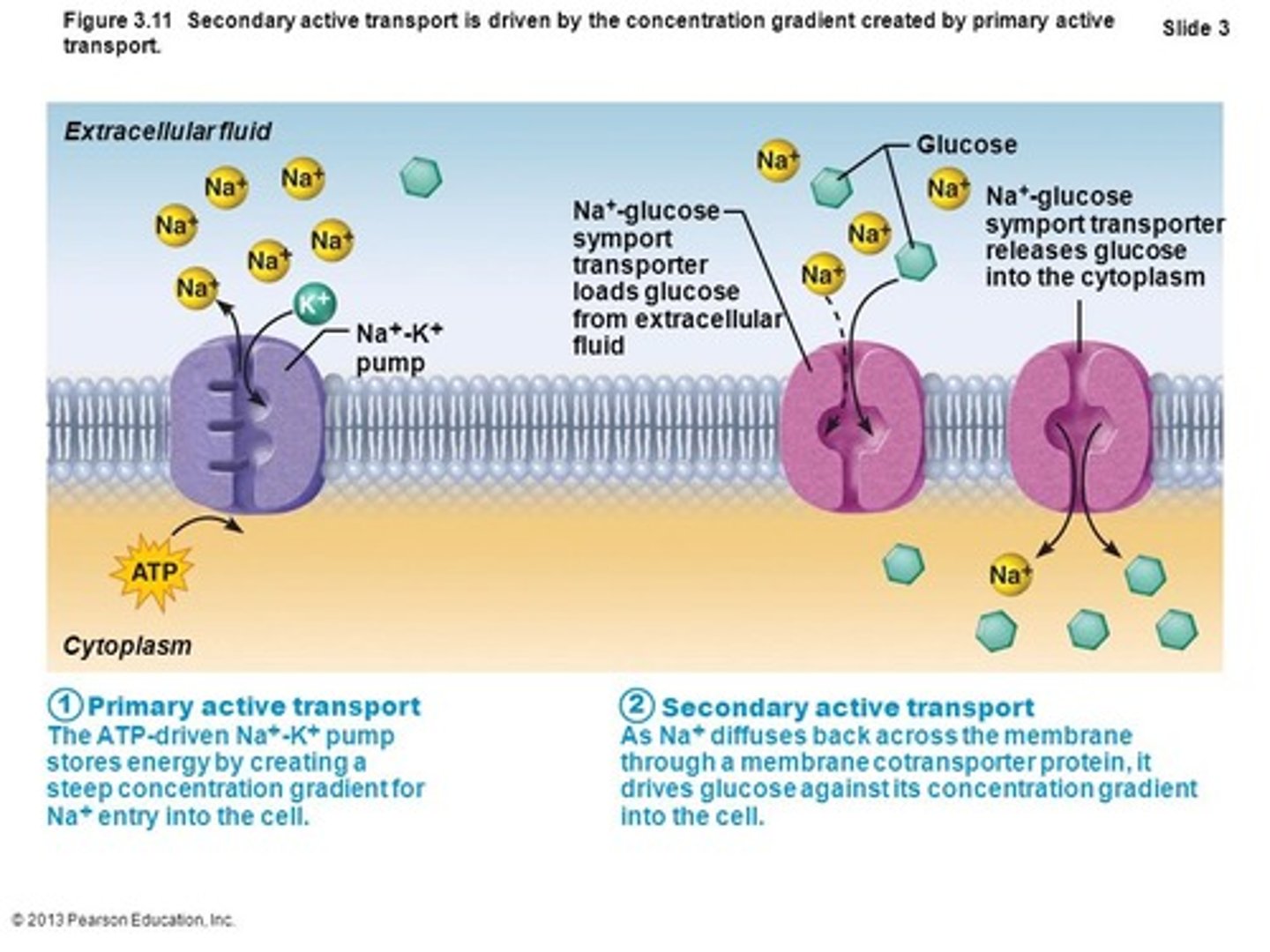
Compare Primary Active Transport to Secondary Active Transport.
Primary Active Transport is when energy is used on a channel, allowing transport of the substance of interest.
Secondary Active Transport is when energy is used to transport certain substances that will create a gradient, allowing the substance of interest to transport without energy input.
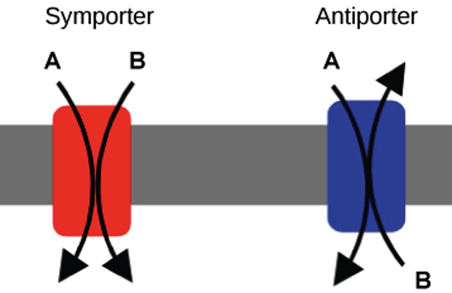
Compare a Symport with an Antiport.
Symports transport two different substances in the same direction.
Antiports transport two different substances in the opposite direction.
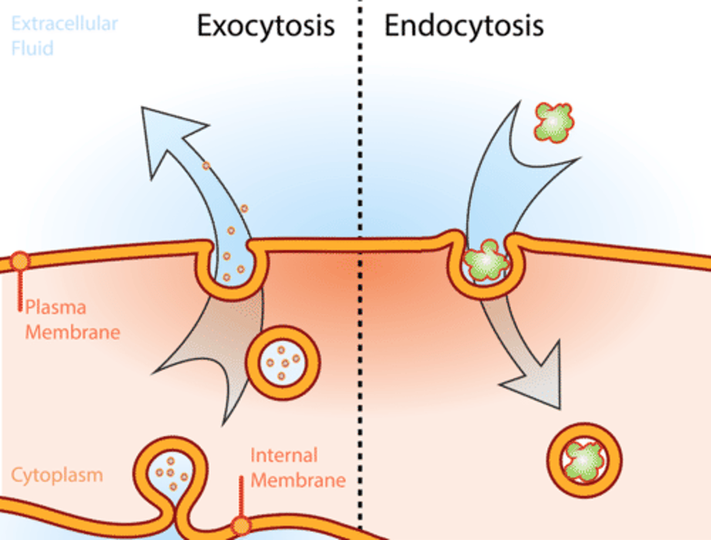
Compare Endocytosis and Exocytosis.
Endocytosis is the transport of a vesicle into a cell.
Exocytosis is the transport of a vesicle to the outside of a cell.
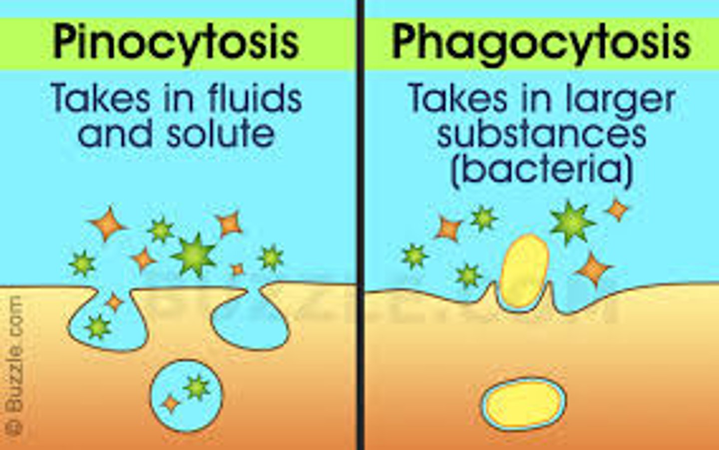
CRB Fill in the blanks: Endocytosis with a fluid and its dissolved particles is called ___________, whereas Endocytosis with large solids is called ___________.
(A) Pinocytosis, Palocytosis
(B) Palocytosis, Pinocytosis
(C) Pinocytosis, Phagocytosis
(D) Phagocytosis, Palocytosis
(C) Pinocytosis, Phagocytosis
Endocytosis with a fluid and its dissolved particles is called Pinocytosis, whereas Endocytosis with large solids is called Phagocytosis.
True or False? Endocytosis is an example of Passive Transport since it simply entails the fusion of a vesicle with a cell membrane.
False. Endocytosis is actually a very energy intensive process.
Draw out the steps involved in the action of a glucose channel that utilizes facilitated diffusion.
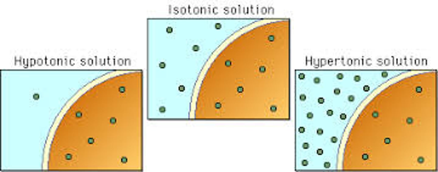
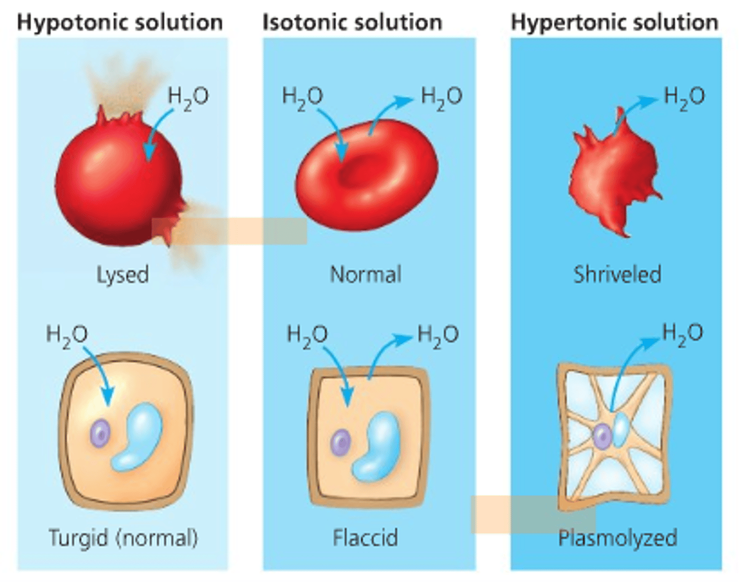
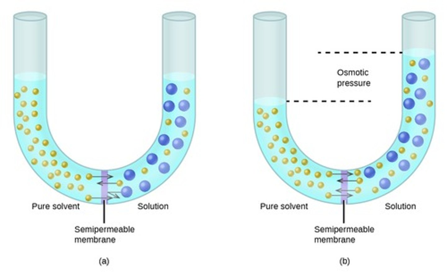
Compare a Hypotonic and a Hypertonic Environment.
A Hypotonic Environment has a lower concentration of solutes as compared to the inside of a cell.
A Hypertonic Environment has a higher concentration of solutes as compared to the inside of a cell.
Diffusion would entail solutes moving from the _________ of a cell to the ____________ of a cell when the cell is Hypertonic.
(A) outside, outside
(B) outside, inside
(C) inside, inside
(D) inside, outside
(D) inside, outside
Diffusion would entail solutes moving from the inside (high concentration) of a cell to the outside (low concentration) of a cell when the cell is Hypertonic.
Osmosis would entail water moving from the _________ of a cell to the ____________ of a cell when the cell is Hypertonic and the semipermeable membrane is impermeable to the solute.
(A) outside, outside
(B) outside, inside
(C) inside, inside
(D) inside, outside
(B) outside, inside
Osmosis would entail water moving from the outside of a cell to the inside of a cell when the cell is Hypertonic and the semipermeable membrane is impermeable to the solute.
CRB True or false? Osmotic Pressure (the driving force behind Osmosis) is a Colligative Property, so Osmosis only depends upon how many solutes there are, not what those solutes are.
True. Osmotic Pressure (the driving force behind Osmosis) is a Colligative Property, so Osmosis only depends upon how many solutes there are, not what those solutes are.
CRB Which of the following is NOT another example of a Colligative Property?
(A) Vapor Pressure Depression
(B) Boiling Point Elevation
(C) Freezing Point Depression
(D) Sublimation Point Elevation
(D) Sublimation Point Elevation
Vapor Pressure Depression, Boiling Point Elevation and Freezing Point Depression are all examples of Colligative Properties.
CRB What is the equation for Osmotic Pressure in terms of Molarity?
π = iMRT
π = Osmotic Pressure (atm)
i = Van't Hoff Factor
M = Molarity (mol/L)
R = Gas Constant (0.0821 L atm/mol K)
T = Absolute Temperature (K)
CRB What is the significance of the Van't Hoff Factor in that Osmotic Pressure equation?
The Van't Hoff Factor is the number of particles that are in solution for each molecule of the substance that dissolved. This is how you account for the number of molecules in solution being different when ionic solutions dissolve into multiple particles!
CRB What would the Van't Hoff Factor be for CaCl2 when it dissolves in water?
(A) 1
(B) 2
(C) 3
(D) 4
(C) 3
CaCl2 dissolves into Ca2+, Cl- and Cl-.
CRB Mitochondria also have two membranes. Compare the permeabilities of each membrane.
The Outer Mitochondrial membrane has many large pores, allowing ions and small proteins to move freely, including electron carriers.
The Inner Mitochondrial Membrane is much less permeable, requiring the integral proteins that are part of the Electron Transport Chain and ATP Synthase for the passage of ions to occur.
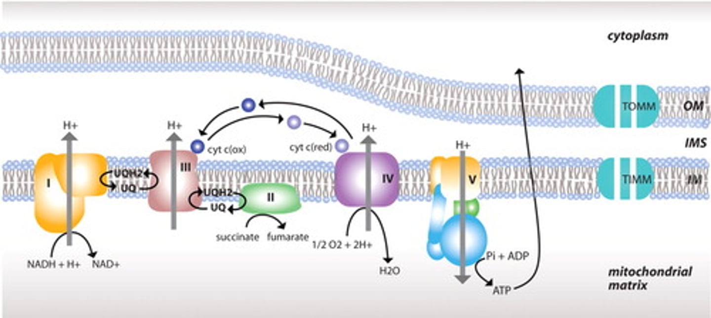
CRB Cristae are the folds in the mitochondria that increase the surface area for the Electron Transport Chain proteins. Would Cristae be folding of the Outer or Inner Mitochondrial Matrix?
Cristae are formed by the folding of the Inner Mitochondrial Matrix.