Anatomy Lab PA: Intestines and Pancreas
 Call Kai
 Learn
Learn Practice Test
 Spaced Repetition
 Match
Match Flashcards
 Knowt Play
1/70

There's no tags or description
Looks like no tags are added yet.
Name | Mastery | Learn | Test | Matching | Spaced | Call with Kai | Chat |
|---|
No analytics yet
Send a link to your students to track their progress
71 Terms
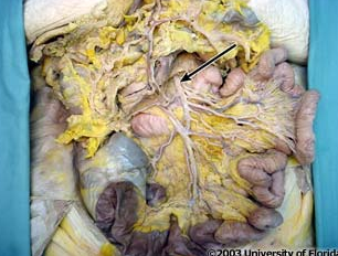
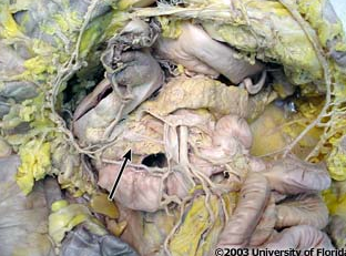
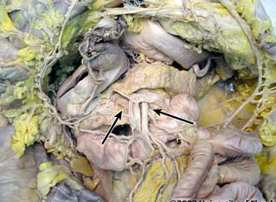
what artery is being indicated to in the image?
superior mesenteric artery
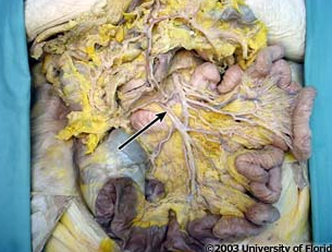
what vein is being indicated to in the image?
superior mesenteric vein
where does the superior mesenteric artery orginate from?
aorta
at what vertebral level does the superior mesenteric artery arise from?
below L1
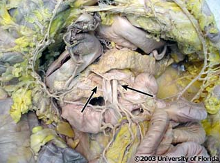
what structure of the pancrease does the superior mesenteric artery pass through, as shown in the image?
uncinate provess
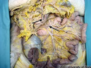
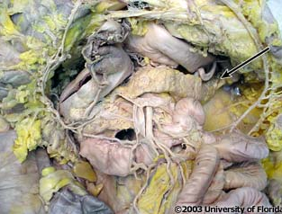
what artery is pictured?
middle colic artery
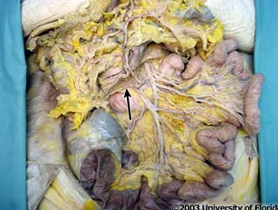
what artery is pictured?
right colic artery
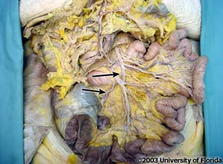
what artery is pictured?
ileocolic artery
what is the name of the artery that is the terminal branch of the superior mesenteric artery?
ileocolic artery
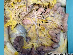
what arteries are pictured?
intestinal arteries
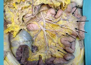
what is the term for loops of arteries that supply the jejunum and ileum (as pictured here)
arcades
the number of arcades should ______ when moving from the jejunum to the ileum
increase
the amount of fat in the distal portion of the mesentery should _______ when moving from the jejunum to the ileum
increase
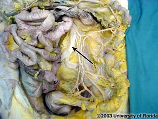
what structure is pictured (context, loops of the small intestine have been removed)
abdominal aorta
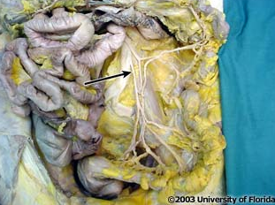
what artery is pictured (context, loops of the small intestine have been removed)?
inferior mesenteric artery
the inferior mesenteric artery arises from the _____ aspect of the abdominal aorta at the level of the __ vertebra
anterior; L3
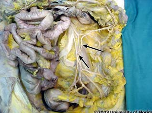
what artery is pictured (context, loops of the small intestine have been removed)?
left colic artery
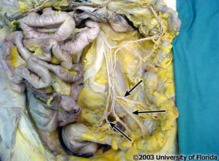
which arteries are pictured (context, loops of the small intestine have been removed)?
sigmoidal arteries
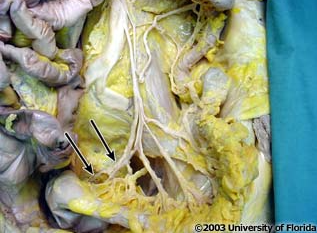
what artery is pictured (context, loops of the small intestine have been removed)?
superior rectal artery
what is the terminal branch of the inferior mesenteric artery?
superior rectal artery
the _______ arteries enter the root of the sigmoid mesocolon and supply the sigmoid colon
sigmoidal
the ______ _____ artery passes into the pelvic cavity and supplies the rectum and superior portion of the anal canal.
superior rectal
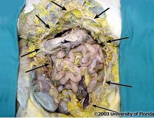
which artery is being indicated by the arrows?
marginal artery
The anastomosis of the ileocolic artery, right colic artery, middle colic artery, left colic artery and sigmoidal arteries around the internal margin of the cecum and colon forms a single arterial trunk called the _______ ______
marginal artery
the _____ _____ begins at the ileocecal junction and ends on the rectum.
marginal artery
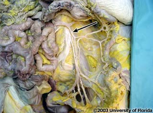
what vein is being pictured (context, parts of the small intestine have been moved)?
inferior mesenteric vein
the inferior mesenteric vein joins with the ____ ____, usually posterior to the body of the pancreas
splenic vein
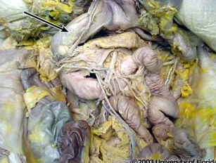
what structure is pictured (be specfic; context: transverse colon and stomach have been reflected)?
superior part of duodenum
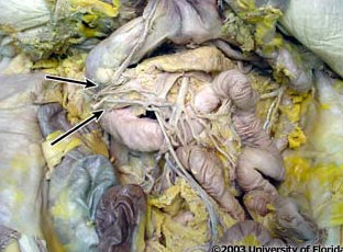
what structure is pictured (be specfic; context: transverse colon and stomach have been reflected)?
descending part of duodenum
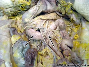
what structure is pictured (be specfic; context: transverse colon and stomach have been reflected)?
horizontal part of duodenum
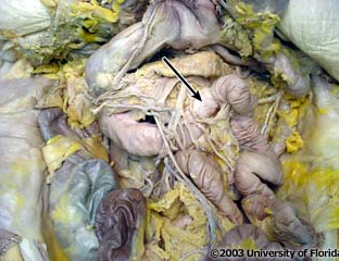
what structure is pictured (be specfic; context: transverse colon and stomach have been reflected)?
ascending part of duodenum
with the exception of its ______ part, the duodenum is a retroperitoneal organ
superior
the acending part of the duodenum is continous with the jejunum and become intraperitoneal at the _______ ______
duodenojejunal flexure
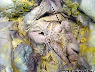
what is the term used to descibe where the duodenum becomes the jejunum (as pictured here)?
duodenojejunal flexure
what vertebral level is the duodenojejunal flexure located at?
L2
the first part of the duodenum has a _______ surface
smooth
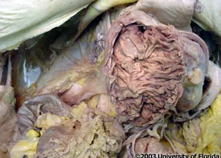
what kind of folds are seen in all parts of the duodenum except the first part (as pictured)?
circular mucosal folds
on the ______ wall and longitudinal fold of the descending duodenum, you can find the _____ ______ _____.
anterior; greater duodenal papilla
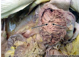
what structure is being indicated to (be specfic)?
greater duodenal papilla
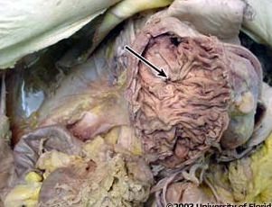
what structure is being indicated to (be specfic)?
lesser duodenal papilla
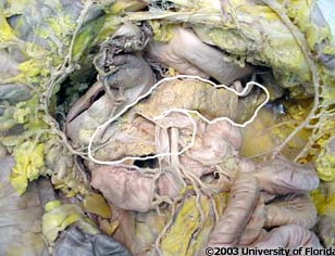
what structure is being highlighted?
pancreas
what structure is being indicated to (be specfic)?
head of the pancreas
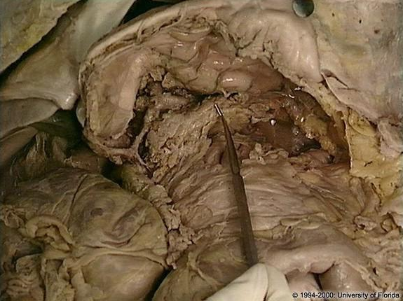
what structure is being indicated to (be specfic)?
body of pancreas
what structure is being indicated to (be specfic)?
tail of pancreas
what structure is being indicated to (be specfic)?
uncinate process
the most inferior portion of the pancreas head is the ______ ______, which “hooks” to the left and lies posterior to the superior mesenteric artery and vein
uncinate process
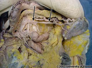
what vein is pictured here?
splenic vein
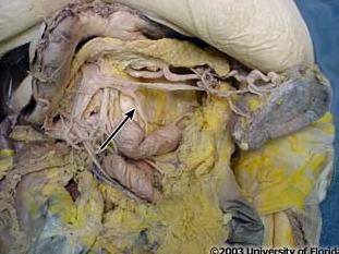
what vein is pictured here?
inferior mesenteric vein
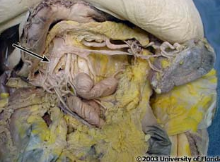
what vein is pictured here?
superior mesenteric vein
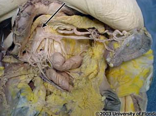
what vein is pictured here?
portal vein
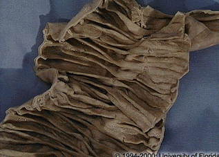
what is the name of the circular mucosal folds found on the internal surface of the jejunum and ileum (as pictured here)?
plicae circulares
the plicae circulares in the proximal jejunum are _____ pronouced and ____ numerous than they are in the ileum.
more; more
in the distal ileum, submucosal aggregates of lymphatic nodules called _____ ______ are visible and are often palpable; they are NOT present in the jejunum.
Peyer’s patches
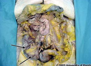
what structure is pictured (be specific)?
cecum of large intestine
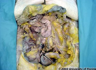
what structure is pictured (be specific)?
vermiform appendix
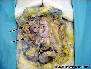
what structure is pictured (be specific)?
ascending colon
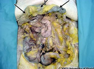
what structure is pictured (be specific)?
transverse colon
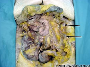
what structure is pictured (be specific)?
descending colon
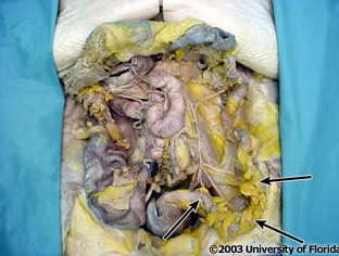
what structure is pictured (be specific)?
sigmoid colon
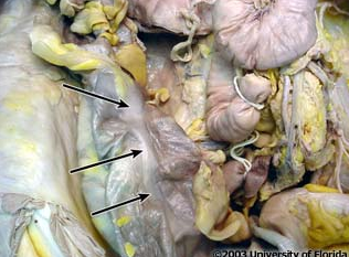
what structure on the large intestine is this (be specfic)?
taeniae coli
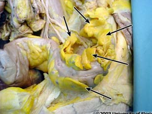
what structure on the large intestine is this (be specfic)?
epiploic appendages
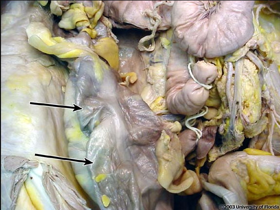
what structure on the large intestine is this (be specfic)?
haustration
the _____ ____ are three narrow but distinct longitudinal bands of smooth muscle which run the entire length of the cecum and colon
taenia coli
the three taenia coli converge at the base of the _______ _____ on the cecum
taeniae coli
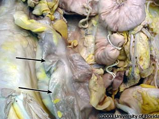
what structure on the large intestine is this (be specfic)?
haustra coli
the longitudinal constriction of the taeniae coli force the wall of the large intestine between the bands to pouch out into a longitudinal series of sacculations called the ____ _____
haustra coli
______ _____ are fat-filled pouchings of the visceral peritoneum; found along the large intestine expect on the surface of the rectum, anal canal, and vermiform appendix
epiploic appendages
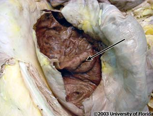
what structure in the large intestine is this (be specfic)?
ileocecal orifice
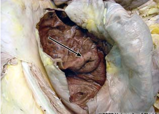
what structure in the large intestine is this (be specfic)?
superior fold of the ileocecal valve
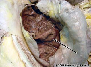
what structure in the large intestine is this (be specfic)?
inferior fold of the ileocecal valve
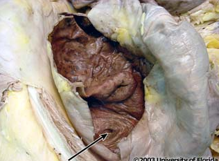
what structure in the large intestine is this (be specfic)?
orifice of the vermiform appendix