(E2) Intro to cross sectional anatomy
 Call Kai
 Learn
Learn Practice Test
 Spaced Repetition
 Match
Match Flashcards
 Knowt Play
1/43

There's no tags or description
Looks like no tags are added yet.
Name | Mastery | Learn | Test | Matching | Spaced | Call with Kai | Chat |
|---|
No analytics yet
Send a link to your students to track their progress
44 Terms
advantages of cross sectional anatomy
eliminates overlapping structures
helps to differentiate between adjacent structures
offers view of anatomy in a variety of planes
Sagittal plane or median =
left to right
axial plane or transverse =
inferior to superior
coronal plane or frontal=
posterior/anterior
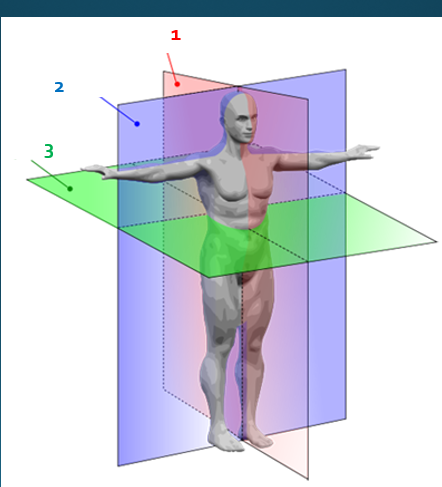
what plane is 1?
sagittal
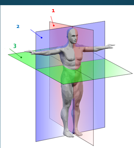
what plane is 2?
coronal

what plane is 3?
axial
thickness of slice is dependent on ____
technical settings
thinner slices = ____ detail
more
slices are determined by _____ and procedure protocols
institutional
slices are adjusted by
technologist/protocols
slices are annotated on scout images to provide ______ (scanogram)
regional location
____ patterns may change location of anatomy
breathing
specific arrangement of structures varies from _______
one person to the next
determining location of the slice helps to identify
anatomy
what is computed tomography
generates a 3D image of the body from a large series of 2D x-ray images taken around a single axis rotation
when was CT developed and publicized
in the 1970s
when was the first patients brain scan and how long was it
1971- London Atkinsons Morley Hospital
2.5 hours
hounsfield unit for bone is
1,000 HU (bright white)
hounsfield unit for water is
0 HU (gray)
hounsfield unit for air is
-1,000 HU (black)
hounsfield units represent the ____ of each tissue
densities
an ____ and ____ image is obtained ot designate where the slices will be imaging
AP and Lateral
slice thickness may very from _____ to _______
millimeters to centimeters
scans are taken on one plane and reconstructed to create the ____ and ____ planes
sagittal and coronal
Contrast use in CT:
Iodine based and barium-sulfate type contrast can be used in CT
this is used to highlight different structures of the body to evaluate pathologies and disease
advantages of CT
visualization of anatomy without superimposition of structues
axial imaging
ability to differentiate small differences in density of anatomic structures and abnormalities
CT: indications
diagnose muscle and bone disorders
detect internal injuries and internal bleeding
pinpoint the location of a tumor, infection, clot
guide procedures such as surgery, biopsy, Rad Therapy
detect and monitor diseases and conditions
CT: contraindications
higher doses
contrast allergies, extravasation
claustrophobia
Magnetic resonance is what
utilizes a powerful magnet field to align the magnetization of some atoms in the body
magnetic properties of hydrogen atoms
MR was discovered in
1973

how does MRI work?
a “coil” is placed around the area of interest to obtain the info of that area. the magnet aligns the single protons in the hydrogen nuclei within the field
radio waves enter the patient from another angle, the radio waves cause the nuclei to flip positions and either align with the magnet or against it
at termination of radio signals, nuclei return to original position by spinning like a top back into their varied position
energy that is released from the relaxation is used to generate images
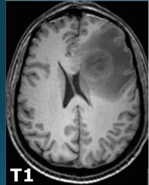
MRI T1 weighted images
fat appears bright
water appears dark'
best for anatomy
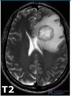
MRI T2 weighted images
water appears bright (white)
fat appears dark
best for pathology
Contrast use in MRI:
gadolinium is contrast used in many MRI scans to improve the clarity of the images and to improve the diagnostic accuracy of the MRI scan
Gandolinium is a safer contrast due to
less adverse reactions and eliminated through kidneys easier
advantages of MRI
no exposure to ionizing radiation
superior contrast resolution
superior tissue characterization
multi-planar capabilities
possibly safer intravenous contrast agents
visualization of angiography without using contrast
MRI: indications
a noninvasive way to examine organs, tissue and the skeletal system by producing high-resolution images that help diagnose a variety of problems
MRI: contraindications
possible contrast allergy, extravasation
metal
take longer
claustrophobia
patient size (larger)
human body = ___% hydrogen
80
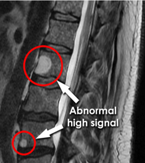
is this a T1 or T2 image?
T2
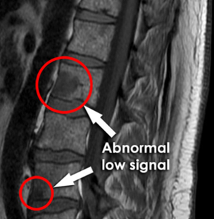
is this a T1 or T2 image?
T1
How does MRI and diagnostic work togther?
x-ray can screen for metal before going to MRI
metal, pacemaker, abdomen
fluoro and arthrograms
how does CT and diagnostic work together?
better detail on fractures
myelograms and arthrograms
kidney stones
abdomen x-ray looking for prior contrast before CT