HEMONC 1.1 Midterm
 Call Kai
 Learn
Learn Practice Test
 Spaced Repetition
 Match
Match Flashcards
 Knowt Play
1/36

There's no tags or description
Looks like no tags are added yet.
Name | Mastery | Learn | Test | Matching | Spaced | Call with Kai | Chat |
|---|
No analytics yet
Send a link to your students to track their progress
37 Terms
RBC structure + function?
biconcave, anucleate (glycolysis for ATP), central pallor on smear, lipid bilayer membrane, cytoskeleton (deform)
loss of deform → splenic sequestration
membrane defect → hemolysis
O2 delivery via hemoglobin
dec. ATP → membrane fragile → hemolysis
RBC lifespan? recycling?
120 days, loss of membrane + enzyme depletion = tag for removal via RES (spleen + liver MO)
erythropoiesis in BM (DNA, globin, heme synthesis; Fe) via EPO in kidney stroma → reticulocytes (bluish, larger) w/ residual RNA = indicator of BM response
in MO = globin → aa, heme → Fe (stored w/ ferritin or transported w/ ferritin) + protoporphyrin (bilirubin → liver → bile)
hemoglobin synthesis requirements?
globin chain (in cytoplasm of developing red nuclei)
Chr. 16 (a-like), Chr. 11 (B-like)
heme synthesis (in RBC mitochondria, O2 binds + relies on Fe)
Fe availability (absorbed in duodenum via ferroportin; transport via transferrin)
hemoglobin types?
HgbA → 2a + 2B chains (a2B2)
HgbA2 → 2a + 2o chains (a2o2); inc. B thalassemia
HbF (fetal) → 2a + 2y chains (a2y2)
inc. O2 affinity, tissue hypoxia in adult
iron metabolism? iron absorption location + regulation? transport? storage?
plasma Fe → BM → RBC
duodenum, upper jejunum via ferroportin (low Fe = high FP) → regulated via hepcidin (high Fe = high hepcidin → dec. FP/ (-) feedback)
inactivating HPE mut. = Fe overload (bronze skin, arthralgia, amenorrhea, weight loss, organ damage (liver, pancreas, heart))
transferrin → blood to liver + BM (via low hepcidin/ no Fe absorption downregulation)
IC Fe bound to ferritin = prevent free radicals
anemia mechanism? symptoms? smear?
RBC or Hb loss/destruction/impaired production → deficiency → dec. O2 carrying capacity of blood
weakness, dyspnea, fatigue, pale skin, headache, lightheadedness, MI, reticulocytosis, splenomegaly, Hx pigmented gallstones
types of microcytic anemia?
thalassemia, anemia of chronic disease, iron def., lead poison (TAIL)
dec. production of Hb = RBC progenitor cells undergo extra division to maintain Hb (e.g conserve)
thalassemia mechanism? geographic distribution?
defective globin synthesis → imbalanced globin chain → insoluble unpaired/individual chains precipitate → RBC destruction → inadequate Hb → hypoxia
mediterranean, middle east, african, southeast asian
absence/mut of a/B globin = dec. HbA → tissue hypoxia → EPO inc
a-thalassemia mechanism? B- thalassemia mechanism?
gene deletion on Chr. 16 = absent a chains
absent (B0) or mutated (B+) B chains affecting HBB on Chr. 11
silent a-thalassemia? a-thalassemia minor?
¾ a globin gene normal → asymptomatic, slight microcytosis (-a/aa)
loss of 2/4 a globin genes (- -/aa) or (- a/- a)
microcytosis w/ minimal or absent anemia, HbF levels low or normal
affected parents w/ (-/-) haplotype = risk
peripheral smear → target cell, spherocytes, hypochromasia
hemoglobin H disease (HbH)?
loss of ¾ a globin genes (- -/-a)
B tetramers (4 B subunits) form w/ very high affinity for O2 → tissue hypoxia disproportionate to Hb level
HbH prone to oxidation → splenic red cell sequestration + phagocytosis → moderate severe anemia
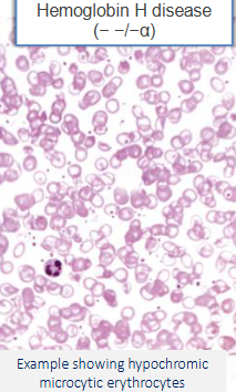
peripheral smear → hypochromic, microcytic erythrocytes
homozygous/Bart’s hemoglobin (hydrops fetalis)?
loss of all 4 globin genes (- -/ - -)
y-tetramers (4 y-subunits) form w/ extremely high affinity for O2 → anoxia → fetal IU death
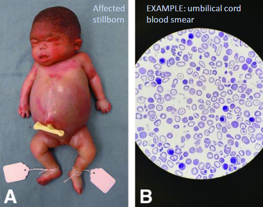
if baby born → severe pallor, generalized edema, massive hepatosplenomegaly
peripheral smear → severe hypochromic microcytic anemia, anisopoikilocytosis, nucleated erythroid forms (normoblasts), target cell
B-thalassemia minor/trait (heterozygous)? B-thalassemia intermedia?
1 absent + 1 normal (B0/B) OR 1 mutated + 1 normal (B+/B)
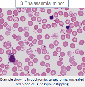
peripheral smear → hypochromasia, microcytosis, basophilic stippling, target cells, nucleated RBC
BM → mild erythroid hyperplasia
mutation on both B chains (B+/B+) OR 1 mutated + 1 absent (B+/B0)
variable, ranges form mild/asymptomatic to severe/transfusion dependent
B-thalassemia major (homozygous)?
loss of both B chains (B0/B0)
severe transfusion-dependent anemia → associated 2ndary hemosiderosis + hemochromatosis organ damage (heart, endo, liver, pancreas)
marked hemolysis, erythroid hyperplasia w/ marrow expansion + extramedullary hematopoiesis → cortical bone remodel (maxillary, frontal facial) + organomegaly
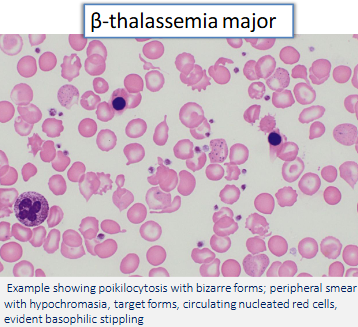
peripheral smear → marked anisocytosis, poikilocytosis, microcytosis, hypochromia, target cell, basophilic stippling, fragmented red cell, nucleated red cell (poorly hemoglobinized)
anemia of chronic disease mechanism? effect of inflammatory mediators? how can iron be trapped? what chronic conditions contribute to iron deficiency?
chronic disease → APR prod. (hepcidin) → traps Fe in storage → high ferritin + low serum Fe/ TIBC
e.g IL6 → trigger hepcidin prod. → inhibit ferroportin (no uptake)
Fe trapped via limiting Fe transfer from MO to erythroid precursor OR suppress EPO → no erythropoiesis
TNF-a, IL1, INF → suppress erythroid colony formation (EPO resistance) → no erythropoiesis
HF, CKD, ACE I or angiotensin R blocker (inhibit EPO)
chronic blood loss → via menorrhagia, GI bleed, intestinal parasites
iron deficiency populations? iron absorption inhibitors? stages? bone marrow? blood smear?
nursing infants, ppl w/ poor diet/absorption issues, plummer-vinson syndrome, inc. requirement, hypothyroidism, chronic blood loss
tannates (tea), carbonates, oxalates, phosphates
low Fe, + TSAT, Hb, serum ferritin; high TIBC, soluble transferrin receptor + platelets
stainable Fe from MO gone
poikilocytes (pencil cell), anisocytosis, microcytic/hypochromic RBC → enlarged pallor zone
lead poisoning (sideroblastic anemia) mechanism? heme pathway? causes? labs?
defective heme/protoporphyrin synthesis → Fe trapped in mit. → high TSAT
SCA (via B6 + ALAS) → a-ALA ( via ALAD) → porphobilinogen → hydroxymethylbilane → uroporphyrinogen III → coproporphyrinogen III → protoporphyrin (via ferrochelatase) → heme
lead (inhibits ALAD + ferrochelatase) + ALAS2 mut., alcoholic damage to mit., vit B6 def. (via TB isoniazid)
basophilic stippling (pappenheimer bodies), high serum Fe, ferritin, TSAT (Fe overload), microcytic hypochromatic RBC
BM → erythroid hyperplasia + rigned sideroblasts (from Fe PO4-enorged mit.)
normocytic anemias (MCV = 80-100)?
non hemolytic → aplastic, acute blood loss
hemolytic
intrinsic → damage affecting RBC membrane, metabolism, of Hb
extrinsic → RBC damaged externally via mechanical, immunological, infectious or metabolic/oxidant damage
aplastic anemias?
renal failure (low EPO prod.), hepatocellular disease (folate + Fe def.), damage to BM RBC precursor cell
parvovirus B19 → attaches to P antigen on erythroid progenitor cell → lysis → reticulocytopenia (slapped cheek, arthralgia)
chronic conditions contributing to aplasia → thymoma, glandular lymphocytic leukemia, neutralizing Ab against EPO, autoimmune
acute blood loss mechanism? RBC appearance?
internal blood loss = Hb recycled
external blood loss = Hb lose = iron deficiency
RBC → normocytic + normochromic initially → production inc. → reticulocytosis
hemolytic intrinsic anemias?
RBC membrane or cytoskeleton defect → hereditary spherocytosis, hereditary elliptocytosis, end-stage liver or kidney disease (burr cells), PNH
metabolic abnormalities → G6PD deficiency + pyruvate kinase deficiency
defects of hemoglobin → sickle cell + hemoglobin C
hereditary spherocytosis mechanism? complication? lab? blood smear?
mut. of spectrin, ankyrin, band 3 or 4.2 → affects assembly of inner cytoskeleton to outer lipid layer
aplastic crisis (parvovirus kills all RBC progenitor cells), folate deficiency, pigmented gallstones
anemia, reticulocytosis, serum bilirubin, LDH, urine urobilinogen, +osmotic fragility test, -direct coombs test
spherocytes small, dark-stain, spherical cells lacking central pallor, polychromasia
paroxysmal nocturnal hemoglobinuria mechanism? how to test for it? findings?
acquired spontaneous mutation in phosphatidylinositol glycan complementation group A gene (PIGA)
PIGA → enzyme for synthesis of membrane-associated complement regulatory proteins (GPI-linked protein)
deficient = RBC susceptible to complement induced lysis (C5b-C9 MAC)
test via flow cytometry → check CD55 + CD59 deficiency
paroxysmal ep. dark urine at night (hemoglobinuria), partial clearing during day, vasospasm, thrombosis
lab → inc. bilirubin, LDH, hemosiderinuria; dec. Hgb, serum Fe, ferritin or folate
peripheral smear → schistocytes (IV hemolysis)
G6PD deficiency-related hemolysis mechanism? geographic distribution? triggers? labs?
deficient G6PD = dysfunctional hexose monophosphate path → rigid spectrin + oxidation of Hgb
african, mediterranean or middle eastern
asymptomatic until trigger → acute episodes, self-limited, 2-4 days after exposure
oxidative stress (fava beans), infections, meds (ciprofloxacin, metronidazole)
labs → inc. bilirubin, urobilinogen, LDH; reticulocytosis, hemoglobinuria, pallor, scleral icterus; dec. Hgb, haptoglobin
peripheral smear → bite cell, blister cell, heinz bodies, polychromasia, anisocytosi, spherocytes
hemoglobin C disease cause? sickle cell disease mechanism? geographic distribution?
hereditary B globin (HBB) gene pt. mut. (G → A; Glu → Lys) on Chr. 11 → HbC production = HbC crystallization in RBC → hemolysis, target cells, dec. flexibility
hereditary B globin (HBB) gene pt. mut. (A → T; Glu → Val) on Chr. 11 → HbS
west + central africa, w/ mediterranean, middle east, india
sickle cell disease mechanism? higher expression of what?
alignment/polymerization of deoxygenated HbS → sickling (dehydrated, dense, rigid RBCs)
higher level adhesion molecules → microvascular occlusions + thrombosis → hypoxia (affects bones, lungs, liver, brain, spleen, penis; e.g acute chest syndrome)
peripheral smear → sickled cell, reticulocytes, target cells, howell-jolly bodies, small nuclear remnants
sickle cell disease symptoms? treatments?
severe hemolytic anemia, marrow expansion (erythroid hyperplasia), hyperbilirubinemia (jaundice, pigmented gallstones)
adults → leg ulcers; children → dactylitis + priapism
Tx = hydroxyurea → inc. HbF production to interfere w/ polymerization of HbS + dec. sickle cell formation
sickle cell disease genotype? sickle cell trait? sickle cell anemia? HbSC disease? HbSB-thalassemia?
both HBB alleles mut. + at least 1 BS allele
HbAS → HBB/BS
SCA → BS/BS
BS/BC
BS/HbB0
non-immune mediated anemias?
oxidant injury, mechanical trauma, infectious disease, toxin/poison, hypertonic saline, hypersplenism, thrombi
mechanical → prosthetic valve, microangiopathies (TPP, HUS, DIC)
infectious → malaria (RBC rupture), clostridium perfringens (release enzymes that digest RBC membrane)
immune mediated anemia?
Ab and/or complement bind to RBC membrane components, flagging RBC for destruction via extra or intravascular hemolysis
types of immune mediated hemolysis → autoimmune, alloimmune, drug induced
intravascular hemolysis mechanism? labs?
complement mediated lysis in circulation → free Hbg released w/in circulating plasma
unique labs → hemoglobinuria, hemosiderinuria, fragmented RBCs (schistocytes)
both IV + EV hemolysis → inc. reticulocytes, LDH, bilirubin; dec. haptoglobin
extravascular hemolysis mechanism? complications? labs?
MO in reticuloendothelial system of spleen, liver, BM recognize C3b opsonized RBC
incomplete phagocytosis results in spherocyte
splenomegaly via inc. MO + damaged/deformed RBC → inc. congestion in splenic cords of billroth
indirect hyperbilirubinemia, risk of gallstones (chronic hemolysis)
autoimmune hemolysis types? mechanism?
warm AIHA: IgG Ab → (+) indirect coombs = detectable circulating Ab directed against RBC
cold AIHA: IgM Ab → (+) direct coombs = detectable Ag bound on RBC surface
ass. w/ mycoplasma pneumonia, EBV, CMV
paroxysmal cold hemoglobinuria
alloimmune hemolysis types?
hemolytic disease of fetys/newborn: Rh(D) incompatibility → maternal IgG cross placenta + attack fetal RBC = anemia, hydrops, kernicterus
prevent w/ Rh(D) immunoglobulin prophylaxis + kelihauer-betke test (fetal-maternal hemorrhage)
hemolytic transfusion rxn: rapid IV hemolysis; IgM-mediated complement fixation
fever, renal failure (=flank pain + hematuria), hypotension, DIC
delayed hemolytic transfusion rxn: IgG mediated EV hemolysis; dys-wks post transfusion
anemia, jaundice, spherocytosis
drug induced hemolysis mechanism?
direct toxicity
hapten mediated production
autoantibody induction
complement fixation or opsonization
folate processes? megaloblastic anemia (folate deficiency) cause? findings?
leafy greens + absorbed in jejunum
purine synthesis, conversion of homocysteine to methionine, dTMP synthesis (req for DNA syn)
dec. intake (diet, hemodialysis), impaired absorption (tropical sprue), inc. reqs (pregnancy), anticonvulsant phenytoin + OOC, folate antagonist (methotrexate)
macrocytic RBC w/ hypersegmented neutrophils, normal methylmalonic acid, glossitis, low serum folate, high serum homocysteine
megaloblastic anemia (cobalamin/B12 deficiency) cause? labs?
impaired division + enlarged RBC precursor
impaired gastric absorption (gastrectomy, ZES), ileal damage (fish, tapeworm, IBD), pancreatic insufficiency, low intake (vegans)
B12 binds intrinsic factor (parietal cells), absorbed in ileum → pernicious anemia = parietal cell destruction
Vit B12 needed for CNS dev/myelination/fxn, healthy RBC formation, DNA syn. → B12 injection cause inc. MMA
macrocytic RBC w/ hypersegmented neutrophils, low reticulocytes, BM hyperplasia, inc. MMA + homocysteine