Ch.1 Intro to DMS pt.2
 Call Kai
 Learn
Learn Practice Test
 Spaced Repetition
 Match
Match Flashcards
 Knowt Play
1/41

There's no tags or description
Looks like no tags are added yet.
Name | Mastery | Learn | Test | Matching | Spaced | Call with Kai | Chat |
|---|
No analytics yet
Send a link to your students to track their progress
42 Terms
What device converts energy from one form to another?
Transducer
Most transducers today are a combination of elements that form an..
array
Annular array
Has a large diameter with multiple rings of focus
Electronically scanned arrays may be ____ or ______
Curved, or linear
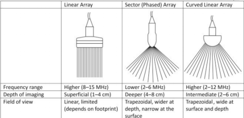
Phased/Sector Array
Has multiple small elements within the transducer face.
Type of transducer selected depends on several factors:
Type of examination, size of patient, amount of fatty or muscular tissue present.
This transducer is used for smaller structures like thyroid, scrotum, breast, etc.
High frequency linear array probes
Abdomen is usually scanned with..
A curved array and/or sector array.
Frequency for abdomen depends on
The size of the patient (lower frequency than linear)
Which transducer is used to scan between ribs in echocardiography?
Phased array
What probe is used for intercavity areas like the prostate?
Transvaginal (Intraluminal) probe.
Transesophageal studies are obtained with a..
Transesophageal probe to image the detailed anatomy of cardiac structures.
Obstetric and Gynecologic scans are usually performed with..
Curved array Transducer
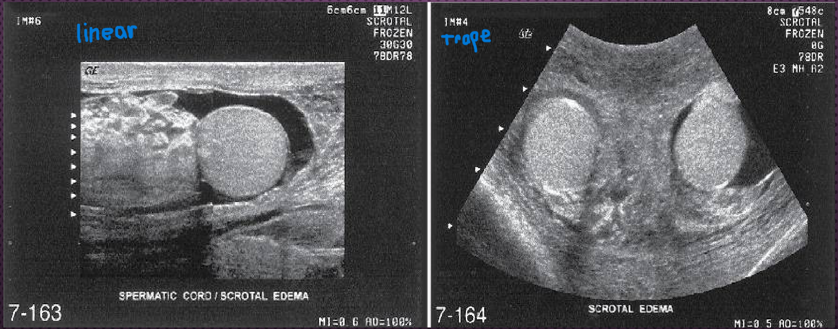
What type of image can a linear-array transducer be steered to produce?
Trapezoidal image
What is the field of view range for a transvaginal probe?
90 to 120 degrees
Which transducer is typically used for abdominal scans?
Curved Array
What axes are used to reconstruct 3D ultrasound images?
x, y, z
3D reconstruction is useful for imaging the fetal face in the second and third trimesters.
True
Power output
Determines the strength of the pulse transmitted into the body
What should you do first if the image is too bright?
Turn down output power
Returning echoes are ____ when the transmitted pulse is _____. This means the image is _____
Stronger, Stronger, Brighter
Power output is displayed as
Decibles (DB) or as a percent of maximum
ALARA Principle
As Low As Reasonably Achievable Less Than 100% Output Power
Receiver Gain
Allows the sonographer to amplify or boost echo signals.
It’s done to compensate for the attenuation of sound in deeper tissues.
Acoustic exposure to the patient increases when receiver gain is increased.
False. Acoustic exposure to the patient remains unchanged when receiver gain is increased.
What happens if the gain setting is too high?
Artificial echo noise will be displayed throughout the image
Time Gain Compensation
TGC control allows the sonographer to amplify receiver gain gradually at specific depths.
Time Gain Compensation is also reffered to as
Depth Gain Compensation (DGC)
Focal Zone
Allows transducer to focus transmitted sound at different depths. Where the sound beam is the thinnest creating optimal image quality.
Where should the focal point be set for optimal image quality?
At or just below the region of interest
Region of Interest
The specific area or structure you want to examine.
What happens to the frame rate as multilevel focusing is used?
Frame rate decreases
Field of View (depth)
Allows the sonographer to adjust the depth and width of an image (how much of the body you can see on the ultrasound)
Depth is displayed in _____ on the side of the image
Centimeters
According to the 2-3 finger rule, where should the ROI be?
2-3 fingers from bottom of the image.
The sound beam is thinnest at the focal point.
True
Multiplanar imaging
Can “follow” the path of a tortuous vessel, moving cardiac structure, or fetus.
What technique allows evaluation of blood flow?
Doppler, allows qualitative and quantitative evaluation of blood flow.
How does the cost of ultrasound compare to other imaging systems?
Costs less
What are today's ultrasound contrast agents made of?
Gas microspheres injected intravenously
Contrast agents facilitate the detection or diagnosis of specific
pathologic conditions
How are curved-array transducer pulses typically scanned to produce their characteristic image format?
They produce a blunted sector field of view from curved scanning surface