Urinary system
 Call Kai
 Learn
Learn Practice Test
 Spaced Repetition
 Match
Match Flashcards
 Knowt Play
1/87

There's no tags or description
Looks like no tags are added yet.
Name | Mastery | Learn | Test | Matching | Spaced | Call with Kai | Chat |
|---|
No analytics yet
Send a link to your students to track their progress
88 Terms
The urinary system consists of?
- 2 kidneys
- 2 ureters
- 1 bladder
- 1 urethra
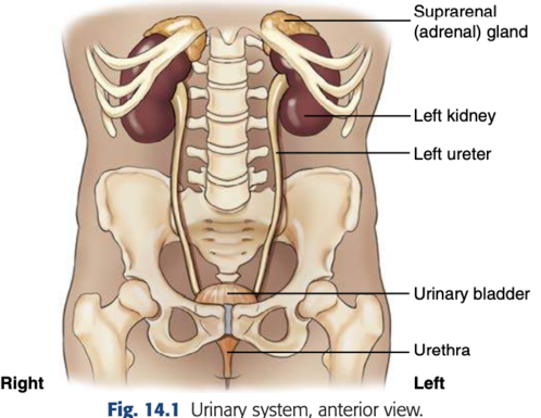
Where are the kidneys located?
beside the spine, between T12- L4
Which kidney is more superior (sits up higher)?
the left kidney. due to the liver located in the RUQ
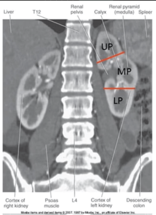
The kidneys lay In what kind of plane?
an oblique plane
What radiographic positions best demonstrate the kidneys?
30° LPO (Left Posterior Oblique) or RPO (Right Posterior Oblique).
In a 30° LPO or RPO position, how does the lower pole of the kidney lie in relation to the IR?
Perpendicular to the IR.
In a 30° LPO or RPO position, how does the upper pole of the kidney lie in relation to the IR?
Parallel to the IR.
Where does filtration occur in the kidney?
In the renal cortex.
What is the functional unit of the urinary system?
The nephron (located in the renal cortex)
Where do the ureters begin?
At the ureteropelvic junction (UPJ) of the renal pelvis.
How do the ureters descend through the body
Anteromedially to the psoas muscle, anterior to the lumbar vertebrae
In males and females, what structure do the distal ureters pass behind?
male: ductus deferens
female: uterine artery
What is the purpose of contrast media in urology?
To help visualize the urinary system during imaging exams.
How is contrast media administered in an antegrade exam?
It is injected intravenously.
What is another name for an intravenous urogram?
Pyelogram.
How is contrast media administered in a retrograde exam?
Through a catheter.
functional study showing bladder and urethra while voiding is
Voiding Cystourethrogram (VCUG), syudy of the bladder and urethra
what contrast does intravenous urography (IVU) use
water-soluble, iodinated contrast agent
common side effects
temporary hot flash, metallic taste in teh mouth
patient medication history
metformin and glucophage
Name three types of retrograde contrast procedures.
- VCUG/CUG
- cystography
- retrograde pyelogram.
What is the purpose of an intravenous urogram?
To visualize the anatomy of the urinary system's collecting portion.
What functional aspect does an intravenous urogram assess?
The functional ability of the kidneys
Is an intravenous urogram a timed procedure?
Yes, it is timed to evaluate how well the kidneys function over time.
What information should be included in a patient history for contrast media use?
- Clinical complaints
- consent form
- allergies
- past reactions
- asthma/hay fever/hives, and lab values.
What key question should be asked to diabetic patients before a contrast procedure?
"Are you currently taking Glucophage or other medication for diabetes mellitus?"
What must be done with metformin meds before and after iodinated contrast procedures?
They must be withheld for 48 hours after the procedure.
What is the general eGFR threshold below which contrast media should not be used?
Less than 60 mL/min/1.73 m².
What are some contraindications (where a treatment should not be used because it could harm the patient.) for contrast media in IVU?
- Graves' Disease (radioactive iodine therapy)
- Pheochromocytoma (adrenal tumor)
- Sickle Cell Anemia (can trigger crisis)
- Renal Failure (risk of death if kidneys can't filter contrast)
- eGFR < 60 mL/min/1.73m² (poor kidney function)
What is the normal creatinine level for adults?
0.6 to 1.3 mg/dL
What is the normal BUN (blood urea nitrogen) level for adults?
6 to 20 mg/dL
What is the normal GFR (glomerular filtration rate) for adults?
Greater than 60 mL/min/1.73 m²
What happens to ionic contrast media when injected?
It dissociates into separate ions.
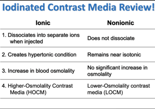
What happens to nonionic contrast media when injected?
It does not dissociate.
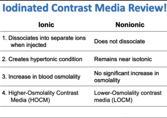
What type of condition does ionic contrast create?
A hypertonic condition.
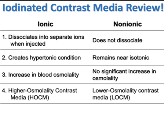
What type of condition does nonionic contrast maintain?
It remains near isotonic.
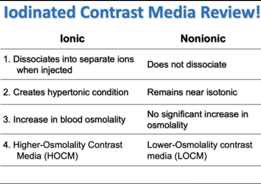
How does ionic contrast affect blood osmolality?
It increases blood osmolality.
How does nonionic contrast affect blood osmolality?
It causes no significant increase in osmolality.
pre-medication protocol
pre treated with benadryl and prednisone over 12 or more hours before procedure
if there a history of allergic reaction, only adminnister contrast to patient with radiologist approval
no guarantee that there will be no reaction
In an RPO (Right Posterior Oblique) position, the kidney that is best demonstrated parallel to the image receptor (IR) is:
Left kidney
What is Extravasation? what is the first step in treating it?
IV has come out of the vein and contrast is injected directly in to the tissue
Notify the department nurse and/or physician.
Why should the affected extremity be elevated above the heart?
To decrease capillary pressure and promote reabsorption of contrast media.
What compresses are used in treating extravasation, and in what order?
Cold compresses first to relieve pain, followed by warm compresses to promote reabsorption.
What is the first image taken in an IVU exam?
a Scout radiograph.
What must be noted at the beginning of the IVU injection?
The exact time of the injection.
What is the imaging routine at 1 minute post-injection?
Nephrogram or nephrotomography basically a KUB
What is the imaging routine at 5 minutes post-injection?
AP supine.
What is the imaging routine at 10-15 minutes post-injection?
AP and posterior obliques.
What is the final imaging step in the IVU routine?
Postvoid (urinated) image (either prone or erect).
What is the purpose of an AP nephrotomogram?
to demonstrate the renal parenchyma or functional portion of the kidney.
nonionic iodine contrast agents differs from anionic agents in that nonionic
hey do not break down or dissociate into charged particles (cations and anions) when dissolved in blood plasma
which contrast agent contain positively charged cation
which contrast agent contain positively charged cation
How do you estimate the kidney level for tomography?
Divide the total A-P measurement by 3 (kidneys are in the posterior 1/3 of the abdomen).
How many tomo cuts are typically taken for the kidneys?
Three tomo cuts at the calculated level and posterior.hw
what is the CR for AP nephrotomogram
CR perpendicular to IR directed to MSP midway between xiphoid process and iliac crest
SID 40wh
at is being demonstrated in an AP nephrogram
renal parenchyma
what is the CR for AP IVU
perpendicular to IR, directed to MSP and iliac crestwh
what are critics needed to include in AP IVU
entire urinary system visuazlied from upper renal shadows to distal urinary bladder. symphysis pubis should be included
what is the CR for IVU posterior oblique (RPO LPO)
perpendicular to IR, directed to MSP and iliac crest
rotate body 30 degrees
what are critics needed in IVU posterior obliques
upside kidney = parallel to plane of IR
downside ureter = free of superimposition
whats the CR for AP/PA post void
perpendicular to IR, directed to MSP and iliac crest
How is contrast media delivered in cystography?
Through a urinary catheter using gravity flow of CM
150-500 mL
what is the CR for AP axial bladder
perpendicular, directed to MSP and symphysis pubis
for voiding study CR has 15 degree caudad angle (for VCUG akla voiding cystourethrography)
purpose of Custourethrography
functional study of bladder and urethra
whats the CR of axial posterior oblique bladder
perpendicular to IR at 2’’ superior to upper border of pubicx symphysis and 2’’ medrial to upper ASIS
voiding; perpendicular to level of pubic symphysis
what are the patietn position and anatomy shown on axial posterior oblique bladder
45-60 posterior onlique
urinary bladder not superimposed by pubic bones or lower lumbs
When should AP nephrotomogram (IVP) images be taken?
At 1 minute post-contrast injection.
benign prostatic hyperplasia (BPH)
enlargement of the prostate
bladder calculi
stones that form in the urinary bladder
bladder carcinoma
tumor usually at old age. symptoms hematuria and frequency in urination
oliguria
low urine output
anuria
almost no urine production
polyura
excessive urine output
urinary incontinence
loss of bladder control
micturition
act of urinating/voiding urine
uremia
waste products build up in the blood because kidneys are failing e
ectopic kidney
kidney that remains in the pelvis and failed to descend into the abdomen
horseshoe kidney
fusion of kidney in fetal developmennt
cystitis
inflammation of the urinary bladder caused by bacterial or fungal infection glo
merulonephritis
inflammation of the capillary loops of the glomeruli of the kidneys
hydronephrosis
distention of renal pelvis and calyces of the kidney that results from obstruction of ureter or renal pelvis
polycystic kidney disease
cysts scattered througout kidney. most common cause of enlarged kidneys
pyelonephritis
inflammation of kidney and renal pelvis caused by pyogenic
renal calculi
calcification that occur in the uminal aspect of urinary tract reba
renal cell carcinoma (hypernephroma)
common malignant tumor of kidney
renal hyperntension
increased blood pressure to kidney through the renal artery due to atherosclerosis
whats the routine of IVU
AP (scout and series)
nephrotomogram
RPO and LPO
AP
voiding cystourethography routne
Male - RPO
female - AP