Biology:Exchange Surfaces and Breathing
1/49
There's no tags or description
Looks like no tags are added yet.
Name | Mastery | Learn | Test | Matching | Spaced |
|---|
No study sessions yet.
50 Terms
7.1 Surface area to volume ratip
7.1 Surface area to volume ratio
What material do organisms need to exchange
Living organisms need to exchange materials like oxygen, glucose, excretory products like urea, and heat with their environment.
This exchange occurs across plasma membranes.
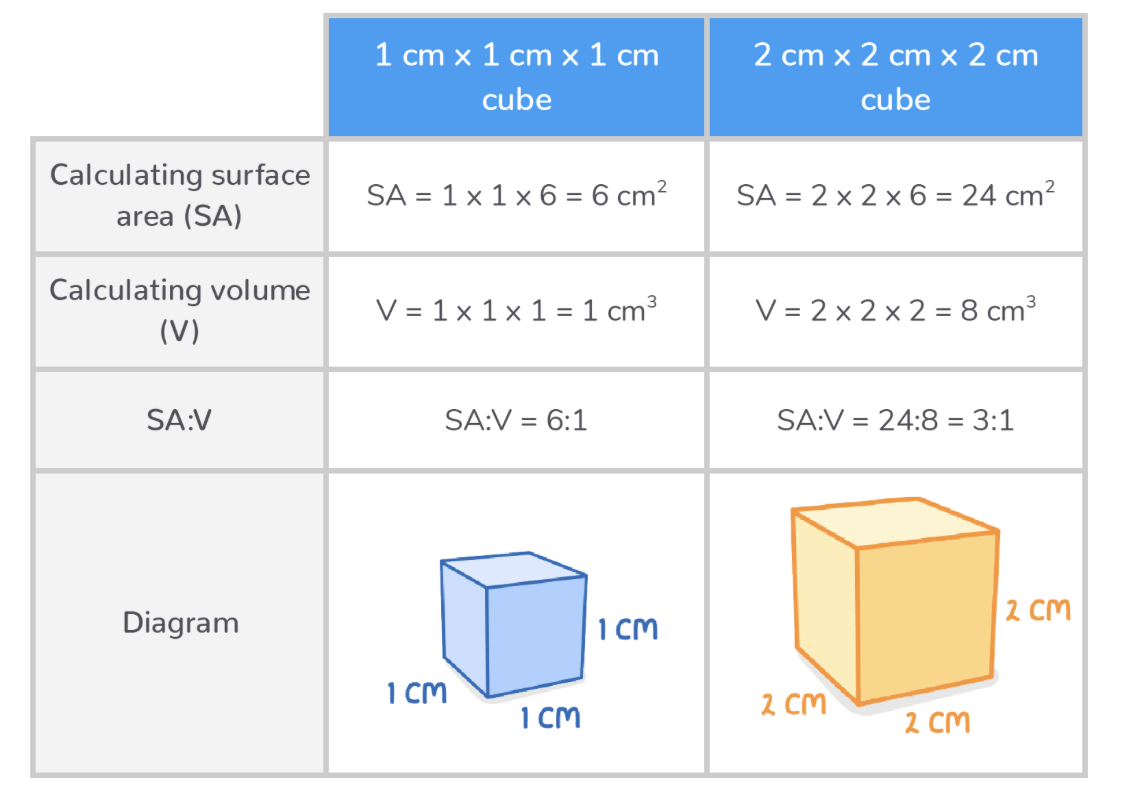
How does surface area to volume ratio affect diffusion
How SA:V affects rate of diffusion:
High SA:V - These organisms have a large surface area relative to their volume so the diffusion of substances is fast.
Low SA:V - These organisms have a small surface area relative to their volume so the diffusion of substances is slower.
Generally, smaller organisms have a higher SA:V while larger organisms have a lower SA:V.
How to calculate SA:VOL
7.2 Specialised Exchange surfaces
7.2 Specialised Exchange surfaces
Why doo multicellular organisms require specialised surfaces?
In single-celled organisms, substances diffuse directly across the cell membrane.
This cannot happen in multicellular organisms because:
Cells are not in direct contact with the external environment.
Diffusion distances between cells and their environment are large.
Larger organisms have higher metabolic rates, so they need more oxygen and glucose.
To solve this problem, multicellular organisms have evolved specialised exchange surfaces.
Key features of specialised exchange surfaces
A large surface area - This provides a larger area across which substances can be exchanged.
Thin walls - These minimise the diffusion distance.
An extensive blood supply and/or ventilation - This maintains steep concentration gradients.
Being surrounded by selectively permeable plasma membranes - This controls what substances are exchanged.
7.3 Human gas exchange system
7.3 Human gas exchange system
What is the human gas exchange system
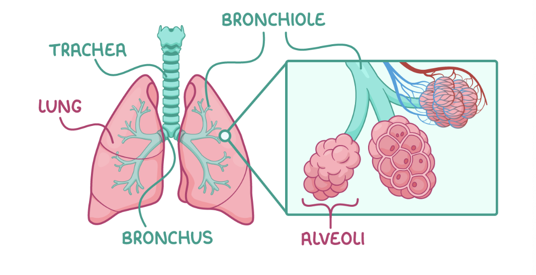
The human gas exchange system consists of the lungs and air passages. This system allows oxygen to enter the blood and carbon dioxide to leave the blood through gas exchange surfaces called alveoli. The lungs are located inside the chest in the thoracic cavity (thorax), protected by the ribcage.
The gas exchange system is located inside the body because:
Air is not dense enough to support and protect these delicate structures.
The body would otherwise lose water and dry out.
Pathway of air stages
Air first enters the trachea.
Air travels into the two bronchi, with one bronchus going to each lung.
Air travels into smaller airways called bronchioles.
Air travels into clusters of air sacs called alveoli at the end of the bronchioles.
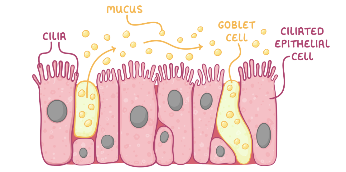
Cilliated epithelium
The ciliated epithelium contains mainly goblet cells and ciliated epithelial cells:
Goblet cells - These produce and secrete mucus that traps dust and microbes.
Cilia on ciliated epithelial cells - These waft the mucus upward to the mouth so it can be swallowed.
structure and adaption of trachea
The trachea is the large tube that carries air from the throat down to the lungs.
Adaptations:
Rings of cartilage keep the airway open.
Smooth muscle can contract or relax to constrict or dilate the airway and change airflow.
Elastic tissue contains elastic fibres with elastin that allows stretching and recoiling.
Lined with ciliated epithelial cells and goblet cells.
structure and adaptation of bronchi
The bronchi are two main branches extending from the trachea that carry air into each lung.
Adaptations:
Reinforced with cartilage to keep the airway open.
Smooth muscle can contract or relax to constrict or dilate the airway and change airflow.
Elastic tissue contains elastic fibres with elastin that allows stretching and recoiling.
Lined with ciliated epithelial cells and goblet cells.
structure and adaptations of bronchioles
The bronchioles are smaller airways branching from the bronchi that carry air to the alveoli.
Adaptations:
No cartilage, can change shape.
Smooth muscle can contract or relax to constrict or dilate the airway and change airflow.
Elastic tissue contains elastic fibres with elastin that allows stretching and recoiling.
Simple squamous epithelium (only larger bronchioles have a ciliated epithelium).
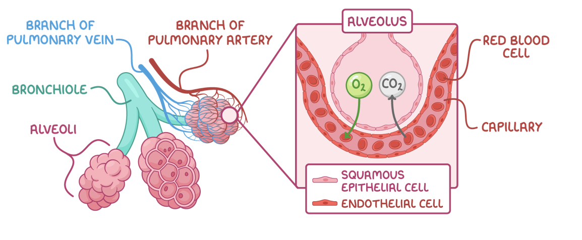
Alveoli and blood vessels
Alveoli are tiny air sacs clustered at the ends of the bronchioles. They are surrounded by a network of capillaries so gases can be exchanged between the air in the alveoli and the blood.
Gas exchange occurs across the alveolar membrane of alveoli.
How alveoli carries out gas exchange
Oxygen diffuses from the alveoli into the pulmonary capillaries where it binds to haemoglobin in red blood cells.
Carbon dioxide dissociates from haemoglobin and diffuses from the blood into the alveoli.
Adaptations of alveoli for gas exchange
Wall consists of one layer of squamous epithelial cells - This allows rapid diffusion.
Large surface area - This increases rate of gas exchange.
Partially permeable - This means that only certain gases can move across the wall.
Surrounded by dense network of capillaries - These bring blood close to air for gas exchange.
Ventilation of air - This maintains steep diffusion gradient.
Elastic fibres - These allow stretching and recoiling.
Collagen fibres - These contain strong collagen that prevents alveoli from bursting and limits overstretching.
Moist inner surface - This allows gases to dissolve, and lung surfactant helps alveoli remain inflated.
Pulmonary blood vessels
The pulmonary blood vessels are those involved in circulation of the lungs.
They include:
The pulmonary artery - This delivers deoxygenated blood from heart to pulmonary capillaries.
The pulmonary vein - This delivers oxygenated blood from capillaries to heart.
The pulmonary capillaries - These are the site of gas exchange between blood and alveoli.
Adaptation of pulmonary capillaries
Thin walls (one endothelial cell thick) - This maintains a short diffusion distance.
Red blood cells pressed against capillary walls - This reduces diffusion distance.
Large surface area - This increases diffusion speed.
Movement of blood - This maintains steep diffusion gradient.
Slow blood movement - This allows more time for diffusion.
7.4 Ventilation
7.4 Ventilation
What is ventilation?
Ventilation, or breathing, is the constant movement of air into and out of the lungs. It consists of inspiration (breathing in) and expiration (breathing out).
It allows air to enter and leave the lungs, providing the body with oxygen and removing carbon dioxide.
Muscles involved in ventilation
The ribcage is made up of bones called ribs that enclose the thorax - the cavity where the lungs are located (thoracic cavity).
In mammals, ventilation is controlled by specific muscles. When the muscles attached to the ribcage contract and relax, they move the ribs to change the volume of the thoracic cavity. This affects the pressure in the lungs and controls ventilation.
What are the muscles that act on the ribcage?
The diaphragm - This is a sheet of muscle that moves the ribcage up and out when it contracts.
The external intercostal muscles - These are found between the ribs and pull the ribcage up and out when they contract.
The internal intercostal muscles - These are found between the ribs but pull the ribcage down and in when they contract.
The external and internal intercostal muscles have opposite effects on the ribcage.
The external muscles expand the ribcage during inspiration, while the internal muscles shrink it during expiration.
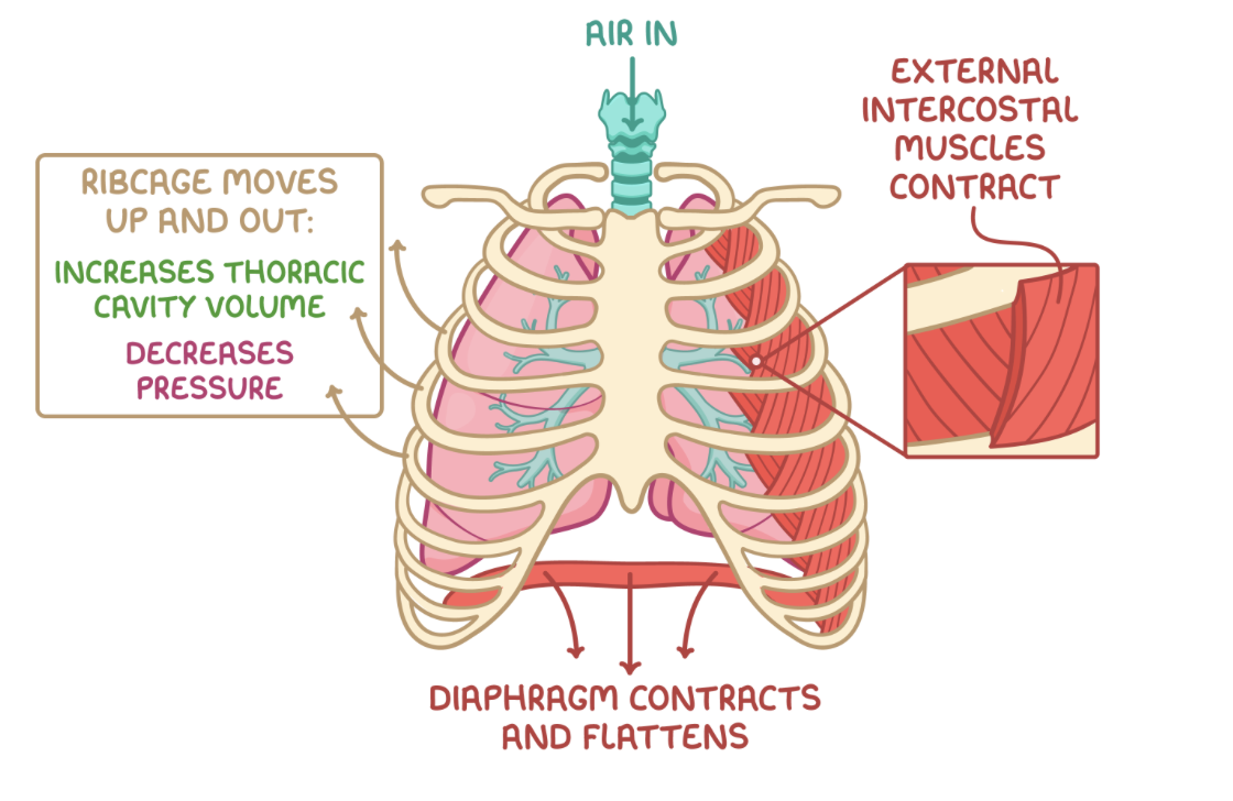
Inspiration
Inspiration is an active process requiring energy for muscle contraction
During inspiration:
The external intercostal muscles contract while the internal intercostal muscles relax, moving the ribcage up and out.
The volume of the thoracic cavity increases.
The diaphragm contracts and flattens, further increasing the volume of the thoracic cavity.
The lung pressure decreases below atmospheric pressure.
Air flows into the lungs down the pressure gradient.
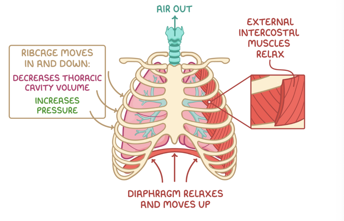
Expiration
Normal expiration at rest is a passive process so it does not require energy.
However, expiration can be forced by contracting the internal intercostal muscles to actively pull the ribcage down and in, forcing more air out.
During expiration:
The external intercostal muscles relax, moving the ribcage down and in.
The volume of the thoracic cavity decreases.
The diaphragm relaxes and unflattens, further decreasing the volume of the thoracic cavity.
The lung pressure increases above atmospheric pressure.
Air is forced out of the lungs down the pressure gradient.
Elastic fibres in the alveoli also shrink and recoil back to their original shape when the thoracic cavity volume decreases.
This increases the pulmonary pressure and helps to push air out of the lungs.
7.5 Measuring ventilation
7.5 Measuring ventilation
Measuring data on lung function, volume and capacity
There are various instruments that can measure the flow of air into and out of the lungs, providing data on lung function and capacity.
Some examples of instruments that provide data on lung function include:
Peak flow meter - This measures the maximum speed of expiration through a mouthpiece by tracking the movement of an indicator.
Vitalograph - This records a graph showing the volume and rate of forced expiration through a mouthpiece.
Spirometer - This calculates different lung volumes using a chamber containing a known volume of gas connected to a mouthpiece and recorder.
Spirometer traces
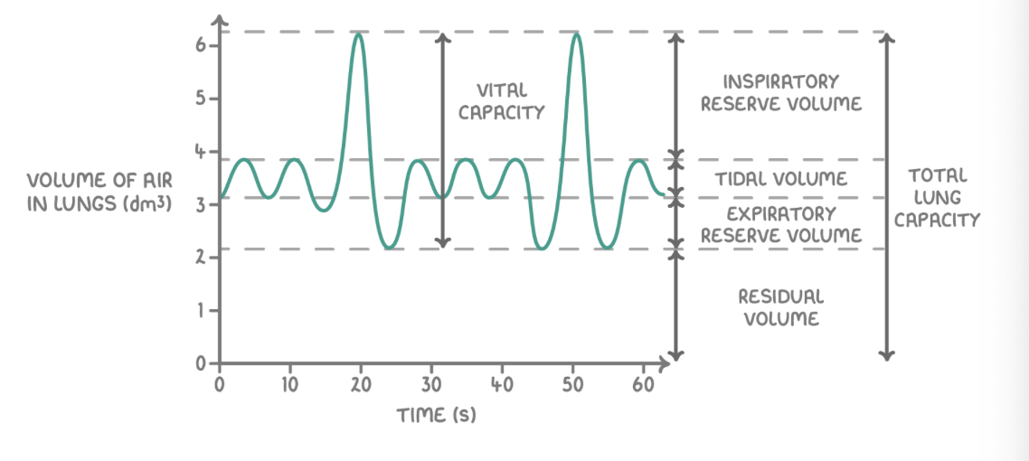
Use spirometer graph calculations
Breathing rate - This is the number of breaths taken per minute, measured by counting the number of peaks in a minute.
Tidal volume - This is the volume of air breathed in or out in an average breath during rest, measured from the height of each peak at rest.
Vital capacity - This is the maximum volume of air that can be inhaled or exhaled in one deep breath, measured from the maximum peak height.
Inspiratory reserve volume - This is the maximum volume of air that can be inhaled above a normal inhalation.
Expiratory reserve volume - This is the maximum volume of air that can be exhaled above a normal exhalation.
Residual volume - This is the volume of air that remains in the lungs after the largest possible exhalation.
Total lung capacity - This is the vital capacity added to the residual volume.
Calculating oxygen consumption
Oxygen consumption is the volume of oxygen used per minute.
If we had a spirometer trace, we could also measure oxygen consumption. This is the slope of a spirometer trace.
We can calculate oxygen consumption as the change in the volume of gas in the spirometer over a period of time and divide this value by the time taken.
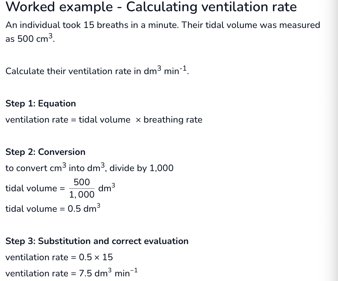
Ventilation rate
Ventilation rate is the volume of oxygen inhaled per minute.
Ventilation rate can be calculated as follows:
Measure tidal volume (dm3).
Measure breathing rate (min-1).
Ventilation rate (dm3 min−1)= tidal volume (dm3)× breathing rate (min−1){3}\text{ min}{-1})=\text{ tidal volume (dm}{3})\times\text{ breathing rate (min}{-1})Ventilation rate (dm3 min−1)= tidal volume (dm3)× breathing rate (min−1)
7.6 Gas exchange in insects
7.6 Gas exchange in insects
Why insects need gas exchange
Insects have high oxygen demands but their tough chitinous external skeleton (exoskeleton) prevents direct gas exchange.
Insects need efficient systems for exchanging gases for two main reasons:
To deliver oxygen to cells - This allows aerobic respiration to occur to release energy for cellular processes.
To remove carbon dioxide from cells - The build up of carbon dioxide produced as a waste product of respiration reduces pH, which can denature enzymes.
Needs that insects as exchange systems have adapted for
Insect gas exchange systems have adapted to balance two conflicting needs:
Maximising gas exchange efficiency
Minimising water loss
The exoskeleton is covered with a waterproof cuticle to help prevent water loss, but the insect gas exchange system itself has additional methods to prevent excess water loss while still being effective at gas exchange.
Structure of insect gas exchange system
Insects have an open respiratory system comprised of tubular structures that transport air
The main structures involved are:
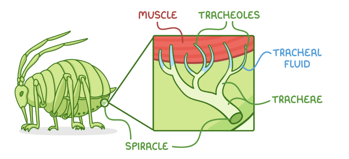
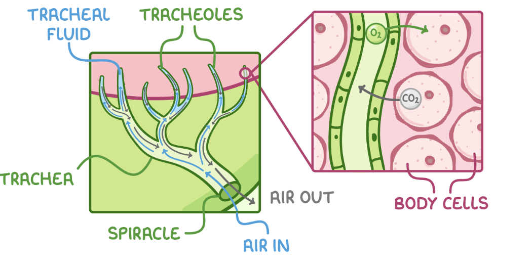
Tracheae - These are air-filled tubes branching throughout the body.
Tracheoles - These are fine branches of tracheae that deliver gases to cells.
Spiracles - These are external openings of the tracheal system on the exoskeleton along the abdomen and thorax.
Adaptations of the structures in the insect gas exchange system
There are some key features that make these structures well adapted for their functions.
Tracheae:
Reinforced with spirals of chitin - This prevents collapsing.
Multiple tracheae - This increases surface area.
Tracheoles:
Penetrate directly into tissues - This reduces the gas diffusion distance.
Thin walls - These reduce the gas diffusion distance.
Highly branched - This maximises the surface area.
Not reinforced with chitin - This allows gas exchange to occur.
Fluid at the ends of the tracheoles (tracheal fluid) - This allows oxygen to dissolve to aid diffusion and reduces water loss.
Spiracles:
Open and close - This allows them to control gas exchange with the atmosphere and minimise water loss.
How gas exchange occurs
These stages are:
Air enters the tracheal system through open spiracles.
Air moves into larger tracheae and diffuses into smaller tracheoles.
Tracheoles branch throughout the body, transporting air directly to cells.
Oxygen dissolves in water in tracheal fluid and diffuses down its concentration gradient from tracheoles into body cells.
Carbon dioxide diffuses down its concentration gradient out of body cells into the tracheoles.
Air is then carried back to the spiracles via the tracheae and released from the body.
How are concentration gradients between tissues and air in tracheal system maintained
Cells using up oxygen for respiration - This keeps oxygen concentration low in cells.
Cells producing carbon dioxide in respiration - This keeps carbon dioxide concentration high in cells.
Continuous ventilation - Fresh air is supplied to the tracheal system via spiracles.
Other ventilation mechanisms
Some insects, particularly active ones, may use additional ventilation mechanisms to drive air through the tracheal system.
Some of these mechanisms include:
More spiracles open - This allows more oxygen to enter the tracheal system.
Mechanical active ventilation - This is when muscles around the tracheae contract and relax, changing the volume and pressure in the abdomen and squeezing the tracheae to pump air in and out of the spiracles.
Movement of tracheal fluid out into tissues - This increases the diffusion rate and surface area for gas exchange.
Enlarged collapsible tracheae, accessory sacs, and air reservoirs - These inflate or deflate to ventilate the tracheal system and can increase the volume of air moved through the system.
Movement of wing muscles connected to sacs - These pump air to ventilate the tracheal system.
Vibration of thoracic muscles - This pumps air to ventilate the tracheal system.
Lactic acid accumulation
Lactic acid accumulates in tissues during activity.
This can affect the rate of gas exchange:
Lactic acid accumulation reduces the water potential in tracheal fluid at the end of tracheoles.
Water leaves the tracheoles via osmosis.
A higher surface area is exposed for gas exchange.
7.7 Gas exchange in fish
7.7 Gas exchange in fish
Respiratory systems in bony fish
Large, active, bony fish have high oxygen needs. These needs exceed simple diffusion across the body surface, which is also covered with scaly skin preventing gas exchange.
Bony fish have evolved specialised respiratory systems to meet the challenges of extracting oxygen from water.
Some of these challenges include:
Water is denser and more viscous than air, resulting in slower diffusion of oxygen.
Water has less oxygen than air.
Bony fish are very active so have high oxygen demands
Structure of the gas exchange system in bony fish
Gills allow bony fish to efficiently take up oxygen from water and to remove carbon dioxide.
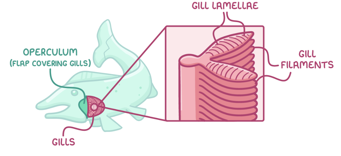
Structure of the gills
Structure of the gills:
Gills are covered by an operculum flap.
Gills consist of stacked filaments containing gill lamellae.
Gill lamellae are surrounded by extensive blood vessels.
Adaptations of the gills for efficient gas exchange
The lamellae provide a large surface area.
The lamellae membranes are thin to minimise diffusion distance.
The gills have a rich blood supply to maintain steep diffusion gradients.
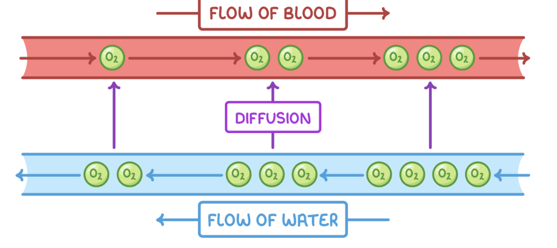
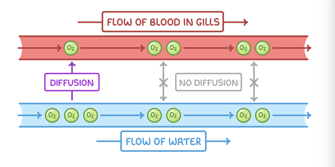
The countercurrent flow of blood and water creates even steeper concentration gradients.
Overlapping filament tips increase resistance, slowing water flow over gills and allowing more time for gas exchange.
Countercurrent exchange principle
The gills of bony fish allow countercurrent flow of blood and water, which is much more efficient than a parallel flow.
Countercurrent flow
Blood and water flow over the lamellae in opposite directions.
This means that oxygen-rich blood meets water that is at its most oxygen rich when it first moves across the gills, maximising diffusion of oxygen into the blood.
Oxygen-poor blood returning from body tissues meets oxygen-reduced water that has had most of its oxygen removed, still allowing diffusion of oxygen into the blood.
This maintains a steep concentration gradient across the entire gill.
Parallel flow
Countercurrent exchange systems enable more efficient gas transfer than parallel flow, because parallel flow reduces the concentration gradient so less oxygen can be absorbed.
Ventilation via thre buccal cavity
The buccal cavity is the mouth and throat area of bony fish.
Bony fish ventilate their gills by opening and closing their mouths, changing the volume of the buccal cavity:
When a fish opens its mouth, this increases the volume of the buccal cavity.
This decreases the pressure, which pulls water into the buccal cavity.
Water flows over the gills.
Water flows out through the operculum.
This drives unidirectional water flow for ventilation, providing freshly oxygenated water and removing carbon dioxide.