IOS1_ANATOMY OF THE NEPHROURINARY SYSTEM
1/44
There's no tags or description
Looks like no tags are added yet.
Name | Mastery | Learn | Test | Matching | Spaced |
|---|
No study sessions yet.
45 Terms
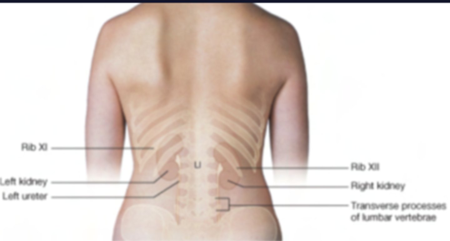
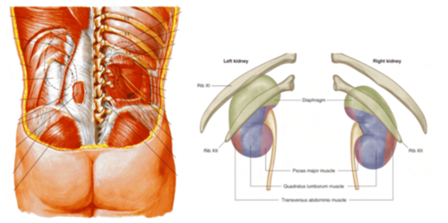
What is the surface projection of the left kidney?
Associated with ribs 11th and 12th
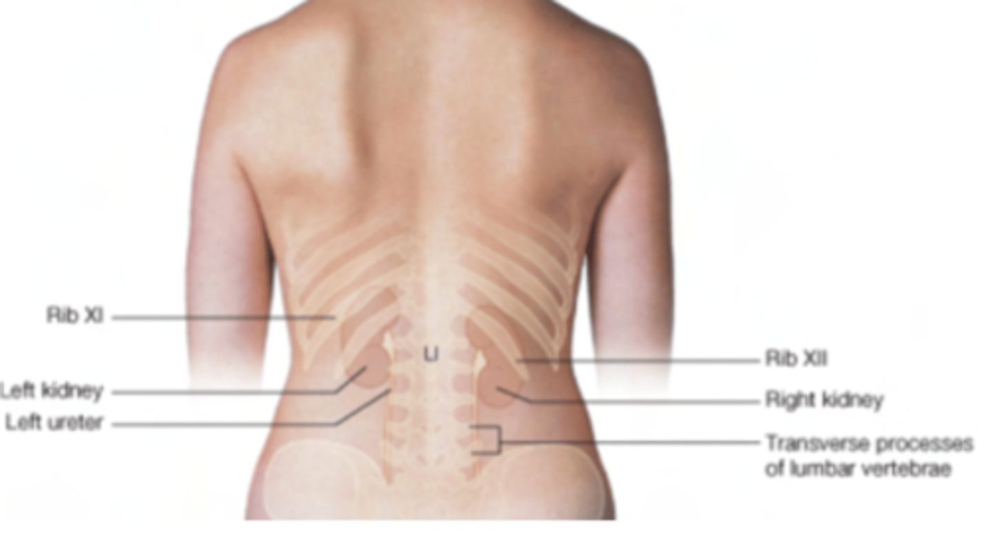
What is the surface projection of the right kidney?
Associate only with the rib 12th
due to the liver pushing down on it.
How are the kidneys vs the peritoneum?
retroperitoneal structures.
- Structures recovered by the visceral peritoneum are called intraperitoneal, (ie stomach and liver) while structures which develop above the parietal peritoneum are called retroperitoneal and are only partially covered by it
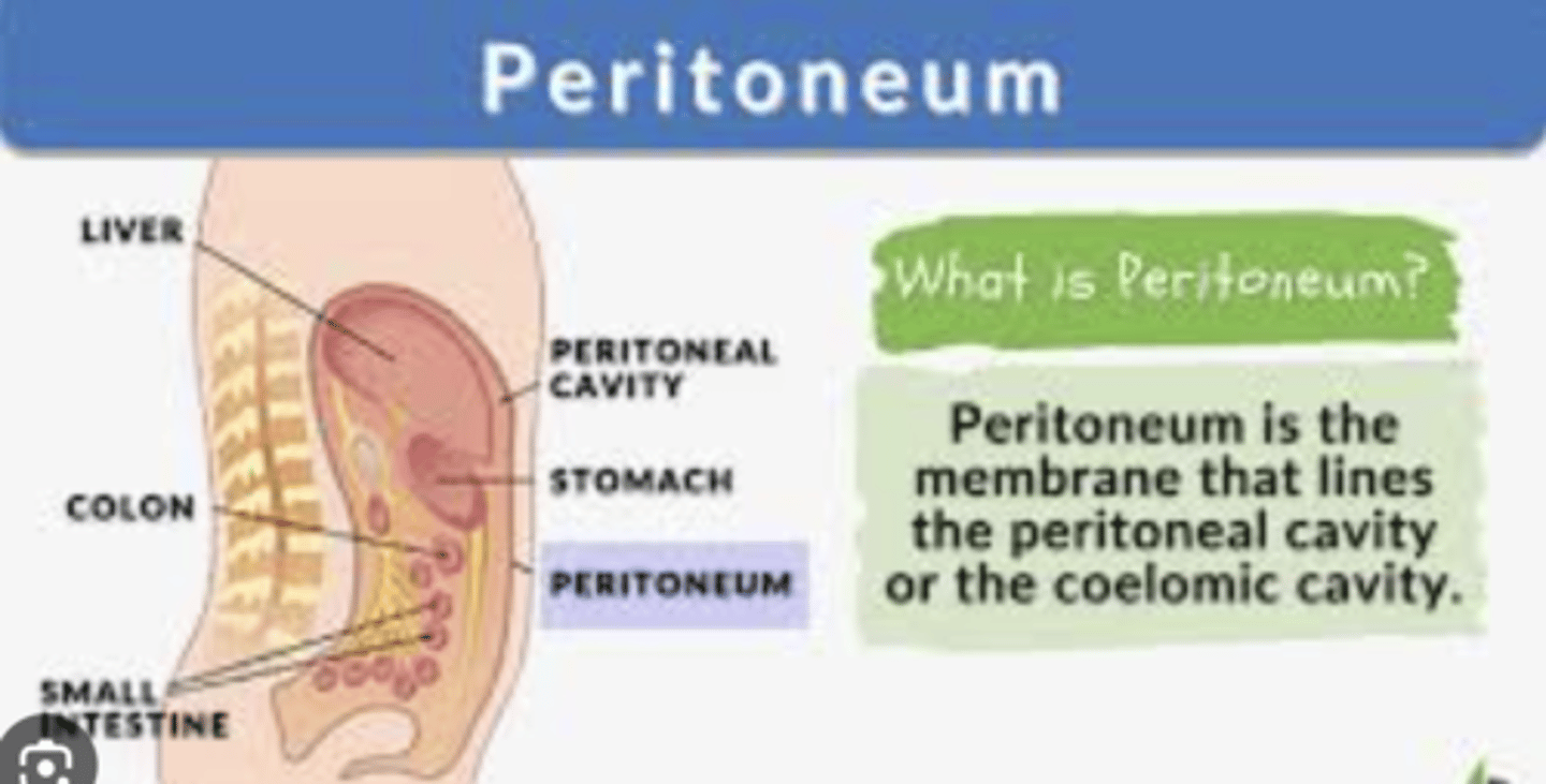
Differences between retroperitoneal and intraperitoneal visceras?
Retroperitoneal viscera will therefore be fixed, attached to the muscles, whilst intraperitoneal may have some mobility
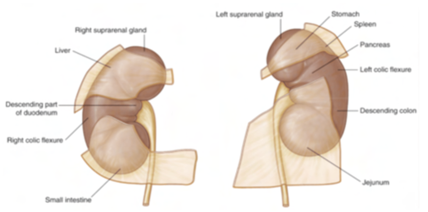
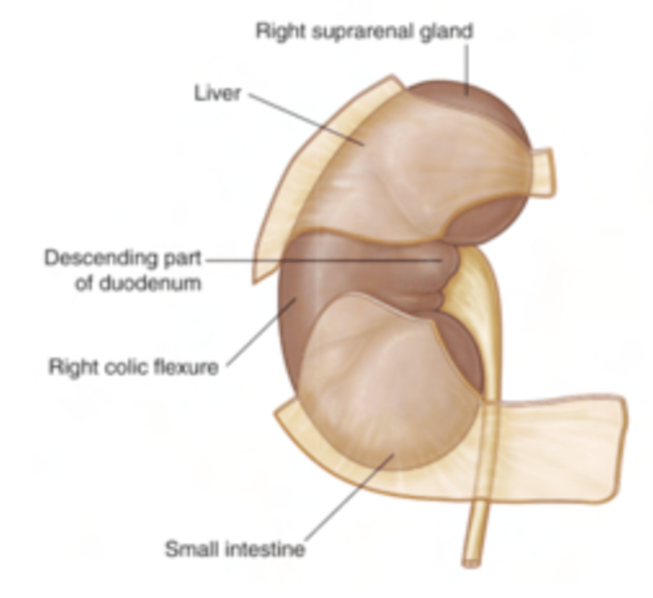
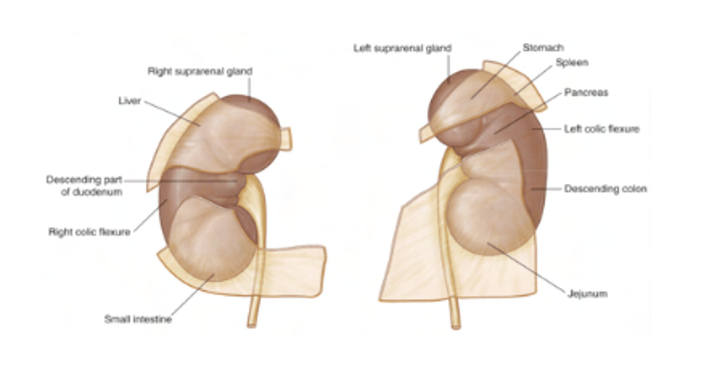
What is the anterior relationship of the right kidney?
the liver separated from the right kidney by peritoneum, onto which it presses against, making it be a little lower.
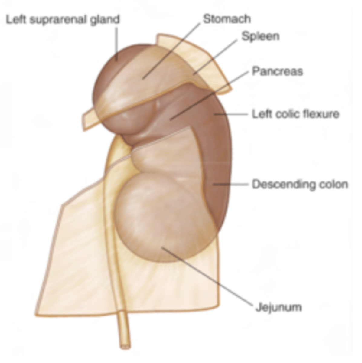
What is the anterior relationship of the left kidney?
directly associated by the suprarenal gland but separated from the stomach and spleen by the peritoneum.
What is the relationship between the organs?
Diseases or conditions (e.g., tumors, infections, or hemorrhages) in the retroperitoneal space can be harder to detect because these structures are not directly visible during abdominal surgeries.
Retroperitoneal infections or abscesses can spread along the space and affect multiple organs.
What are the posterior relationships of the kidneys?
- The diaphragm
- Psoas major
- Quadratus lumborum
- Transversus abdominis
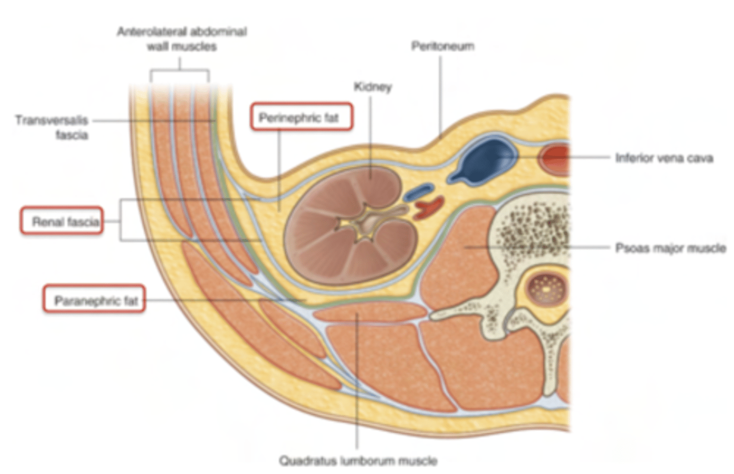
What are the layers protecting the kidneys?
i. Renal capsule, seen in brown. Gives the kidneys their characteristic bean shape. CT capsule, not peritoneum
ii. Perinephric fat: adipose capsule,
iii. Renal fascia: it anchors the kidney and its perinephric fat to the surroundings, such as the muscles we have just mentioned
iv. Paranephric fat: 2nd adipose capsule surrounding the kidneys
What are the regions of the kidney?
Outer portion: renal cortex
Inner portion: renal medulla

What contains the renal cortex?
Nephrons: The functional units of the kidney responsible for filtration and urine formation.
also extended into the medulla (specifically the loop of Henle).
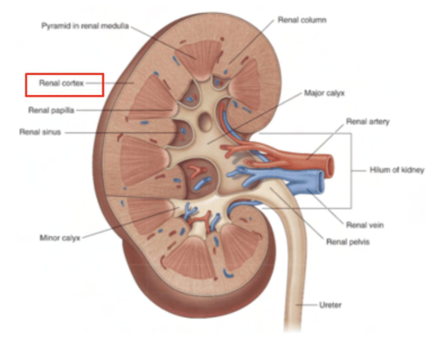
What contains the renal medula?
8–10 renal pyramids:
These pyramids are cone-shaped structures formed by collecting tubules.
They are separated by renal columns, which are extensions of the cortex that project inward between the pyramids.
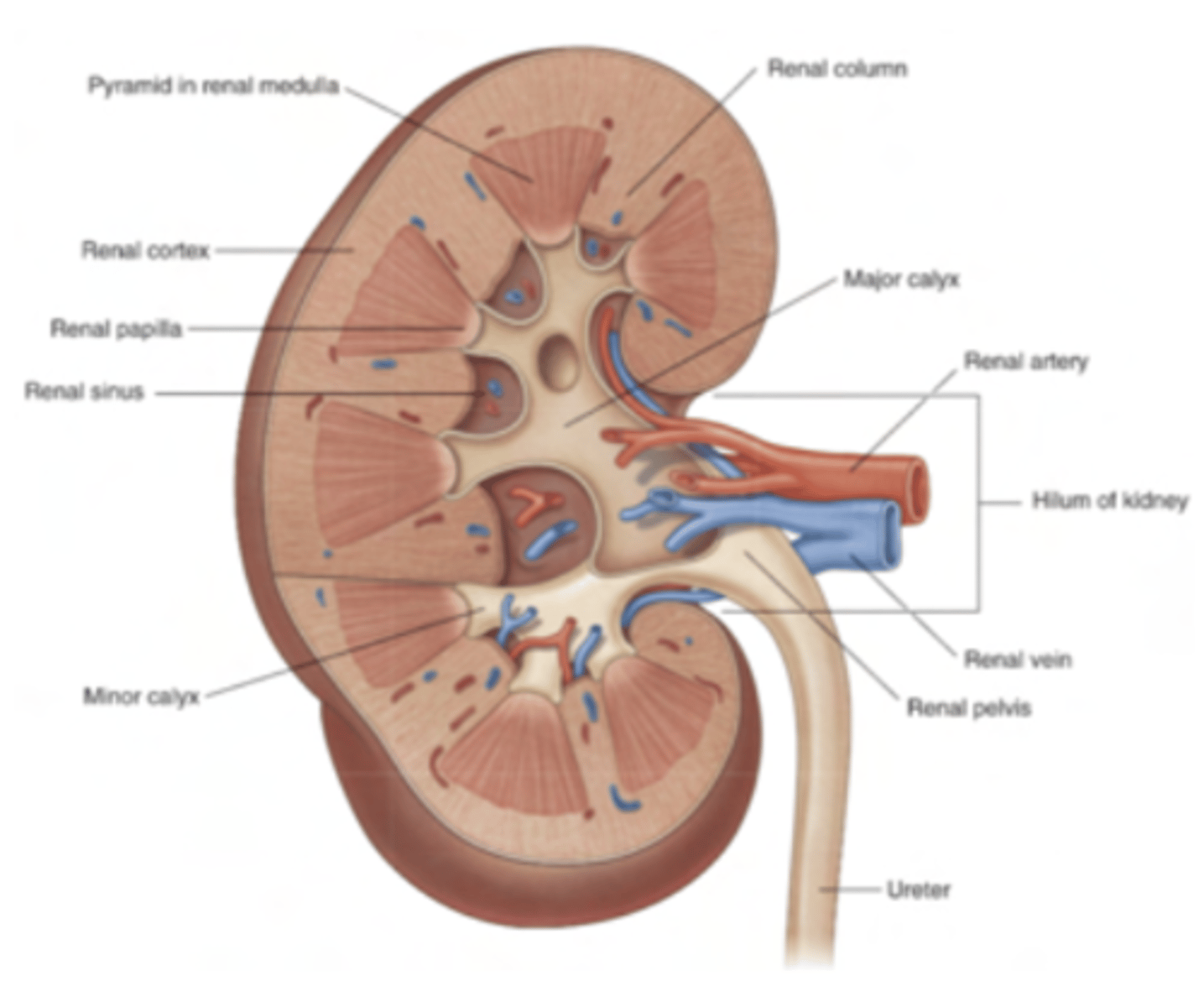
How are formed the pyramids of the renal medula?
collecting tubules which, at the renal papilla, open to the minor calyces, and then into the major calyces, which join to form the renal pelvis and from there to the ureter.

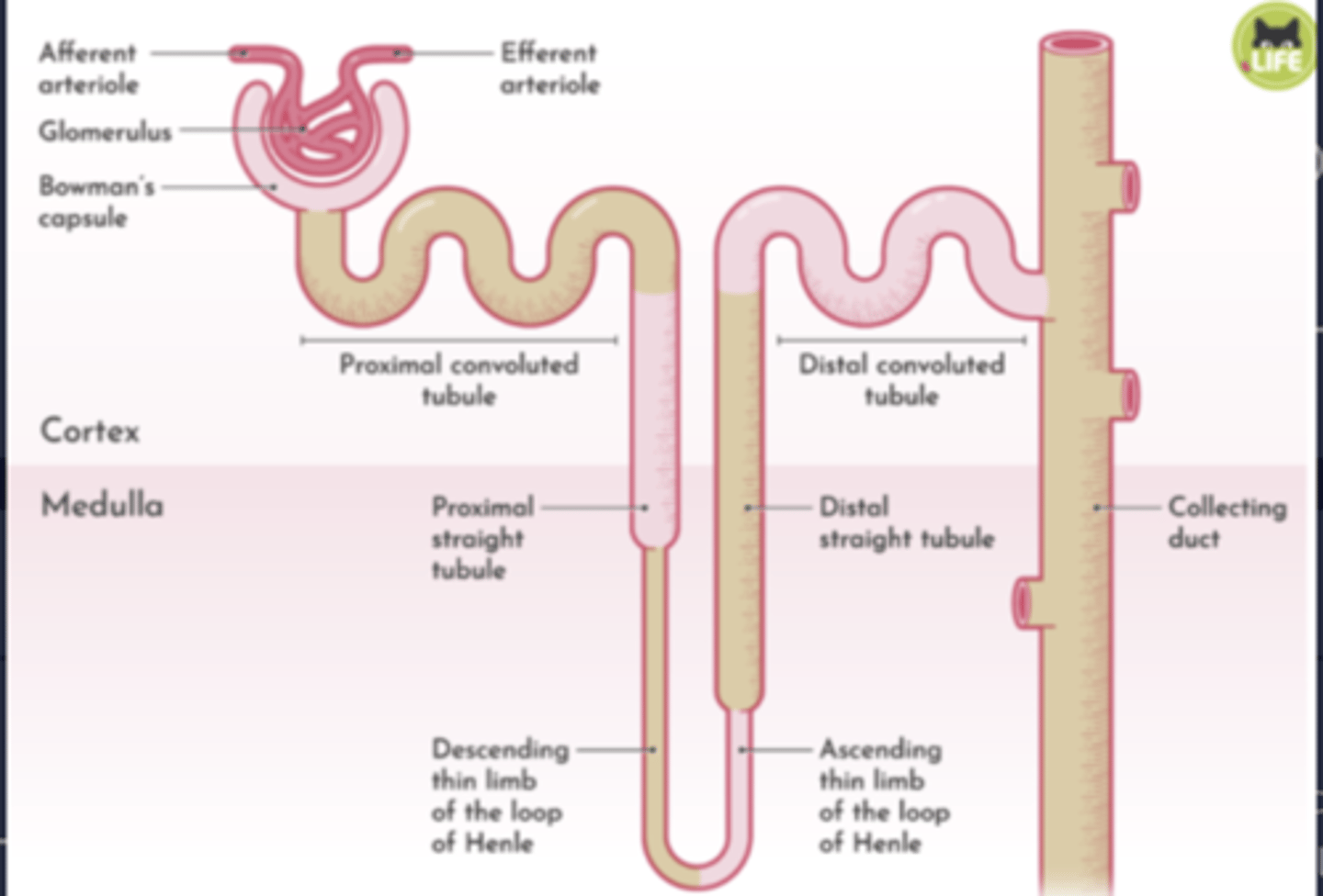
What are made up of the nephrons?
Bowman’s Capsule:
Proximal Convoluted Tubule (PCT):
Henle’s Loop (Loop of Henle):
Distal Convoluted Tubule (DCT):
Collecting Tubule:
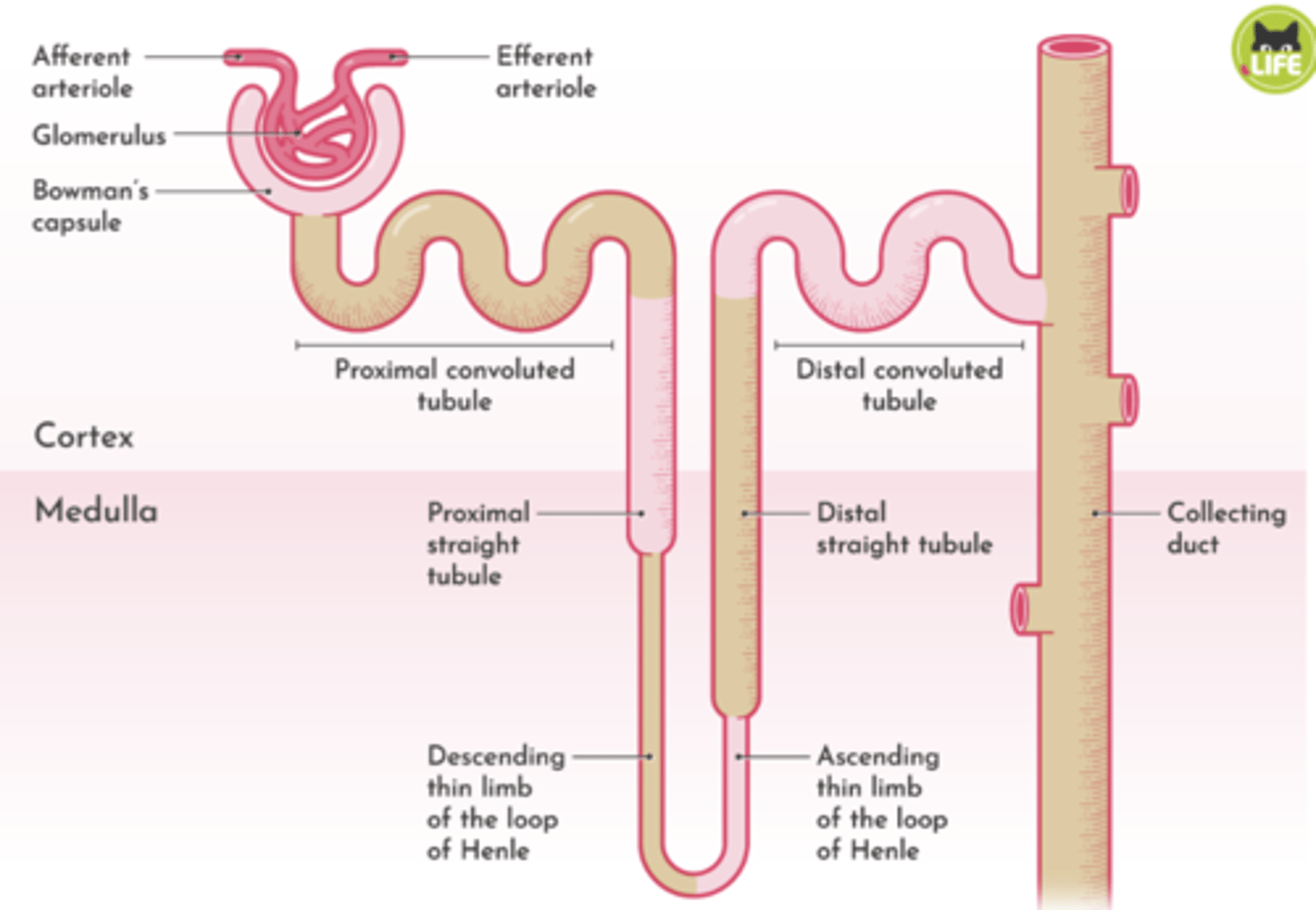
What is the flow of urine in the kidney?
Bowman's capsule → PCT → Loop of Henle → DCT → Collecting tubule → Renal papilla → Minor calyx → Major calyx → Renal pelvis → Ureter.
How is the irrigation of the kidneys?
Anterior: a vein
Posterior: the artery
Behind the artery: renal pelvis (continuations of ureter into
kidney)
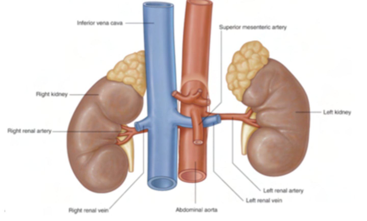
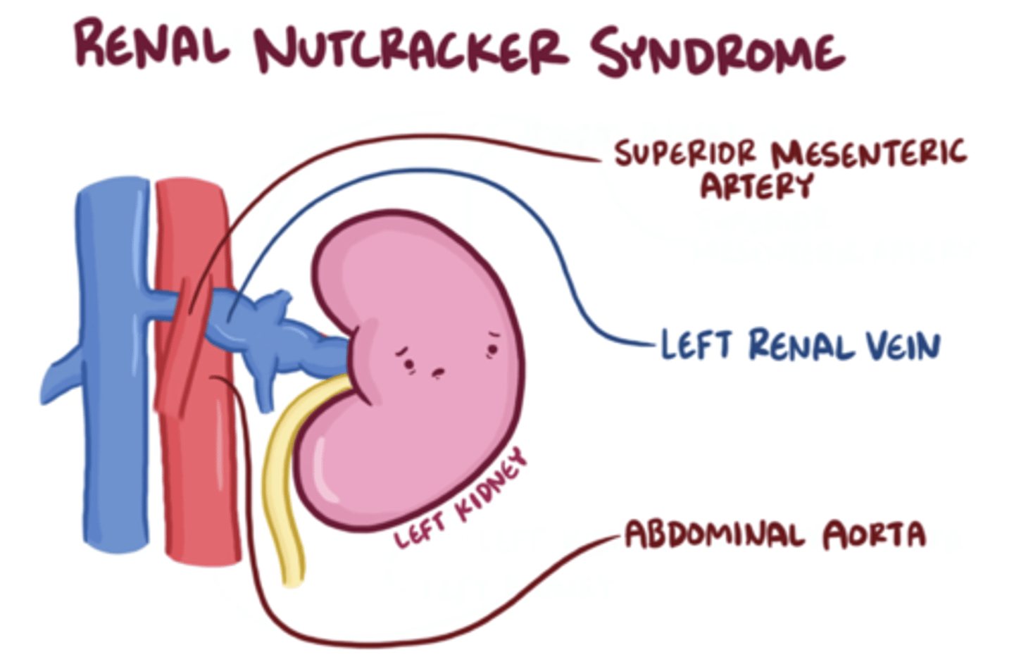
Why this relationship is important (irrigation)?
The position of the SMA and the left renal vein creates a potential site of compression. If the SMA compresses the left renal vein, it can lead to vascular complications:
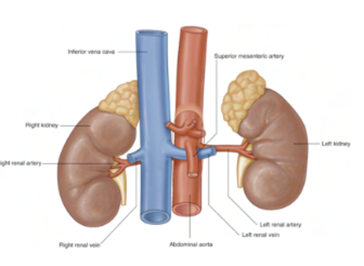
What is the nutcracker syndrome?
left renal vein is compressed between the SMA and the abdominal aorta.
Consequences include:Impaired blood drainage from the left kidney.Increased pressure in the left renal vein, which can cause:Hematuria (blood in urine).Left flank pain.Varicoceles (swelling of veins in the scrotum in males, due to impaired venous return).
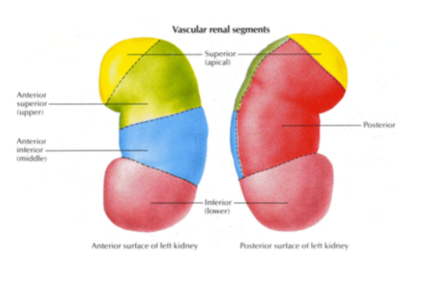
What are the vascular renal segments?
- Posterior: posterior segment
- Anterior:
Superior or Apical segment
Upper segment
Middle segment
Inferior segment
How is the arterial supply?
4 anterior branches and 1 posterior.
SEGMENTAL ARTERIES
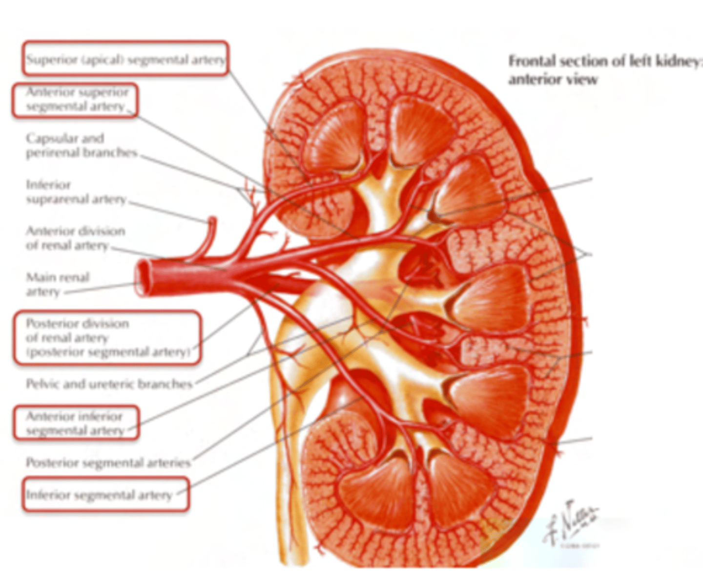
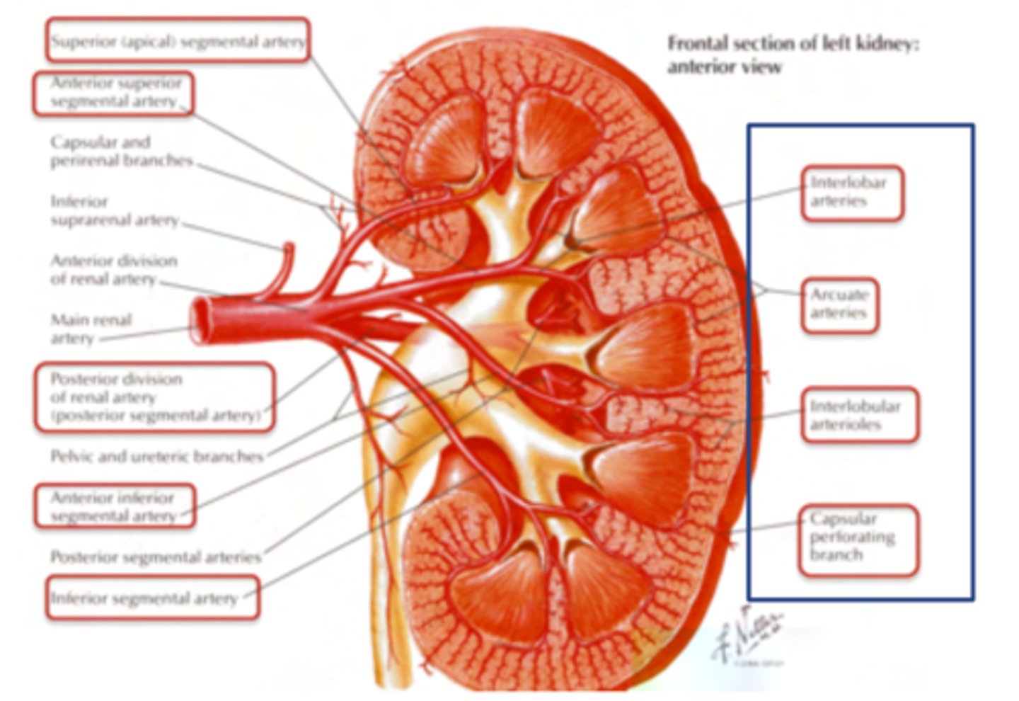
How are the segmental arteries divided?
Interlobar branches
- Arcuate arteries, which surround the pyramids
- Interlobular arteries
- Small arteries which go to form the afferent artery of the glomerulus, and ends in the efferent artery
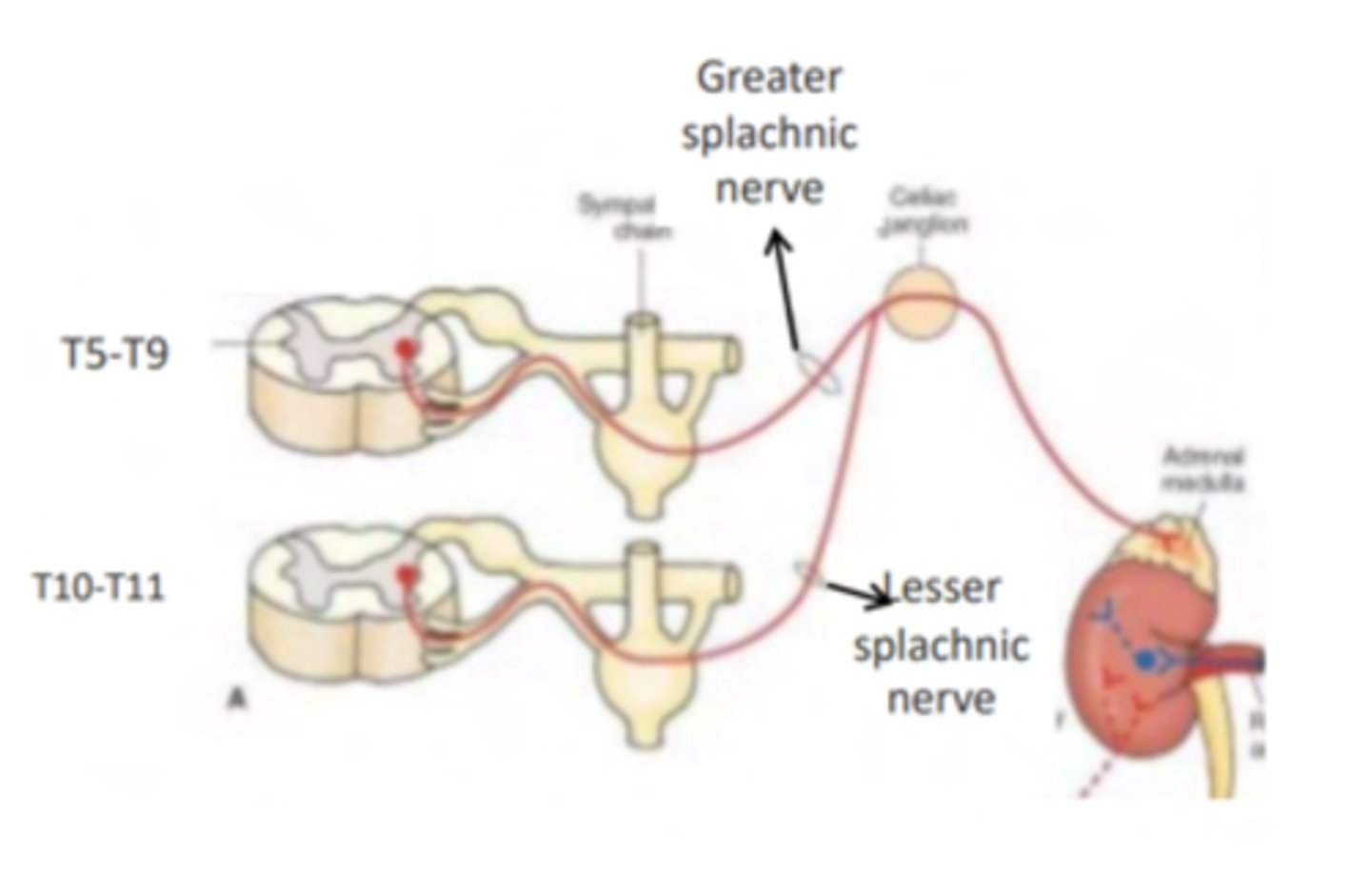
How is the innervation of the kidneys?
Both afferent and efferent nerves.

The efferent nerves of the kidney are siympathetic, PS or both?
Strictly simpathetic

What are the nerves entering the renal plexus?
- The celiac plexus
- Aorticorenal plexus
- Least splanchnic nerve

What are the sympathetic nerves acting on the kidneys?
2 caudal, splanchnic thoracic nerves from T10-T12 (10th, 11th and 12th thoracic splanchnic nerves).

What are the two splanchnic nerves actin on the kidney?
Lesser thoracic splanchnic nerve (T10–T11).
Least thoracic splanchnic nerve (T12).
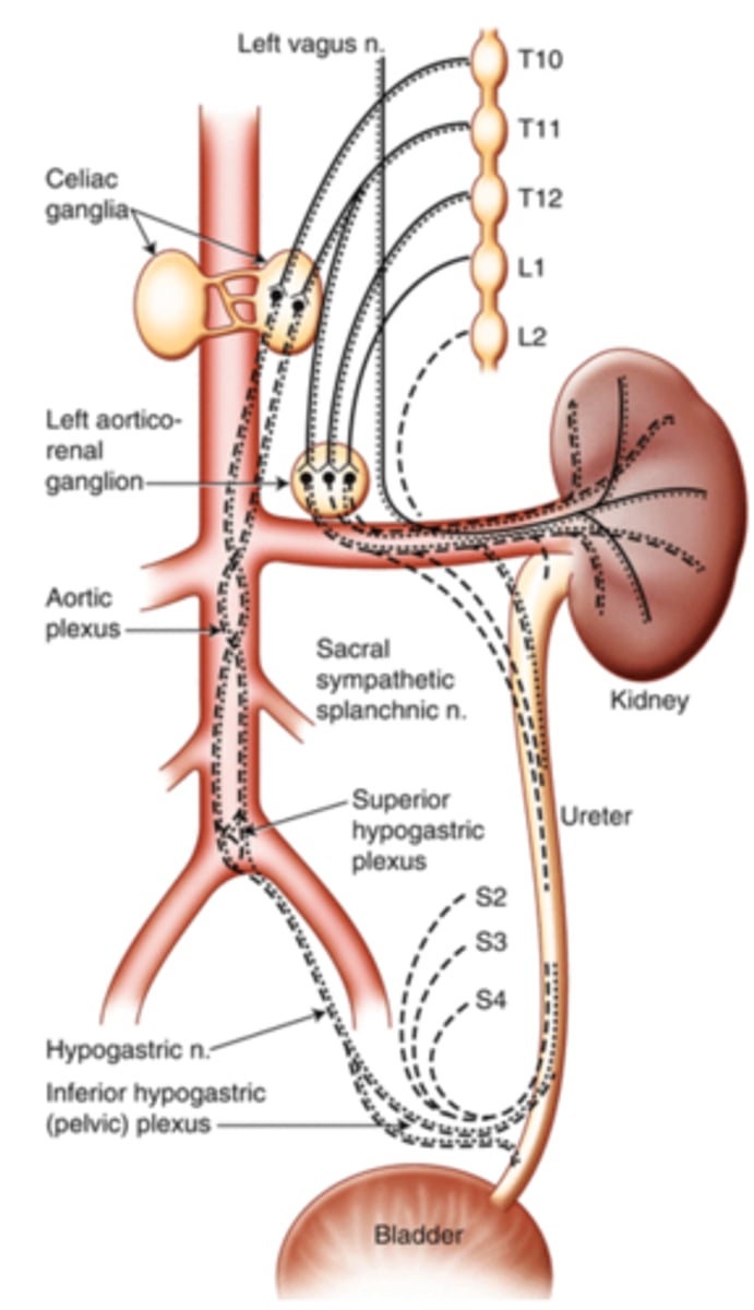
What is the pathway of the thoracic splanchic nerves to the kidney?
Preganglionic Neurons:
Synapse at Preaortic Ganglia:
The preganglionic nerves synapse at the preaortic ganglia, located in front of the abdominal aorta, at approximately the L1 vertebral level.
Postganglionic Neurons:

What are influencing the postganglionic neurons?
Renal blood flow (vasoconstriction or dilation).
Renin secretion (important for blood pressure regulation).

What is the Parasympathetic innervation of the kidneys?
do not receive motor (efferent) parasympathetic input but
but it has sensory (afferent) innervation through vagus nerve providing the NO PAIN info

What is the pathway of the ureters in the body?
Begin at renal pelvis
Retroperitoneal structures which descend anterior to the psoas major muscle and end at the level of the bladder.

How are the ureters supplied with arterial blood?
- 1st branches for the ureter come from the renal arteries
- 2nd are branches from the gonadal arteries
- 3rd branches
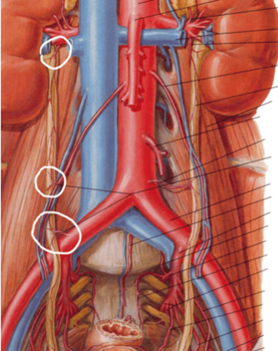
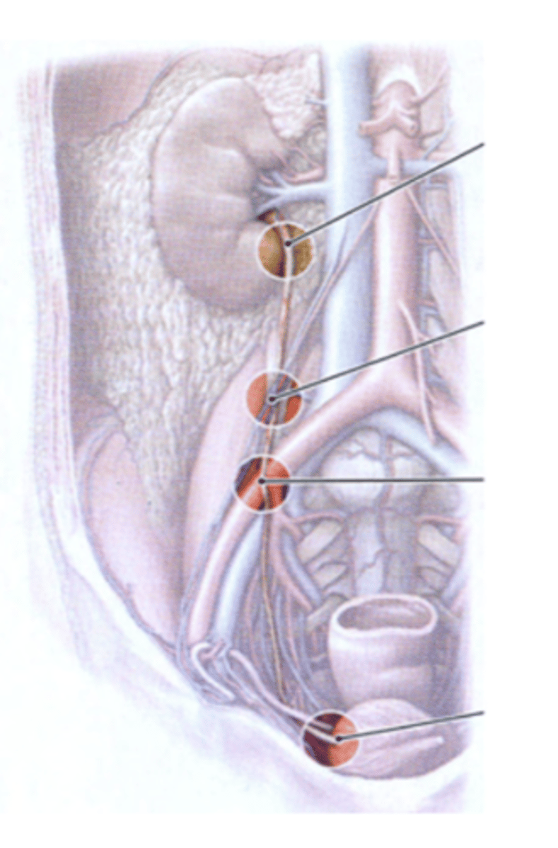
Where can be the ureters physiologically constricted?
1. Ureteropelvic junction (most important)
2. When the gonadal vessels cross anterior to the urethra
3. Pelvic inlet. When the ureters pass anterior to the iliac arteries
4. Entry into the bladder
Important as kidney stones can be lodged in these constrictions!
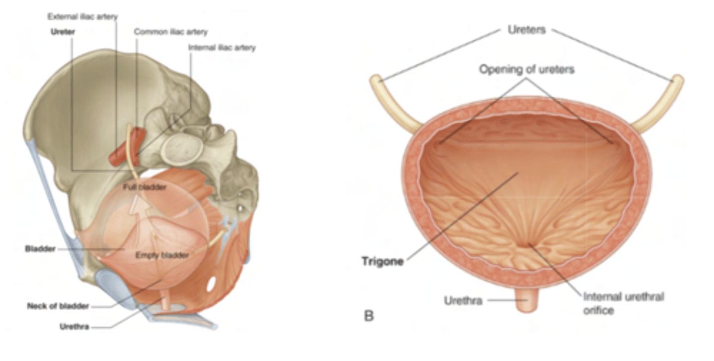
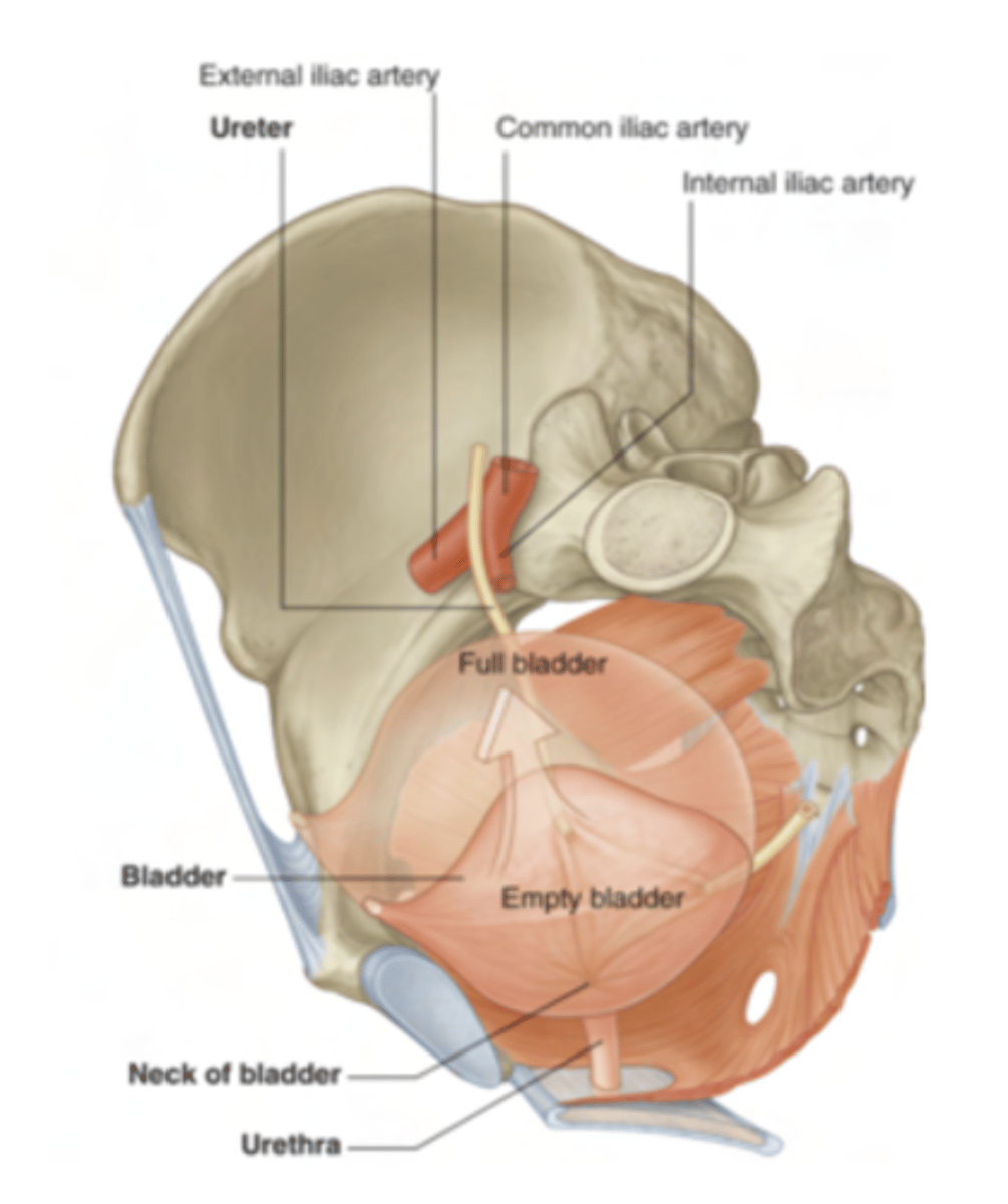
What is the bladder or urethra?
is a muscular balloon: its muscles contract or relax depending on how full the bladder is and the function we want to fulfill.
What is the muscle who contracts the bladder to empty it?
detrusor muscle
contracts to empty and relaxes to allow urine to collect in the
bladder

What is the muscle who contracts the bladder to fill it in?
sphincter
contracts while filling and relaxes when we want to
empty the bladder

What are the rugae?
numerous folds of transitional epithelium that allow the bladder to expand as it fills.
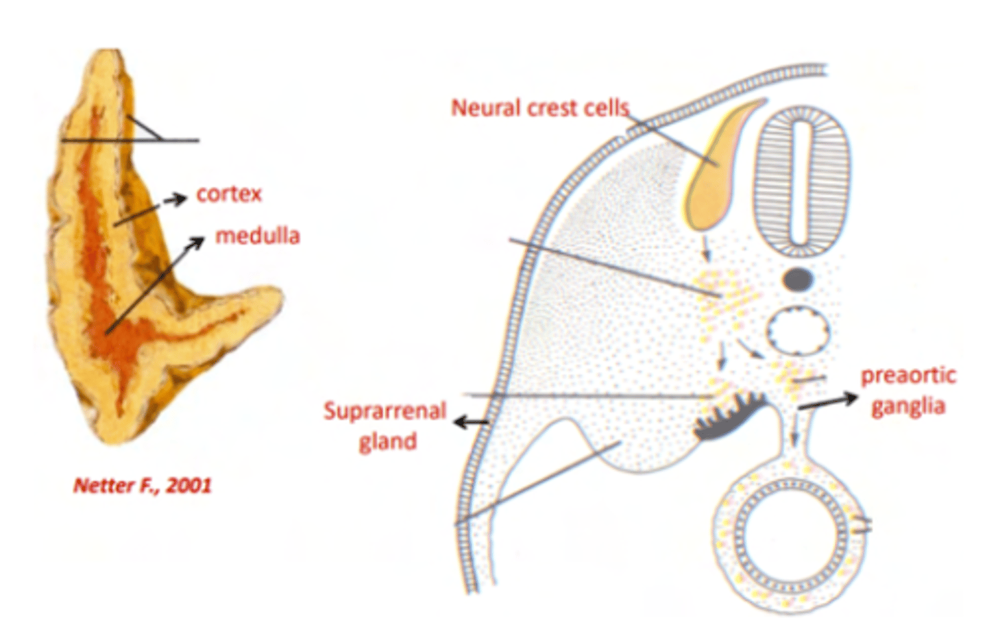
What are the two main structural components of the suprarenal glands?
Cortex (outer layer): Derived from mesoderm.
Medulla (inner layer): Derived from neural crest cells.

What type of cells are found in the medulla, and what do they secrete?
Chromaffin cells are found in the medulla.
They secrete adrenaline and noradrenaline upon preganglionic sympathetic innervation.

How do Chromaffin cells function similarly to postganglionic fibers?
Chromaffin cells release adrenaline and noradrenaline into the bloodstream, acting like postganglionic fibers but as secretory cells rather than neurons.

What is the shape of the suprarenal glands?
They have a triangular or semilunar shape.
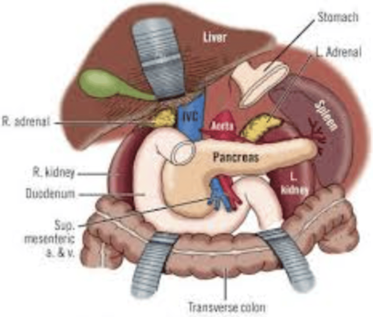
What are the relations of the right suprarenal gland?
in contact with:
Right crus of the diaphragm
Inferior vena cava (IVC)
Liver

What are the relations of the left suprarenal gland?
in contact with:
Left crus of the diaphragm
Spleen
Stomach (more anterior)
Pancreas
What arteries supply blood to the suprarenal glands?
Superior suprarenal arteries: Branches of the inferior phrenic arteries.
Middle suprarenal arteries: Branches of the abdominal aorta.
Inferior suprarenal arteries: Branches of the renal arteries.
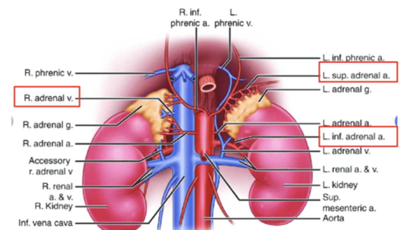
Where do the suprarenal veins drain?
Right suprarenal vein: Drains into the inferior vena cava (IVC).
Left suprarenal vein: Drains into the left renal vein.

How is the suprarenal gland innervated?
Preganglionic sympathetic fibers directly innervate the gland.
These fibers do not synapse in the sympathetic ganglia but synapse in the medulla with Chromaffin cells.