Ribs and Appendicular Skeleton
0.0(0)
Studied by 1 personCard Sorting
1/93
Last updated 3:36 PM on 11/7/22
Name | Mastery | Learn | Test | Matching | Spaced | Call with Kai | Chat |
|---|
No analytics yet
Send a link to your students to track their progress
94 Terms
1
New cards
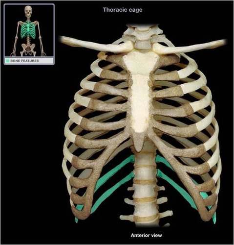
Sternum
chest bone

2
New cards
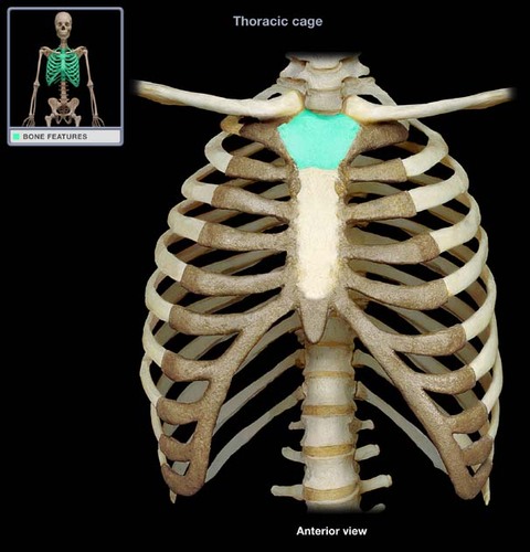
Manubrium
upper portion of the sternum
3
New cards
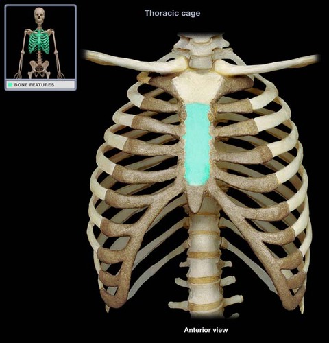
body of sternum
the bony structure that forms the middle portion of the sternum
4
New cards
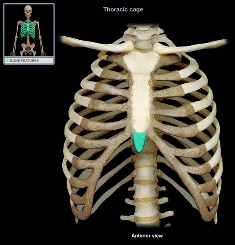
xiphoid process
lower, narrow portion of the sternum
5
New cards
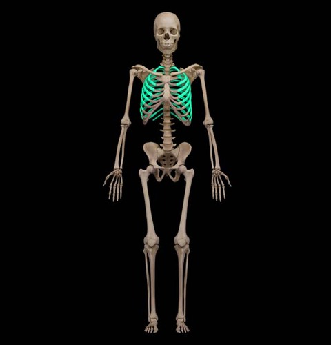
Ribs
12 pairs, attach posteriorly to the thoracic vertebrae
6
New cards
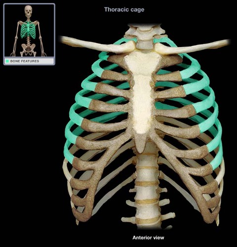
true ribs (vertebrosternal)
first 7 pairs of ribs; attach directly to sternum
7
New cards
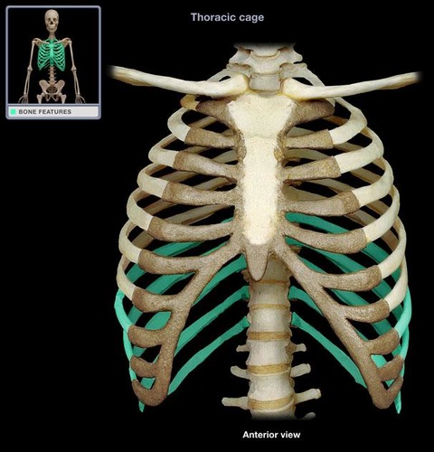
false ribs (8-12)
ribs that do not have a direct attachment to the sternum
8
New cards
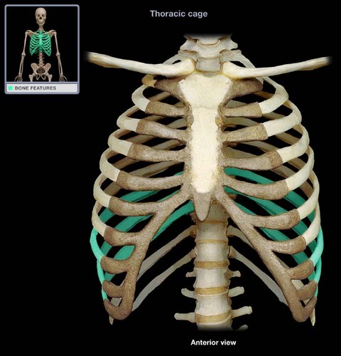
false ribs (vertebrochondral)
next 3 pairs, which join the cartilage of the 7th rib
9
New cards
false ribs (floating/vertebral)
next two pairs, which do not attach to cartilage
10
New cards
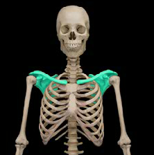
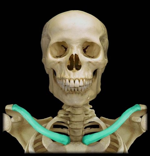
pectoral girdle
clavicle and scapula
11
New cards
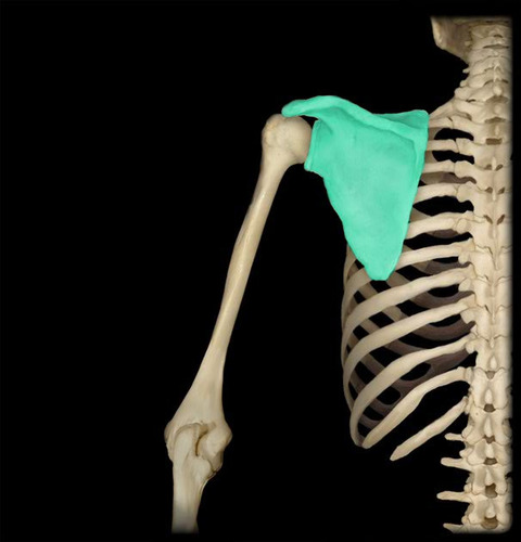
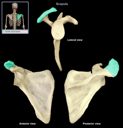
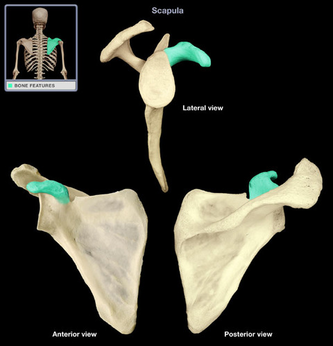
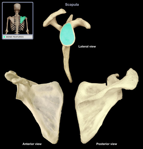
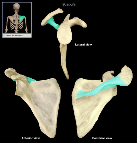
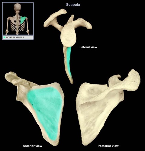
Scapula
shoulder blade
12
New cards
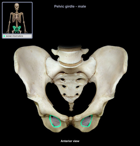
acromium
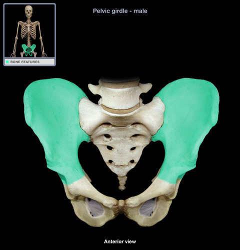
articulates with clavicle
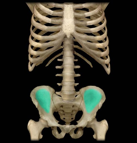
13
New cards
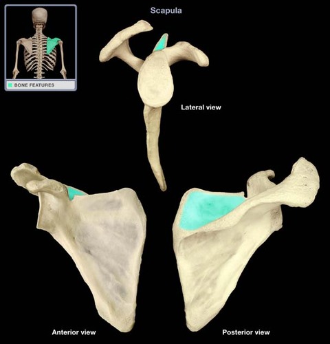
coracoid process
process above the glenoid cavity that permits muscle attachment
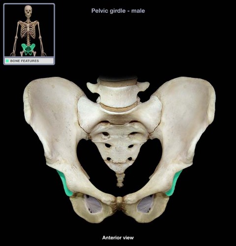
14
New cards
glenoid cavity
socket in scapular that articulates with the head of humerus
15
New cards
spine of scapula
posterior ridge of scapula
16
New cards
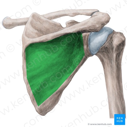
subscapular fossa
anterior surface of scapula
17
New cards
Infraspinous fossa
posterior, below spine
18
New cards
supraspinous fossa
posterior, above spine
19
New cards
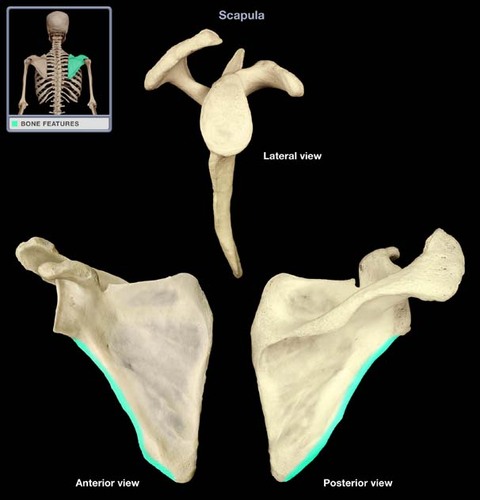
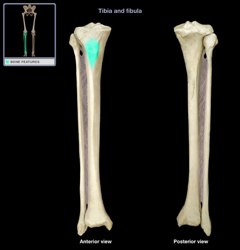
medial border
medial edge of scapula
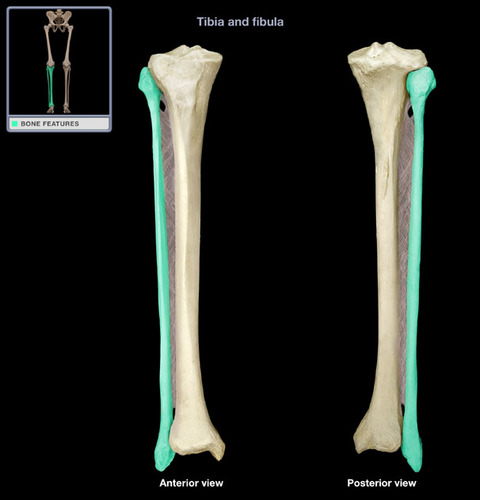
20
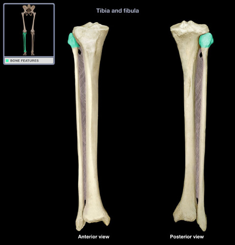
New cards
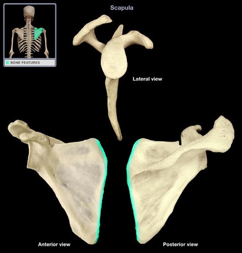
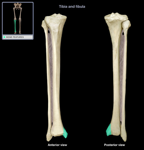
lateral border
lateral edge of scapula
21
New cards
Clavicle
collar bone
22
New cards
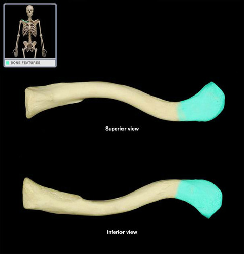
acromial end
lateral end of the clavicle that articulates with the acromion of the scapula
23
New cards
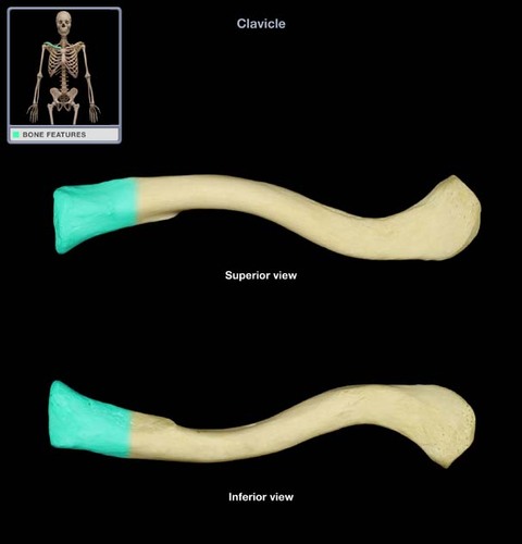
sternal end
medial end of clavicle that articulates with the manubrium of the sternum
24
New cards
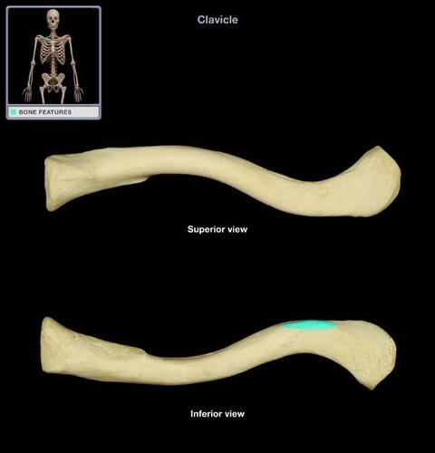
conoid tubercle
A small, cone-shaped projection located on the lateral, inferior end of the bone; serves to anchor ligaments
25
New cards
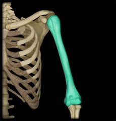
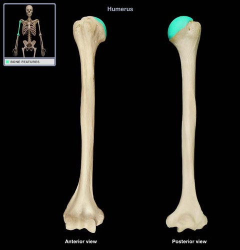
Humerus
upper arm bone
26
New cards
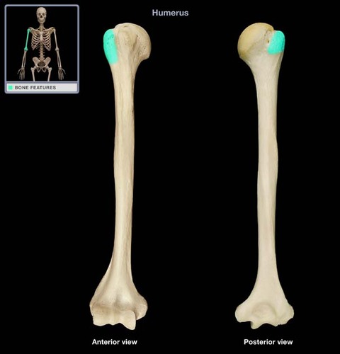
greater tubercle
Large lateral prominence; site of the attachment of rotator cuff muscles
27
New cards
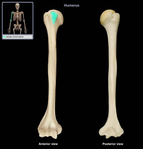
lesser tubercle
more medial and inferior compared to greater tubercle
28
New cards
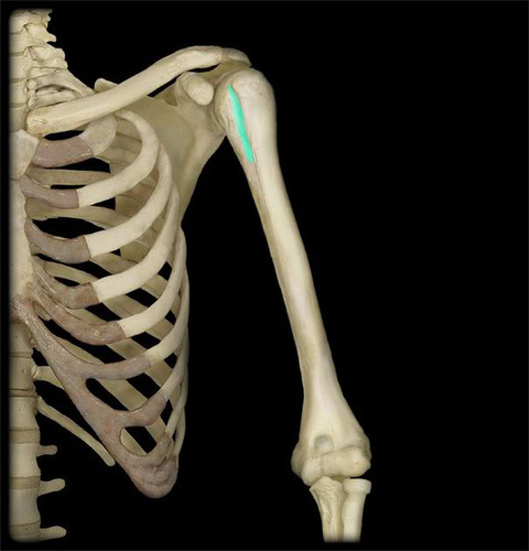
intertubercular groove
groove between the greater and lesser tubercles of the humerus
29
New cards
head of humerus
articulates with glenoid cavity of scapula
30
New cards
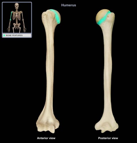
anatomical neck
separates the head from the tubercles
31
New cards
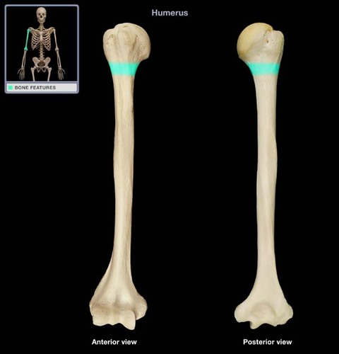
surgical neck
The neck of the humerus that is prone to fractures.
32
New cards
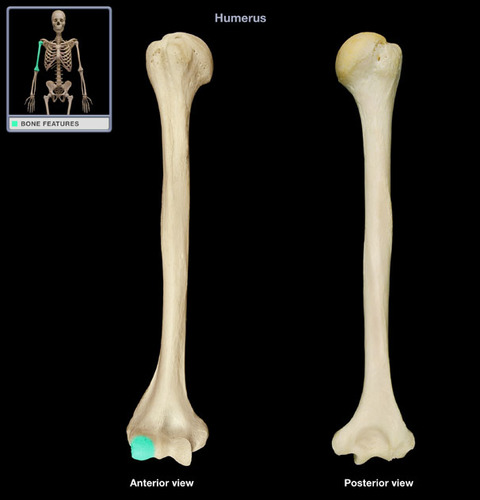
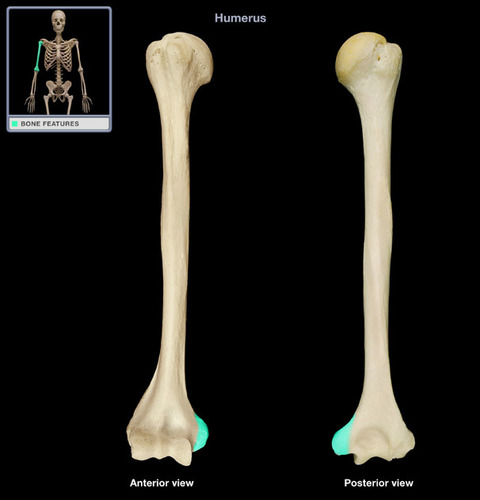
capitulum
lateral condyle on the distal end of the humerus which articulates with the head of radius
33
New cards
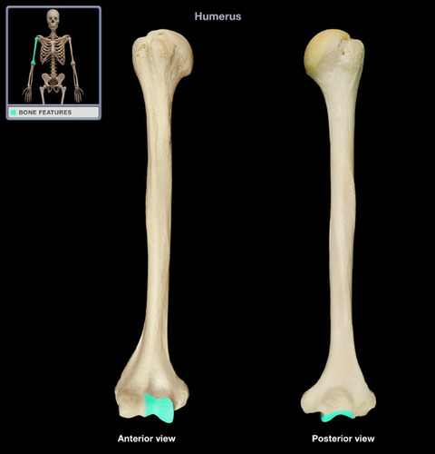
trochlea
medial condyle on the distal end of the humerus which articulates with the trochlear notch of the ulna
34
New cards
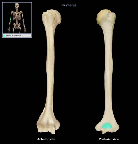
olecranon fossa
located on the posterior side of the distal end of the humerus superior to the trochlea and articulates with the olecranon process of the ulna
35
New cards
medial epicondyle
medial side of bottom that bulges out
36
New cards
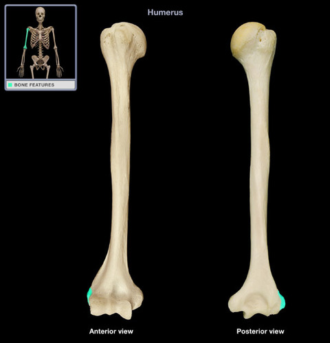
lateral epicondyle
lateral side of bottom of humerus
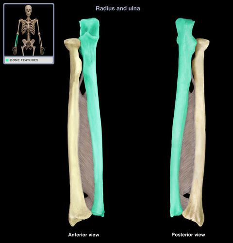
37
New cards
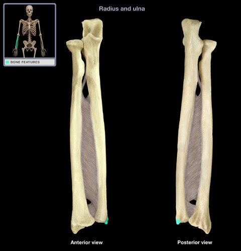
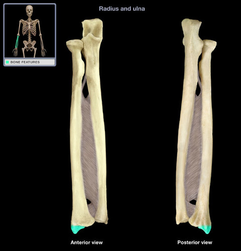
ulna
medial bone of the forearm
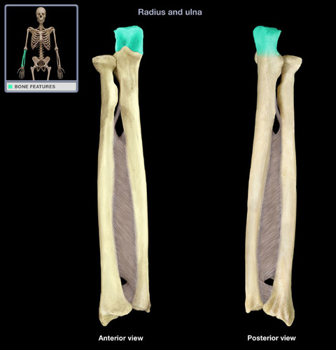
38
New cards
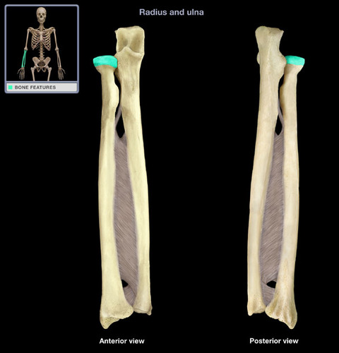
olecranon
The superior end of the ulna, the point of the elbow
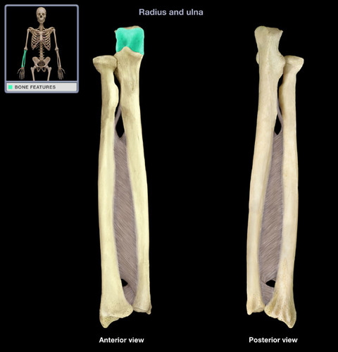
39
New cards
trochlear notch
articulates with trochlea of humerus
40
New cards
coronoid process
inferior lip of the trochlear notch, articulates with the humerus at the coronoid fossa

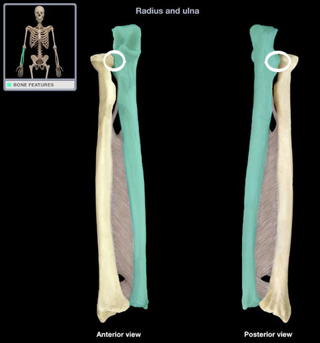
41
New cards
radial notch
on the lateral side of the coronoid process articulates with the head of the radius
42
New cards
head of ulna
distal end of ulna

43
New cards
styloid process of ulna
the thin cylindrical projection on the posterior side of the ulna's head
44
New cards
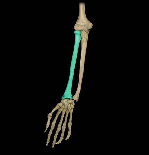
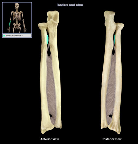
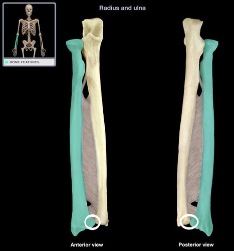
radius
lateral bone of the forearm
45
New cards
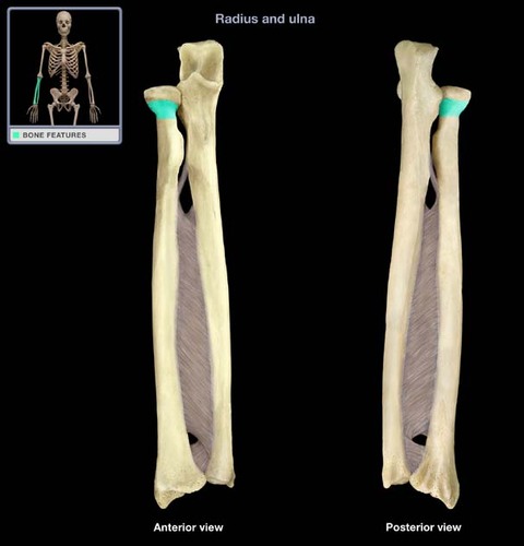
head of radius
articulates with the capitulum and radial notch
46
New cards
neck of radius
narrowed region immediately distal to the head of the radius
47
New cards
radial tuberosity
medial and inferior to neck, attachment site for biceps brachii muscle
48
New cards
ulnar notch
depression on the medial distal end of the radius, articulates with the head of the ulna
49
New cards
styloid process of radius
lateral projection at the distal end of the radius
50
New cards
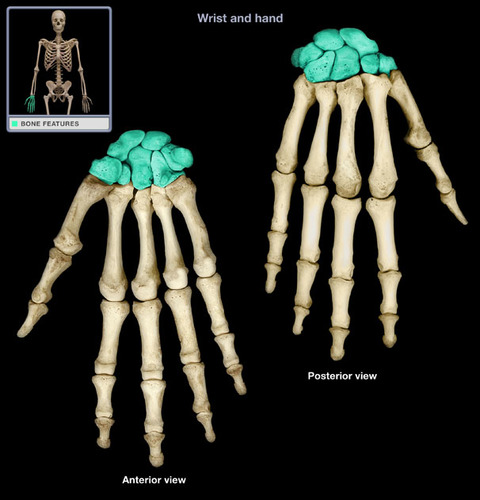
Carpals
8 pairs of wrist bones
51
New cards
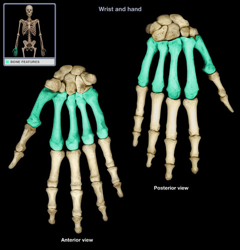
Metacarpals
5 pairs of hand bones
52
New cards
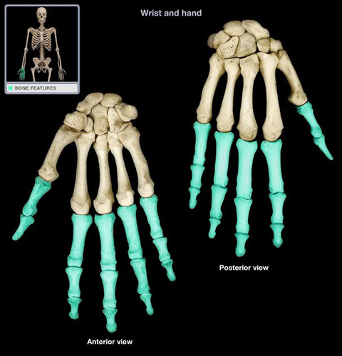
Phalanges (hand)
14 pairs of finger bones; each finger contains a proximal, middle, and distal phalanx. The thumb contains only a proximal and distal phalanx
53
New cards
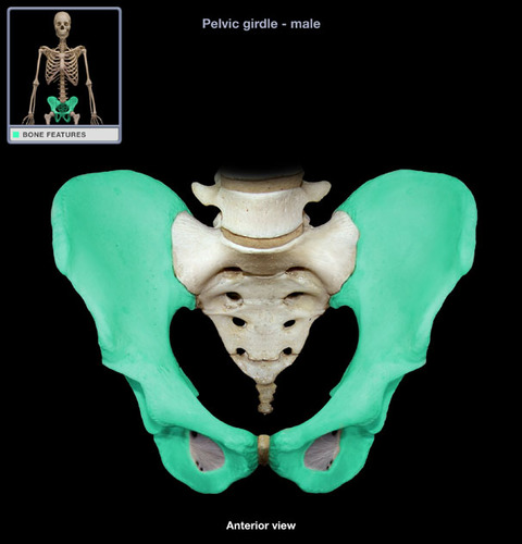
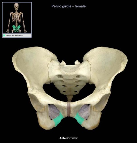
pelvic girdle
coxal bone: composed of ilium, ischium, and pubis
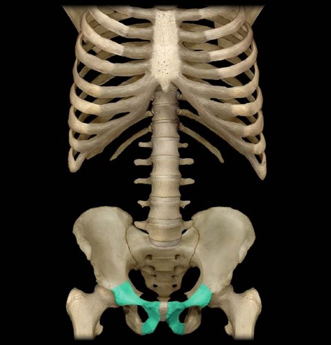
54
New cards
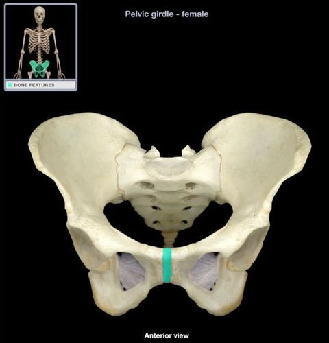
pubic symphysis
Cartilaginous joint that connects the two pubis areas
55
New cards
acetabulum
large socket in the coxal bone for the head of the femur
56
New cards
obturator foramen
opening in hip bone formed by the pubic and ischial rami
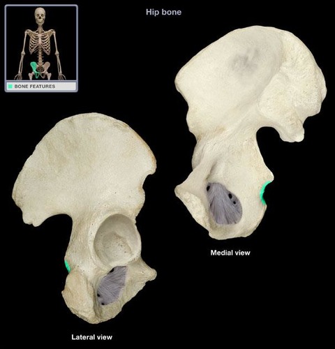
57
New cards
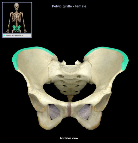
ilium
the superior and widest portion of the pelvis
58
New cards
iliac fossa
The broad, slightly concave inner surface of the ilium.
59
New cards
iliac crest
upper margin of iliac bones
60
New cards
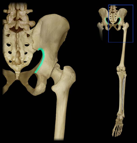
greater sciatic notch
heart shaped notch on medial side of coxal bone
61
New cards
ischium
the lower, posterior portions of the pelvis
62
New cards
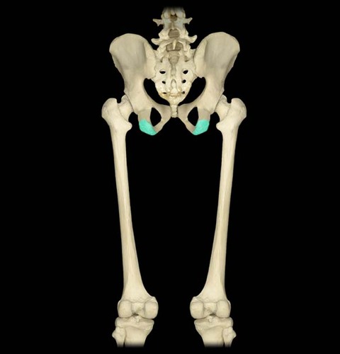
ischial tuberosity
inferior roughened surface of the ischium for ligament and muscle attachment
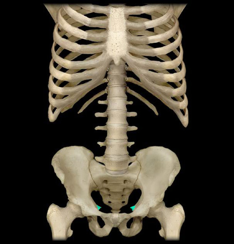
63
New cards
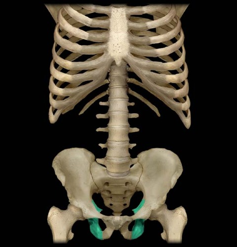
ischial spine
medial projection of ischium at the bottom of the heart
64
New cards
lesser sciatic notch
notch located inferior to the ischial spine
65
New cards
pubis
The medial anterior portion of the pelvis
66
New cards
superior ramus
flattened superior border of the obturator foramen
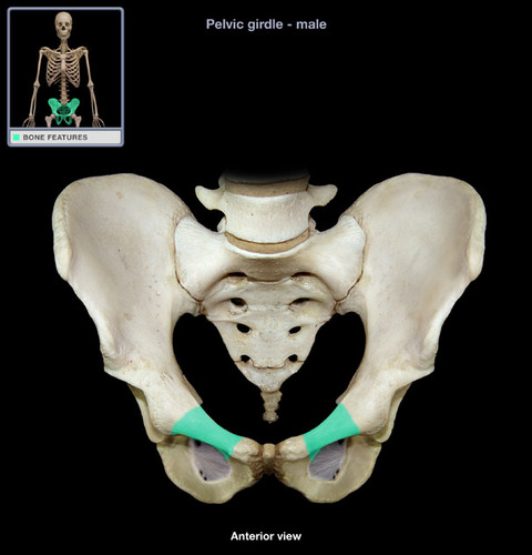
67
New cards
inferior ramus
inferior extension of the body of the pubis; articulates with the ischium
68
New cards
two coxae articulate
anteriorly with each other at the pubic synthesis;
posteriorly with the sacrum
posteriorly with the sacrum

69
New cards
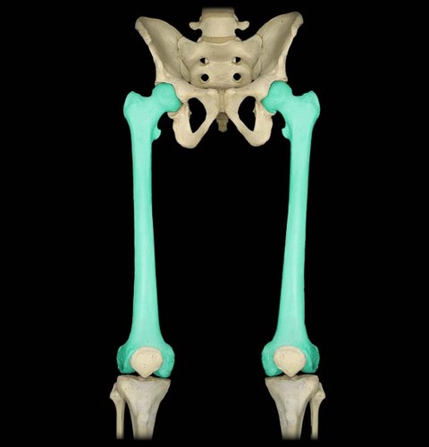
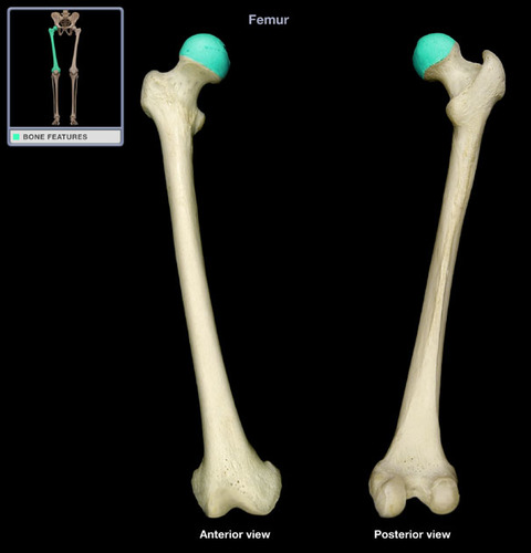
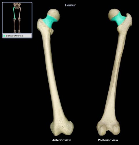
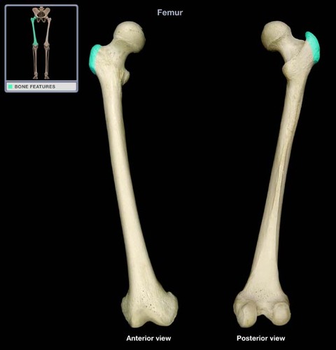
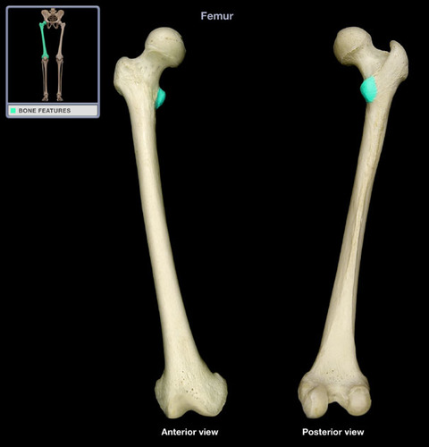
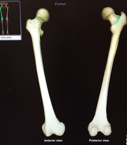
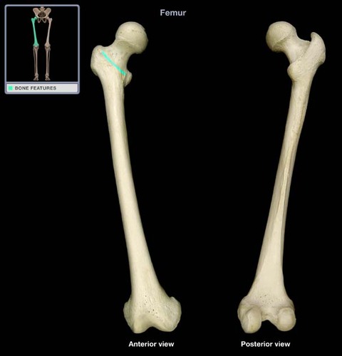
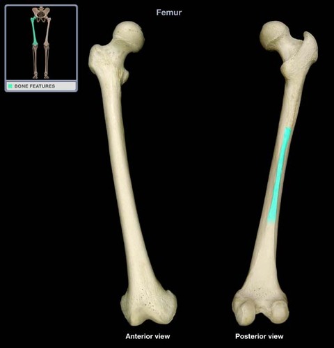
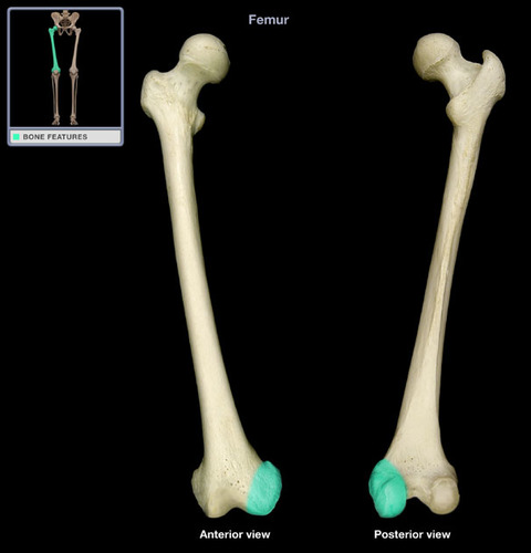
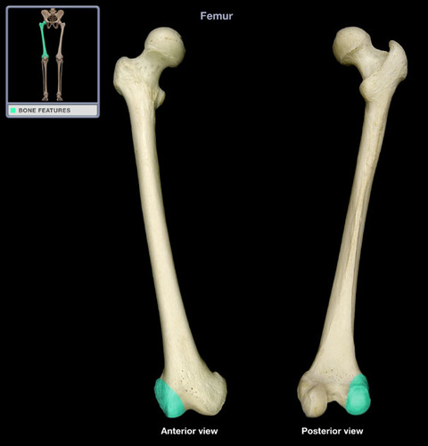
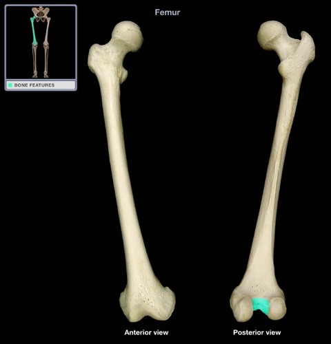
Femur
thigh bone
70
New cards
head of femur
articulates with the acetabulum
71
New cards
neck of femur
between head and greater trochanter
72
New cards
greater trochanter
lateral to the head of the femur
73
New cards
lesser trochanter
medial side of femur, inferior to head
74
New cards
intertrochanter crest
connects the two trochanters posteriorly
75
New cards
intertrochanteric line
region formed anteriorly between the greater and lesser trochanters
76
New cards
linea aspera
on posterior shaft of femur for muscle attachment
77
New cards
medial condyle of femur
medial knob on the distal end of the femur which forms a hinge joint with the medial condyle of the tibia
78
New cards
lateral condyle of femur
lateral knob on the distal end of the femur which forms a hinge joint with the lateral condyle of the tibia
79
New cards
intercondylar fossa
Posteriorly, the femoral condyles are separated by a deep depression called the:
80
New cards
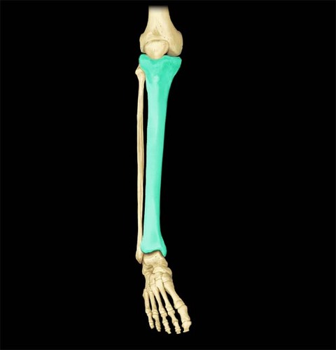
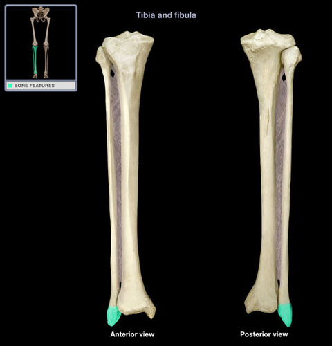
Tibia
the medial and larger bone of the lower leg
81
New cards
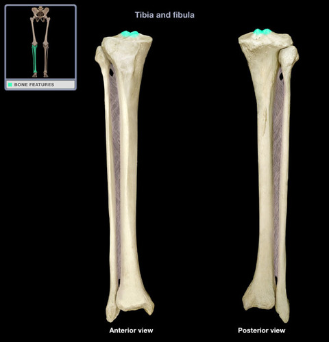
intercondylar eminence
irregular projection located between the two condyles of the tibia
82
New cards
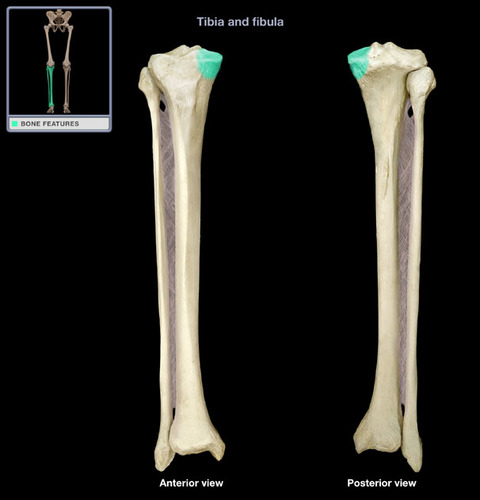
medial condyle of tibia
medial depression on the proximal end of the tibia which articulates with the medial condyle of the femur
83
New cards
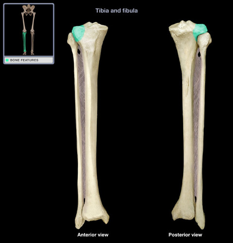
lateral condyle of tibia
lateral depression on the proximal end of the tibia which articulates with the lateral condyle of the femur
84
New cards
tibial tuberosity
anterior portion of tibia inferior to condyles
85
New cards
medial malleolus
medial bulge of tibia; ankle bone
86
New cards
fibula
The lateral and smaller bone of the lower leg
87
New cards
head of fibula
proximal end of the fibula that articulates with the posterior tibia
88
New cards
lateral malleolus
distal end of fibula; forms lateral ankle bone
89
New cards
patella
kneecap; articulates with the distal anterior femur

90
New cards
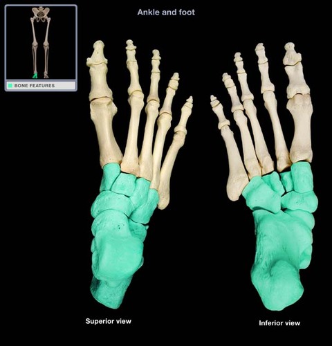
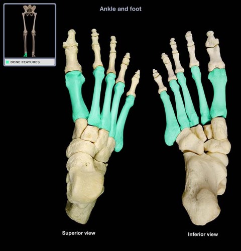
tarsals
7 pairs of ankle bones
91
New cards
talus
superior ankle bone that articulates with distal tibia and fibula
92
New cards
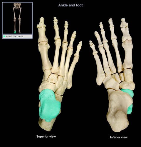
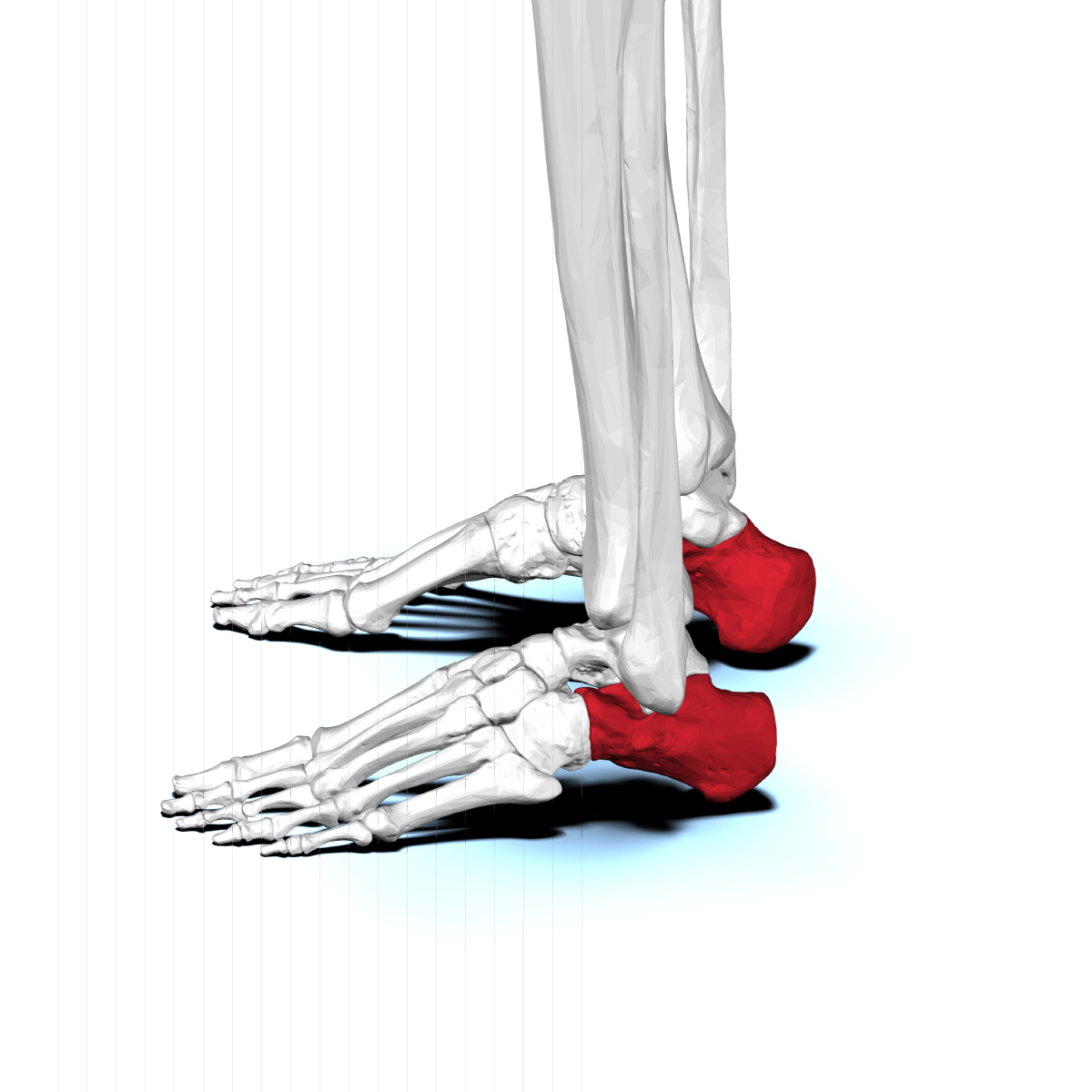
Calcaneous
heel bone
93
New cards
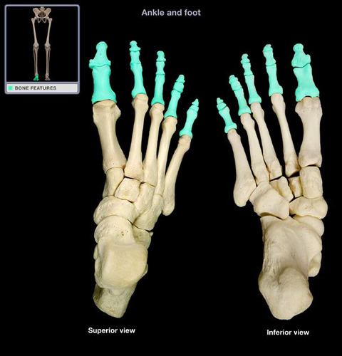
metatarsals
5 pairs of foot bones
94
New cards
Phalanges (foot)
14 pairs of toe bones; each toe contains a proximal, middle, and distal phalanx. The big toe contains only a proximal and distal phalanx