4. Corneal nerves
1/60
There's no tags or description
Looks like no tags are added yet.
Name | Mastery | Learn | Test | Matching | Spaced | Call with Kai |
|---|
No analytics yet
Send a link to your students to track their progress
61 Terms
What is the most innervated tissue in the body?
The corneal epithelium, 300-400 times more dense than skin, with 16,000 nerve terminals/mm2
What types of sensory nerves are at the corneal epithelium?
Mechanical
Chemical
Temperature
What are the corneal nerves derived from?
The ophthalmic branch of the trigeminal nerve (CN V)
Where are the nerves typically found in the cornea?
Only in the upper 2/3 of the stroma.
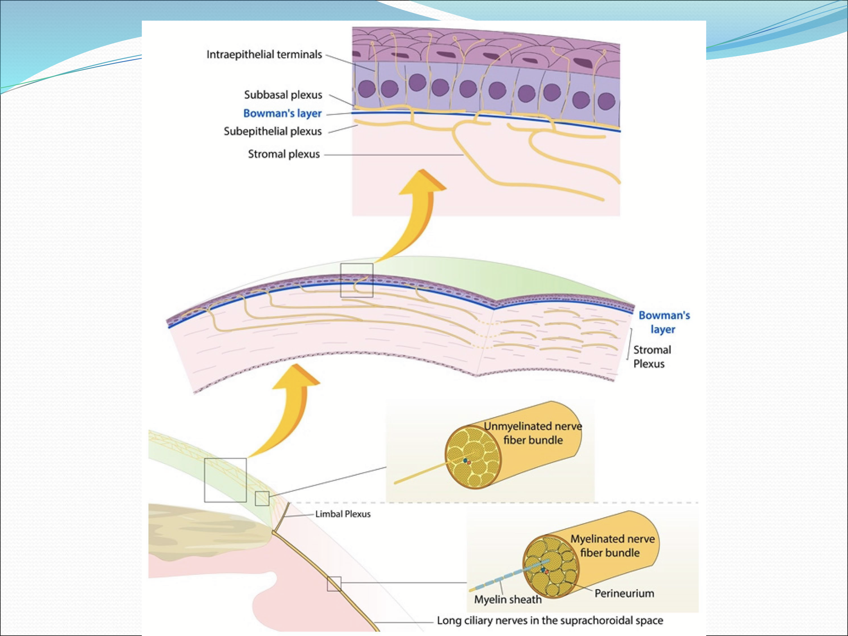
How is the corneal innervation organized?
four levels:
corneal stromal nerves
subepithelial nerve plexus
sub-basal nerve plexus
intraepithelial nerve terminals
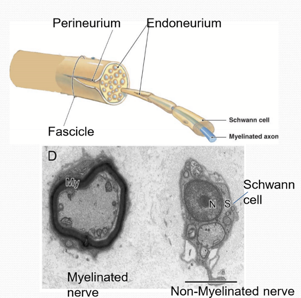
Are the nerves in the cornea myelinated?
No, they are not, but still retain schwann cell
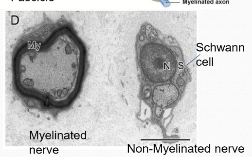
What do the stromal nerve cells lack?
Blood vessels, myelin sheath, and perineurium of fascicle/bundle
What do some of the stromal nerves have within the stroma?
Free nerve endings
What percentage of stromal nerves have a myelin sheath when they penetrate the stroma?
20%, but all is lost within 1 mm.
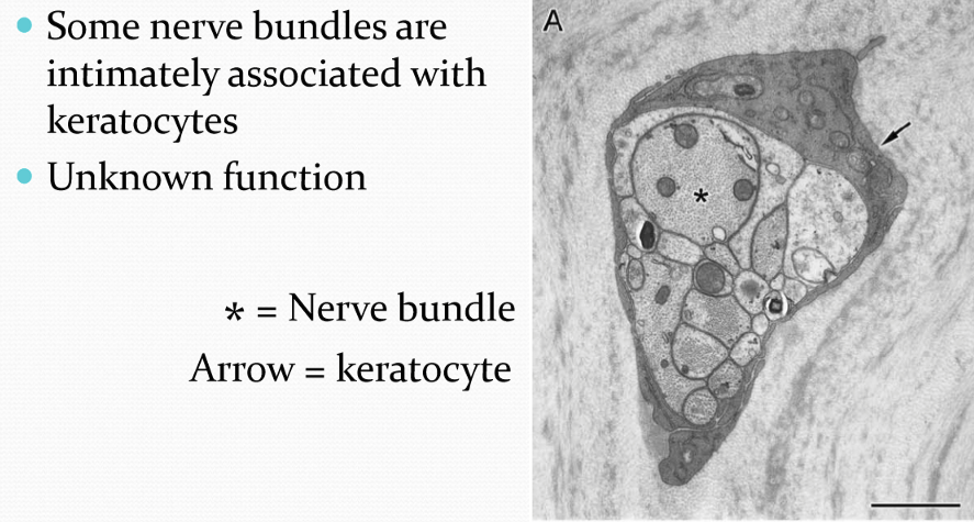
What is the relationship between some nerve bundles and keratocytes?
Some nerve bundles are intimately associated with keratocytes, though their function is currently unknown.
How are the distal branches of the stromal nerves laid out?
The nerves anastomose extensively
Where does the anastomosis of the nerves occur?
Anastomosis occurs more frequently in the anterior 1/3 of the central cornea
What does a more posterior nerve penetration in the corneal stroma indicate?
It correlates with larger sensory fields and innervation of central areas of the cornea.

What does a more anterior nerve penetration in the corneal stroma indicate?
It correlates with smaller sensory fields and innervation of the peripheral areas of the cornea
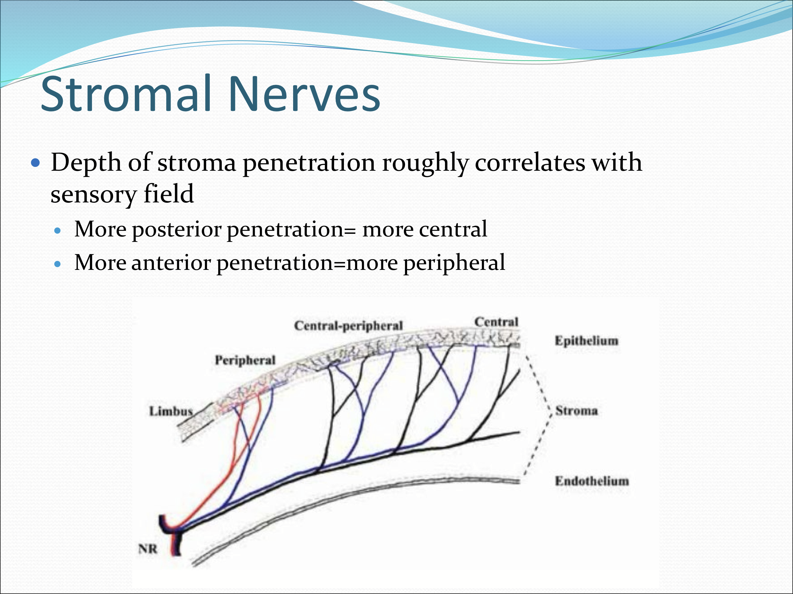
How does the depth of nerve penetration affect sensory field size in the cornea?
Deeper penetration = larger sensory fields; shallower penetration = smaller sensory fields
Where are the subepithelial nerve plexus located?
In the anterior stroma immediately beneath Bowman’s membrane
Where is the subepithelial nerve plexus most dense?
It is greater in the peripheral vs central cornea
What are the two anatomically distinct types of nerve bundles of the subepithelial nerve plexus?
highly anastomotic meshwork of single axons and thin nerve fascicles beneath Bowman’s layer
Bundles that turn 90 degrees and shed their Schwann cells, penetrate bowman’s membrane, and divide into 2-29 thinner nerve fascicles
Where do the nerve bundles penetrate the cornea the most?
Penetration of Bowman’s membrane by nerve bundles occurs mostly peripherally
What happens to the nerve bundles after they penetrate Bowman’s membrane?
A nerve fiber from the subepithelial nerve plexus divides into multiple, thinner nerve fascicles (2-20)

What is an epithelial leash?
When the thinner nerve fascicles run parallel to each other
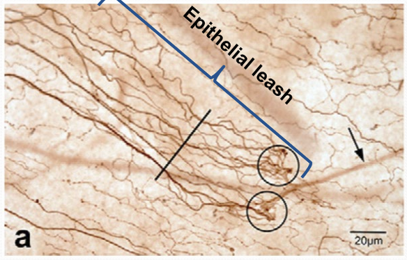
What makes up the sub-basal nerve plexus?
The epithelial leashes

What can the adjacent epithelial leashes do?
The repeated interconnect

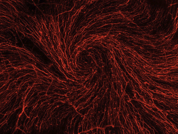
What shape does the sub-basal plexus form?
A whorl-like spiral pattern of fibers

Why does the whorl-like spiral pattern form?
The inward movement of the corneal epithelial cells causes the spiral pattern

Where is the center of the spiral?
Off center, inferior and nasal.
How far do individual axons in the sub-basal plexus travel?
They can travel up to 6 mm
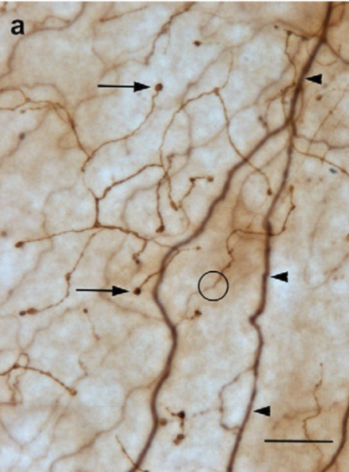
What do the sub-basal nerves do?
They split off and turn 90 vertically and interdigitate between the basal, wing, and superficial epithelial cells.

Each sub-basal nerve fiber give rise to?
10-20 intraepithelial nerve terminals and end as free nerve ending

Where are the endings of the intraepithelial nerve located?
The endings occur in all layers of the epithelium, most common in basal and wing cells

Bulbous nerve endings contain what?
Vesicles filled with excitatory amino acids and neuropeptides
mitochondria
glycogen particles
microtubules
neurofilaments
Where are the cell bodies of the corneal sensory neurons reside in?
The trigeminal ganglion.
Where do the corneal sensory nerves travel to reach the cornea?
They project through the nasociliary nerve, ciliary ganglion, and long and short ciliary nerves
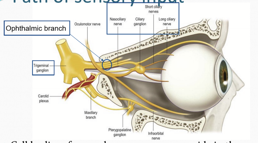
What are the components of the trigeminal brainstem nuclear complex (TBNC)?
Principal nucleus in the pons and the Spinal Nucleus in the medulla.
What can the spinal nucleus be divided into?
Oralis nucleus
Interpolaris nucleus
Caudalis nucleus
Where do the corneal nerves synapse in the TBNC?
They synapse in the caudal portion of the ipsilateral TBNC

Where do nerves of the limbus, conjuntiva, and palpebrae synapse at?
The principal nucleus
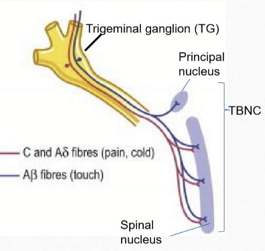
What are the types of fibers that innervate the corneal epithelium?
thin myelinated* A delta type axons
Unmyelinated C type axons
What types of fibers innervate the limbus, conj, palpebrae epithelium?
Thick myelinated A beta axons
Thin myelinated A delta axons
Unmyelinated C type axons
What type of sensations are A beta fibers responsible for?
Touch/pressure
What types of sensation are A delta responsible for?
Touch/pressure, temperature, and pain
What types of sensations are C fibers responsible for?
pain
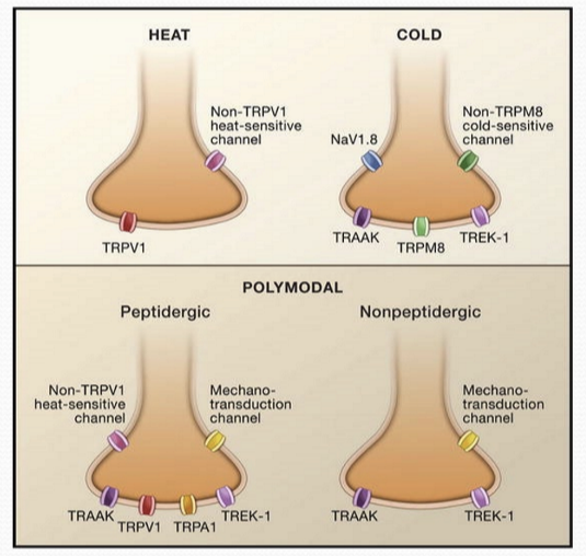
What are the types of stimuli that the corneal nerves respond to?
Polymodal nociceptors
Mechano-nociceptors
Cold thermal receptors
What are the types of stimuli that the nerves of the conjunctiva respond to?
Polymodal nociceptors
Mechano-nociceptors
Cold thermal receptors
Mechano-receptors
Polymodal-receptors
Pruriceptive receptors
What are all A delta fibers?
Mechano-nociceptors
How do mechano-nociceptors respond to any stimulation?
Fire a few nerve impulses with any stimulation, with limited ability to distinguish between strength and duration of stimuli. Their threshold to activate is very low
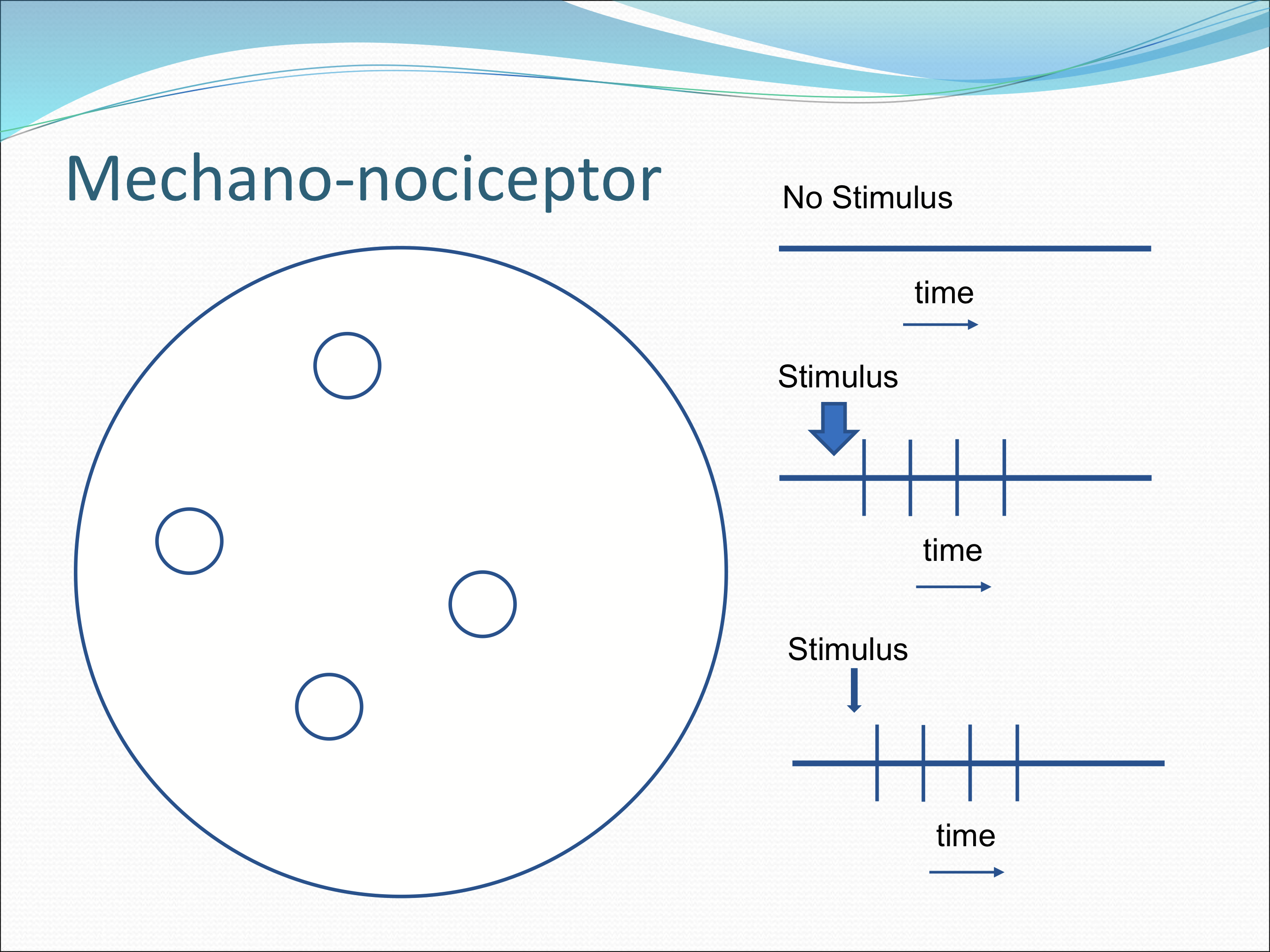
What percentage do mechano-nociceptors make up cornea somatosensory receptors?
15-20% of total receptors
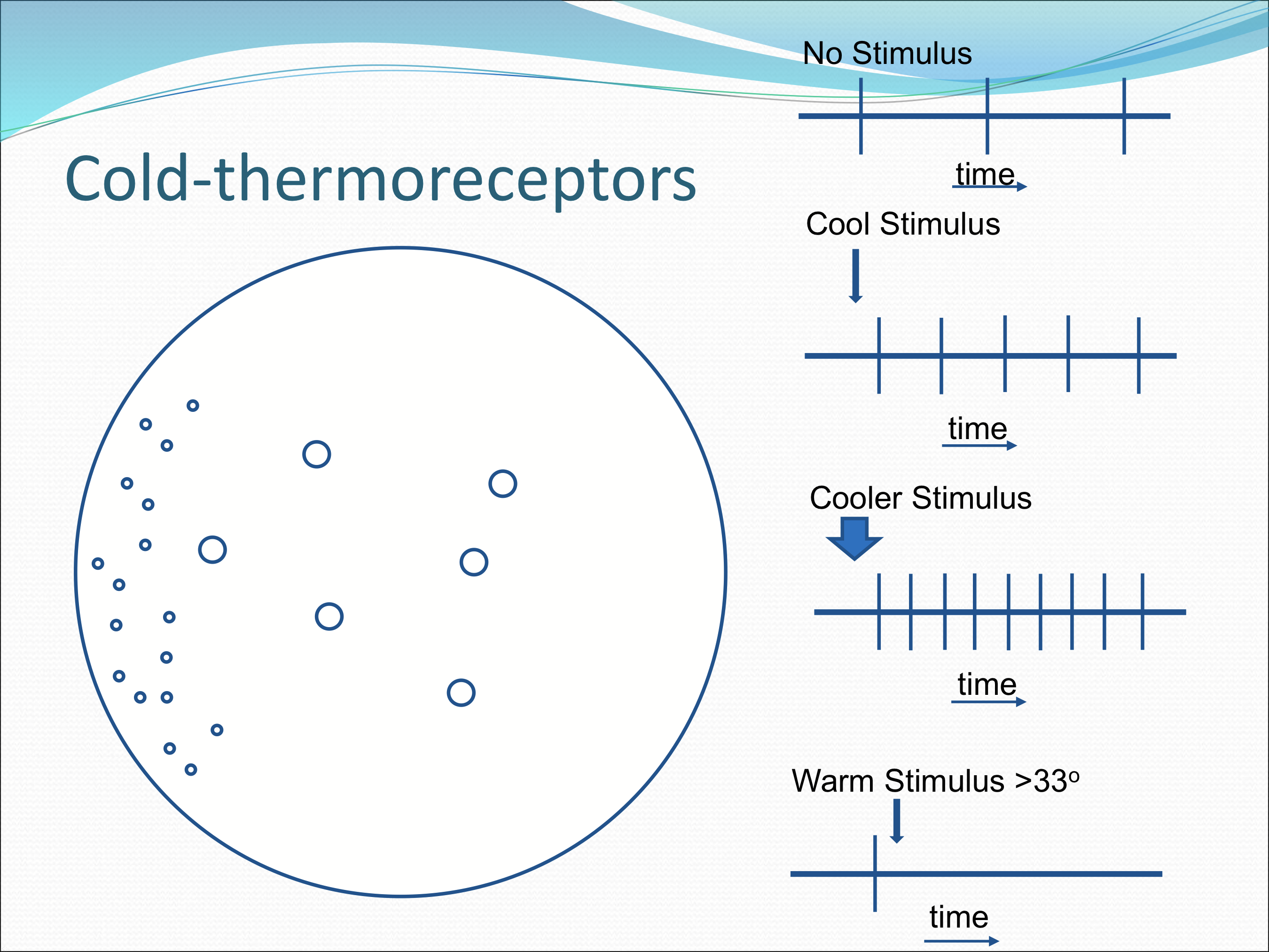
What type of fibers are cold thermoreceptors?
Both A delta and C fibers
How do cold thermoreceptors respond to stimulus?
Without stimulus, low rate of discharges, and changes in impulse frequency with small change in temp. With an increase in frequency with lower temps and a decrease in frequency with higher temps.

Where are these cold thermoreceptors located?
All over cornea, but with smaller fields more abundant peripherally.
What stimulus do polymodal nociceptors respond to?
Heat: >39-40 C
Cold: <29 C
pH: <6.5
Mechanical forces: much more sensitive than mechano-nociceptors
What percentage of corneal nerves are polymodal nociceptors?
70%
What type of fibers are polymodal nociceptors?
Mostly C type fibers, but some A delta too
How are the receptive fields of the polymodal nociceptors organized?
They are large with overlap
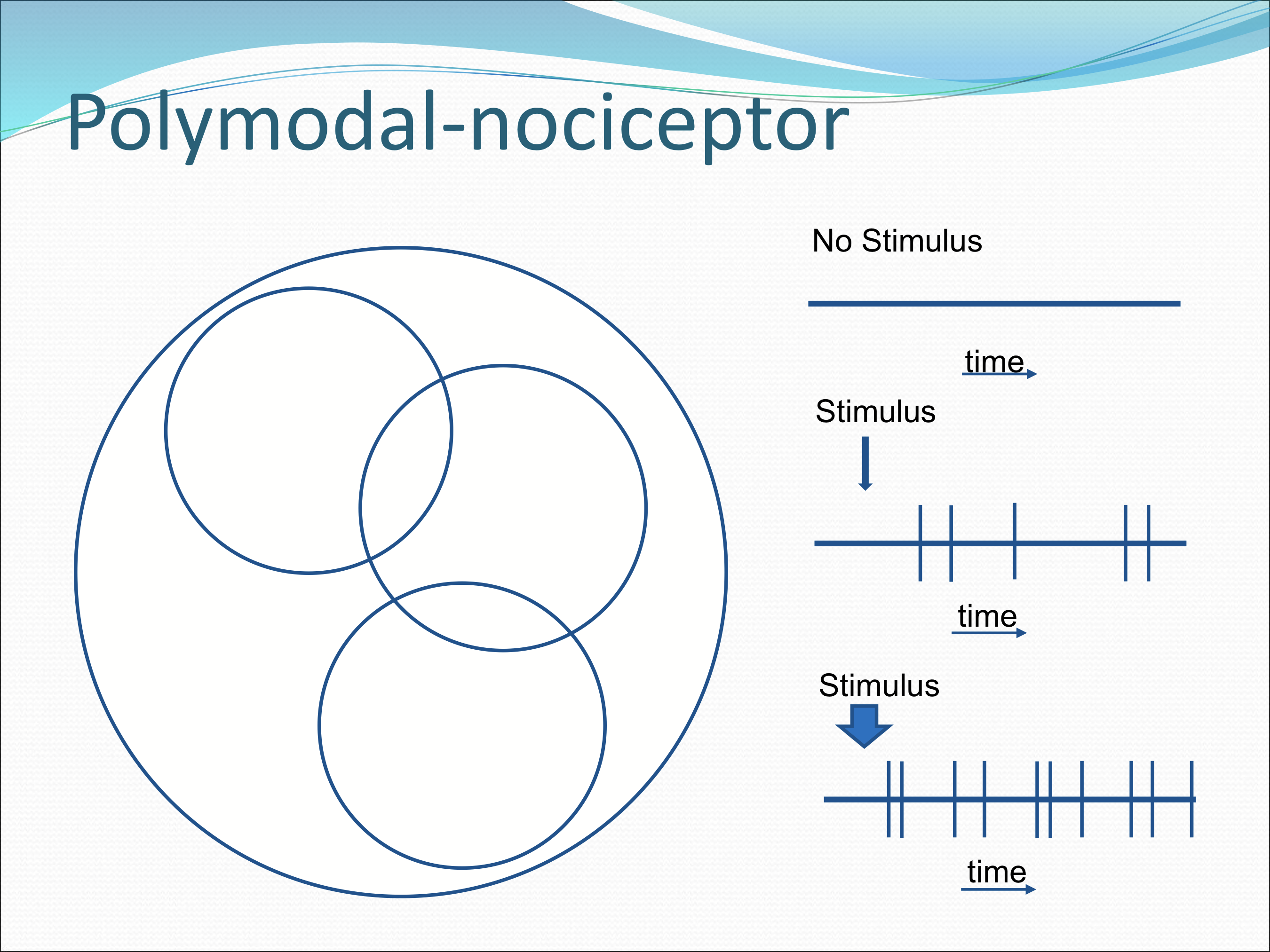
How do the polymodal nociceptors fire in response to stimulus?
Continuous irregular discharge of nerve impulses that persist as long as the stimulus is maintained.

What are transient receptor potential channels?
Numerous channels that determine what type of stimulus to which the sensory receptors respond to, such as membrane stretch, temp, pH, and chemicals such as capsaicin, wasbi, menthol/peppermint.
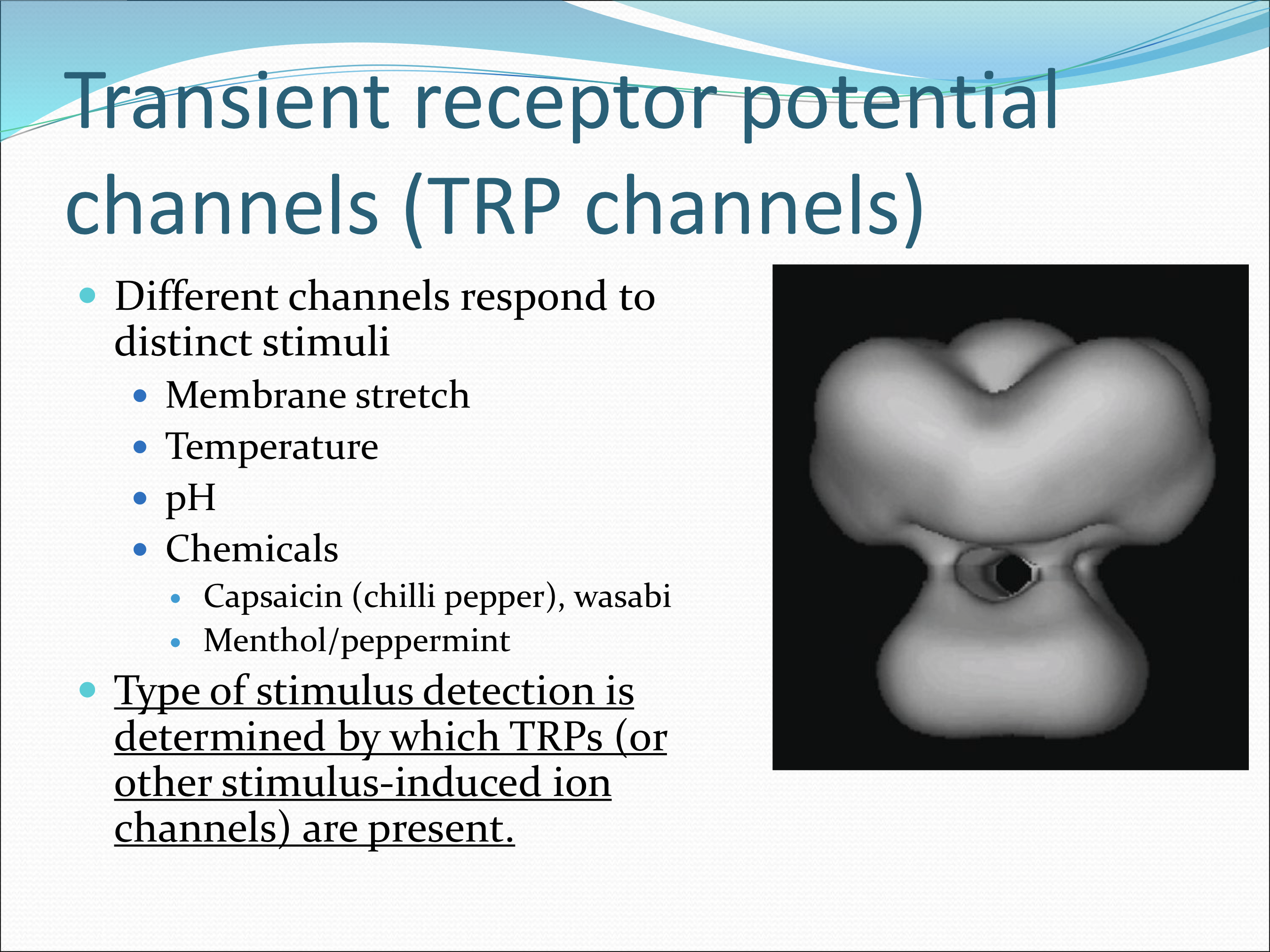
Where are the transient receptor potential channels found?
Embedded int he terminal membranes of nociceptors
How do transient receptor potential channels work?
Stimulus causes conformational change in channel protein complex
Facilitates ion transport across cell membrane
Triggers depolarization and action potential of nerve
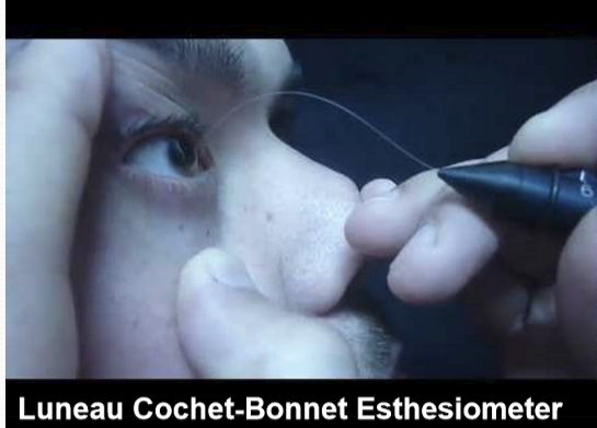
How do you clinically test corneal sensitivity?
Using an esthesiometer, which can be used to diagnose neurotrophic keratopathy
What are major factors that causes large variation in corneal sensitivity?
Age
Dry eye
Acute inflammation
Refractive surgery
Contact lens wear
What are minor factors that affect corneal sensitivity?
Time of day, lowest upon waking
Iris color, chemical sensitivity decreased with more pigmentation
Gender, women slightly more sensitive than men