DAT body systems
 Call Kai
 Learn
Learn Practice Test
 Spaced Repetition
 Match
Match Flashcards
 Knowt Play
1/163

There's no tags or description
Looks like no tags are added yet.
Name | Mastery | Learn | Test | Matching | Spaced | Call with Kai | Chat |
|---|
No analytics yet
Send a link to your students to track their progress
164 Terms
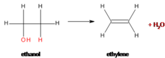
Dehydration Reaction (Condensation)
Creates covalent bond between monomers and releases water
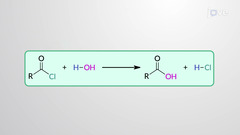
Hydrolysis
Reaction that breaks a covalent bond by using water
Carbohydrates
Used as fuel and structural. Contain: Carbon, hydrogen, oxygen (CHO).
Monosaccharides
Carbohydrate monomers. Ribose, fructose glucose, galactose. Glucose and fructose are isomers (same chemical formulas, different arrangement of atoms)
Disaccharides
Two monosaccharides joined by glycosidic bond. sucrose (glucose+fructose), lactose (glucose+galactose), maltose (glucose+glucose a-1,4)
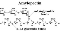
Polysaccharide
Multiple monosaccharides connected by glycosidic bond. Starch (glucose+glucose a-1,4 and a-1,6), glycogen (glucose+glucose a-1,4 and a-1,6), cellulose and chitin (b-1,4)
Proteins
Contain carbon, hydrogen, oxygen, nitrogen (CHON). Combine to form amino acids which link into polypeptides. Amino acid = monomers. Twenty amino acids characterized by unique "R-group"

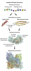
Protein Structure
1. Primary: sequence of amino acids connected through peptide bonds
2. Secondary: intermolecular forces between polypeptide backbone (NOT R GROUPS) due to hydrogen bonding. Form alpha-helices and beta-pleated sheets
3. Tertiary: 3D structure due to interactions between R groups. Can create hydrophobic interactions based on R groups. Disulfide bonds created by covalent bonding between R groups of 2 cysteine. Hydrogen/ionic bonding between R groups
Quaternary: Multiple polypeptide chains come together to form one protein.
Protein denaturation- only higher order structures affected. Primary unaffected
Catalysts
increase reaction rates by lowering activation energy.
Transition state = unstable conformation between reactants and products. Catalysts reduce energy of transition state
Enzymes
Biological catalysts that bind substrates and converts them to products.
Enzymes bind substrates at active site which is specific for the substrate it acts upon
Induced Fit Theory- describes how active site molds itself and changes shape to fit substrate it binds.
Feedback Regulation of enzymes: when end product of enzyme reaction inhibits enzyme activity by bind to allosteric site
More Enzymes
Ribozyme- RNA molecule that act as an enzyme
Cofactor- non-protein molecule that helps enzymes react. Coenzyme = organic cofactor (vitamins) and inorganic cofactors = metal ions
Enzyme inhibition
Competitive- competitive inhibitor binds to active site. Can be outcompeted. Vmax = same Km = increase
Noncompetitive- noncompetitive inhibitor binds in allosteric site that modifies active site. Cannot be outcompeted. Vmax = decrease Km = same
Saturation - when all active sites are occupied, rate of reaction doesn't change even with more substrate

Lipids
Carbon, hydrogen, oxygen. Long hydrocarbon tails make them hydrophobic.
Triacylglyceride- lipid molecule with glycerol backbone (3C 3 hydroxyl group) and 3 fatty acid tails connected by ester linkages
Saturated fatty acids- pack tight, solid at room temp
Unsaturated fatty acid- pack loose. Have double bond that creates kink in fatty acid chain that prevents tight packing and maintains membrane fluidity

Phospholipids and others
Phospholipids- lipid molecules with glycerol backbone, one phosphate group, and two fatty acid tail. Phosphate = polar, fatty acid tail = nonpolar meaning phospholipids are amphipathic (hydrophobic and hydrophilic). They spontaneously form lipid bilayers
Cholesterol- amphipathic lipid that is component of cell membranes. Precursor to steroid hormones. Starting material for vitamin D and bile acids
Lipoproteins- allow transport of lipid molecules in bloodstream
Waxes- simple lipids with long fatty acid chains connected to alcohols
Carotenoids- lipid derivatives containing long carbon chains with double bonds and function mainly as pigment
Sphingolipids- nonaromatic (aliphatic) amino alcohols. Function in structural support, signal transduction and cell recognition
Glycolipids- lipids found in cell membrane with carbohydrate group instead of phosphate. Facilitate cell recognition and adhesion
Nucleic Acid
Nucleotide monomers that build into DNA and RNA polymers.
Nucleosides- 5 C sugar + nitrogen base
Nucleotides- 5 C sugar + nitrogen base + phosphate
Deoxyribose (DNA) have hydrogen at 2' C and Ribose (RNA) has hydroxyl (-OH) at 2'
Phosphodiester bond- condensation reaction where phosphate of one nucleotide (5' end) connects to hydroxyl (3' end). Polymerization proceeds as nucleoside triphosphates are added to 3' end of sugar-phosphate backbone.
Nucleic acid types
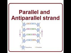
DNA- antiparallel double helix in which two complementary strands with opposite directions twist around each other
mRNA- single stranded after being copied from DNA during transcription
miRNA- small RNA that can silence gene expression
rRNA- formed in nucleolus of cell and helps ribosomes translate mRNA
dsRNA- some viruses carry their code as double strand RNA
tRNA- small RNA that participates in protein synthesis
Miller-Urey & Central Dogma
Experiment simulated early Earth conditions resulting in formation of amino acids and simple organic compounds. Demonstrated that key building blocks an form abiotically
Central Dogma- DNA -> RNA -> Proteins. Drawback of experiment? No O2.
Cell membrane
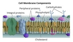
Selectively permeable barrier composed of phospholipid bilayer with embedded proteins. Regulates movement of substances in and out of cell, plays a role in cell signaling and communication
Phospholipid bilayer
Possesses hydrophilic heads and hydrophobic tails that face inward. Barrier to most water soluble molecules
Membrane Proteins
Embedded within the phospholipid bilayer. Facilitate transport, act as receptors for signaling, and provide structural support
Carbohydrates
Attached to proteins and lipids on extracellular surface. Play key roles in cell recognition and adhesion
Cholesterol
Found within eukaryotic cell membrane, helps stabilize membrane fluidity
Integral Proteins
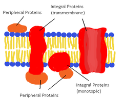
Embedded in phospholipid bilayer. Amphipathic. Some are partially embedded and some are fully embedded (transmembrane)
Transmembrane protein
Integral protein. Traverse entire bilayer. Function as receptors in cell signaling, also function as channels or carrier proteins for transport

Peripheral membrane proteins
Found on surface of bilayer (inside or outside) and are generally hydrophilic. Function as receptors or assist with adhesion and cell recognition
Membrane protein functions
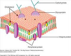
Receptor: transmit signal to cell, trigger secondary responses within cell
- Agonists- molecules that bind to receptors and functionally activate a target
- Antagonist- bind and prevent other molecules from binding, inhibit response
Adhesion- attach cell to other things, anchors for cytoskeleton
Cellular recognition- proteins which have carbohydrate chains (glycoproteins). Used for recognition
Fluid Mosaic model
Describe how components that make up cell membrane can move freely within membrane ("fluid"). Furthermore, cell membrane contains many different kind of structures ("mosaic")
Fluidity impacted by
Temperature- increase temp, increase fluidity and vice versa
Cholesterol- Holds membrane together at high temperatures and keeps membrane fluid at low temp
Degrees of unsaturation- saturated fatty acid = pack tightly, decrease fluidity. Unsaturated = loose, increase fluidity
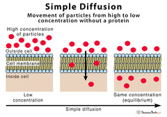
Simple diffusion
Small uncharged molecule. ex) O2, CO2, H2O or lipid soluble (steroids). Down concentration gradient, no energy
Facilitated transport
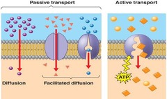
Channel proteins allow diffusion of large (glucose, sucrose) and charged ( NA+, K+, Cl-) molecules down concentration gradient, no energy needed

Active transport
Substances travel against concentration gradient and require energy
Primary- uses ATP hydrolysis to pump molecule against gradient (i.e. sodium potassium pump set membrane potential)
Secondary- Uses energy from one molecule moving down its electrochemical gradient to drive transport of another molecule against concentration gradient
Cytosis
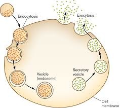
Refer to bulk transport of molecule across cell membrane
Endocytosis- cell membrane wrapping around extracellular substance internalizing it into cell via vesicle or vacuole.
- Phagocytosis- ingest food
- Pinocytosis- ingest water and dissolved materials
- Receptor-mediated endocytosis- require binding of dissolved molecules to peripheral membrane receptor proteins
--Clathrin=protein that aids in receptor mediated endocytosis by forming put in membrane that pinches off as coated vesicle = clathrin mediated endocytosis
Exocytosis
Opposite of endocytosis, material secreted out of cell
Cytosol and Cytoplasm
Cytosol- aqueous intracellular fluid
Cytoplasm- cytosol + organelles
- Only eukaryotic cell contain membrane bound organelles, Prokaryotes contain genetic info in nucleoid
Nucleus
Protect and house DNA. DNA replication and transcription occur here.
Parts:
Nucleoplasm-cytoplasm of nucleus
Nuclear envelope- membrane of nucleus, contains 2 phospholipid bilayers
Nuclear pores- holes in nuclear envelope that allow molecules to travel in and out of nucleus
nucleolus- dense area responsible for producing components of ribosomal subunits
Ribosomes
Not considered organelles. Carry out translation (mRNA to protein). Composed of ribosomal subunits. Eukaryotic = 60S and 40S complete ribosome = 80S in cytoplasm
Prokaryotic = 50S and 30S, 70S in cytoplasm
Endomembrane System
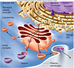
Rough ER- continuous with outer membrane of nuclear envelope. Rough because of ribosomes. Proteins synthesized by embedded ribosomes are sent to lumen for modification. Afterward sent out of cell or become part of membrane
Smooth ER- extension of RER. synthesize lipids, produce steroid hormones, detoxify cell
Golgi apparatus- stores, modifies, and exports proteins that will be secreted from cell. Made up of cisternae that modify and package substances. Vesicle from ER rich cis face and leave from trans face.
Lysosomes- found in animal cells, digestive (hydrolytic) enzymes which break down cellular waste. Play role in apoptosis
Vacuoles- membrane bound vesicles used for storage or transport. Plants have central vacuole that help maintain turgor. Contractile vacuoles in protist pump excess water
Peroxisome
Animal + plant. Perform hydrolysis, break down stored fatty acids, help with detoxification. These processes generate hydrogen peroxide which is toxic since they make ROS which damage cells through free radicals. Peroxisomes contain catalase which breaks H2O2 into H2O and O2

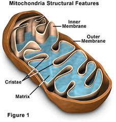
Mitochondria
Powerhouse of cell, produces ATP for energy use through cellular respiration Mitochondrial inheritance is maternal.
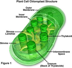
Chloroplasts
Found in plants and some protists, carry out photosynthesis. Type of plastid which is a double membrane organelle found exclusively in plant cells and algae.
Centrosomes
Organelles found in animal cells containing pair of centrioles. Act as microtubule organizing center during cell division
Cytoskeleton
Provide structure and function within cytoplasm

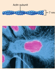
Microfilaments
Smallest structure of cytoskeleton, composed of double helix made of actin filaments. Mainly involved in cell movement and can quickly assemble and disassemble.
1. Cleavage Furrow: during cell division, myosin motors and actin microfilaments form contractile rings that split cell
2. Cyclosis- flow of cytoplasm inside cell. Driven by forces via actin and myosin movement
3. Muscle contraction- actin microfilaments have directionality allowing myosin motor proteins to pull on them for muscle contraction
Intermediate filaments
between microfilaments and microtubules in size. More stable than microfilaments and mainly help with structural support. (ex keratin intermediate fiber in skin, hair, nails)
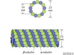
Microtubules
Largest in size and give structural integrity to cells. Made of tubulin protein. Form centrioles and are found in cilia and flagella
Cilia- hair like projections made of tubulin only found in eukaryotes. Line outside of eukaryotic cells and function in locomotion of cell itself or fluids (i.e. cilia in lungs remove debris).
Flagella- found in both pro/eukaroytes. Function in locomotion and fluid movement. Eukaryotic flagella made of tubulin, prokaroytic, flagellin (NOT MICROTUBULE)
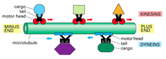
Kinesin and dynein
Transport cargo along microtubules
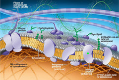
Extracellular matrix (ECM)
Provides extracellular mechanical support for cells.
Proteoglycan- type of glycoprotein that has a high proportion of carbohydrates
Collagen- most common structural protein secreted by fibroblasts organized into collagen fibrils
Integrin- transmembrane protein that facilitates ECM adhesion and signal cells to respond to extracellular environment
Fibronectin- protein that connects integrin to ECM w/ signal transduction
Laminin- behaves like fibronectin. influences cell differentiation, adhesion, movement.
Cell Walls
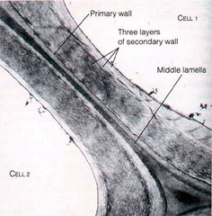
Carbohydrate based structures that act like a substitute ECM. Provide support to cells that either do not have an ECM or a minimal ECM. Present in plants (cellulose), fungi (chitin), bacteria (peptidoglycan), and archaea (non-peptidoglycan polysaccharides)
Cell-matrix junction
Connect ECM to cytoskeleton
1. Focal adhesions- ECM connects via integrins to actin mirofilaments inside cell
2. Hemidesmodomes- ECM connect via integrins to intermediate filaments inside cell
Cell-cell junctions
1. Tight- form water tight seals between cells to ensure substances pass through cells NOT between them. (ex- blood brain barrier, skin epithelial, intestinal barrier, bladder and kidneys)
2. Desmosomes- provide support against mechanical stress. Connect neighboring cell via intermediate fibers (skin, heart, gastrointestinal mucosa, bladder)
Tight- regulate movement of substance Desmosome- regulate structural integrity and anchoring, not watertight
3. Adherens junctions- similar in structure and function to desmosomes but use actin microfilaments
4. Gap junction- allow passage of ions and small molecules betwen cells. Formed from transmembrane proteins known as connexons (ONLY ANIMAL CELLS)
Cell-cell Plant specific
1. Middle lamella- sticky cement similar to tight junction
2. Plasmodesmata- tunnels with tubes between plant cells. Allow cytosol to freely travel between plant cells
Q10 (Temperature Coefficient)
Factor that measures the change in reaction rates for every 10C change in temperature. Ex) Q10 of 1.5 indicates at increase of 10C would increase metabolism by 50%
Reaction coupling
process of powering an energy requiring reaction with an energy release one. It allows unfavorable reaction to be powered by a favorable reaction making Gibbs free energy negative
Aerobic Cellular respiration
Performed to phosphorylate ADP into ATP. Involves four catabolic reactions...
1. Glycolysis
2. Pyruvate oxidation
3. Krebs cycle
4. Oxidative phosphorylation
Glycolysis
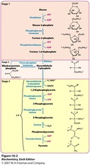
Occurs in cytosol. Does not require oxygen so it is also used in fermentation. Has energy investment and energy payoff phase.
1. Hexokinase uses ATP to phosphorylate glucose into glucose-6-phosphate. Glucose-6-phosphate cannot leave cell.
2. Isomerase modifies glucose 6 phosphate into fructose-6-phosphate.
3. Phosphofructokinase is a major regulatory step. Turns fructose-6-phosphate into fructose-1,6-bisphosphate.
4. Fructose-1,6-bisphosphate into DHAP and G3P which are in equilibrium with one another. G3P continues to energy payoff phase so DHAP is converted into G3P so 2 G3P are made.
6. G3P does redox reactions and produces 4ATP and 2NADH, and 2 pyruvate. 2 net ATP made
Pyruvate Oxidation
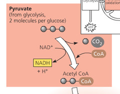
Pyruvate dehydrogenase carries out pyruvate oxidation.
1. Decarboxylation- 3C pyruvate move from cytosol to mitochondria matrix where 1 CO2 released.
2. Oxidation- 2C molecule is converted to acetyl giving electrons to NAD and making NADH.
3. Coenzyme A binds to acetyl group and becomes Acetyl-CoA.
In total... 2NADH, 2CO2, 2 Acetyl CoA made.
Krebs Cycle
Occurs in matrix.
1. Acetyl CoA joins oxalacetate to form citrate
2. Citrate undergoes rearragnements that produce 2 CO2 and 2 NADH, 1 ATP.
3. Molecule transfer electrons to one FAD which is reduced to FADH2
4. Molecule converted back to oxaloacetate producing another NADH.
In total 4 CO2, 6NADH2, and 2 ATP made
Oxidative Phosphorylation
Electron transport chain and chemiosmosis work together to produce ATP. Oxygen acts as final electron acceptor. Mitochondrial inner membrane is the location of the ETC
Complex I ETC
Accept electrons from NADH. NADH produces 3 ATP. If Complex I stops, ETC continues using only complex II, III, and IV. NADH converted to NAD through alternative pathways like fermentation
Complex II ETC
Accepts electrons from FADH2. FADH2 produce 2 ATP. If blocked ETC can continue but at reduced rate, ALSO Krebs cycle would stop at the succinate -> fumerate step
Complex IV
Final complex. Transfers electrons to oxygen. Without it entire chain would back up. No O2, ETC stops because NAD and FAD aren't regenerated, ATP production halts, cells switch to anaerobic
Chemiosmosis
Use proton electrochemical gradient to synthesize ATP. ATP synthase is channel protein that allows protons to flow down their electrochemical gradient. Spontaneous movement of protons generate energy needed to convert ADP to ATP (endergonic). Overall aerobic respiration is exergonic (-G)
Fermentation
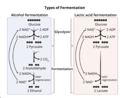
Anaerobic pathway. Only relies on glycolysis by converting pyruvate into different molecules in order to oxidize NADH back to NAD+. Lactic acid and alcohol fermentation
Alternative Sources of energy generation
1. Other carbs. Mostly enter during glycolysis. Glycogenolysis = release of glucose-6-phsophate from glycogen
Glycogenesis = reverse process, conversion of glucose to glycogen. Glycogen stored in liver and muscle cells
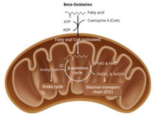
2. Fats- lipases required to digest fats into free fatty acids and alcohols. Adipocytes = cells that store fat and have hormone-sensitive lipase enzymes to help release triglycerides back into circulation as lipoproteins or as free fatty acids bound by a protein called albumin
Free fatty acid undergo beta oxidation
3. Proteins- least desirable because getting them into cellular respiration takes a lot of energy.
The trophs
Heterotrophs- must get energy from food they eat
Autotrophs- make their own food
Photoautotrophs- make their own food using light energy and convert it to chemical energy. Photons are used to synthesize sugars (glucose) in photosynthesis.
Carbon fixation
Process by which inorganic carbon (CO2) is converted into organic molecule (glucose). Photosynthesis takes electrons released from photolysis and excites them using solar energy.
Photosynthesis
Non-spontaneous and endergonic reaction that produces glucose after an input of solar energy
Cellular respiration- spontaneous and exergonic, break down glucose to generate energy in form of ATP
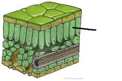
mesophyll cells
Located between upper and lower epidermis of leaves, facilitates gas movement within leaf. Contain chloroplasts
Light Dependent reactions
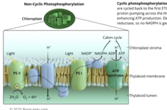
Occur in thylakoid membrane and harness light energy to produce ATP and NADPH which will be used in Calvin cycle (ATP made here is NOT used to power cell, only used in Calvin cycle). Occur in thylakoid membrane of chloroplasts
Photosystems
Contain special pigments such as chlorophyll that absorb photons. Chlorophyll absorb red and blue light and reflect green light giving plants their green color.
Reaction center- special pair of chlorophyll molecules in the center of these proteins. Chlorophyll has a porphyrin ring structure with magnesium atom bound in center. Photosystem II (P680) and photosystem I (P700) are used in photosynthesis
Non-cyclic photophosphorylation
1. Water is split, passing electrons to photosystem II and releasing protons into thylakoid lumen.
2. Photons excite electrons in the reaction center of PS II passing the electrons to primary electron acceptor (pheophytin).
3. Primary electron acceptor sends excited electrons to electron transport chain. Protons pumped from stroma to thylakoid lumen. Electrons deposited to PSI
4. Photons excite pigments in PSI, energizing electrons in reaction center to be passed to another primary electron acceptor (A0)
5. Electrons sent to short electron transport chain that terminate with NADP+ reductase and produce NADPH with electrons and protons.
6. Accumulation of protons in thylakoid lumen generate electrochemical gradient that is used to make ATP
Cyclic photophosphorylation
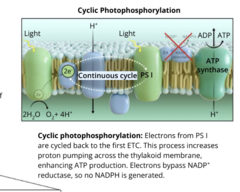
Electrons from PSI are cycled back to first ETC. This process increases proton pumping across thylakoid membrane enhancing ATP production and bypassing NADP+ reductase. NO NADPH MADE, water not needed. ONLY PHOTOSYSTEM I IS USED, PSII SKIPPED
If Non-cyclic produces ATP+NADPH and cyclic produces only ATP, why would plants do non-cyclic over cyclic photophosphorylation?
Cyclic photophosphorylation done to supplement ATP production when Calvin cycle requires more ATP than NADPH. Cyclic photophosphorylation favored under conditions of low CO2 or high light intensity when stomata is closed.
Calvin Cycle
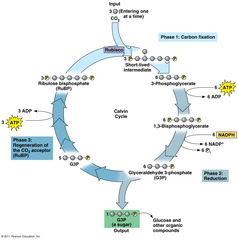
AKA light independent reactions. Only occur if light dependent reactions occur since light-dependent supplies Calvin cycle with ATP and NADPH. Occur in stroma.
RuBisCo
Enzyme that converts inorganic CO2 to six carbon molecule by combining it with 5 carbon RuBP. Six Carbon molecule breaks down to 3-PGA.
Reduction: PGA phosphorylated by ATP and reduced by NADPH to form G3P.
Regeneration- Most G3P converted back to RuBP
Carbohydrate synthesis- Some G3P
Photorespiration (C2 Photosynthesis)
RuBisCo can also cause oxygen to bind to RuBP.
Photorespiration produces two carbon phosphoglycolate and eventually converts into PGA. Process is a net loss of fixed carbon, no glucose made
C3 Photosynthesis
Normal photosynthesis where 3-PGA made
C4 Photosynthesis
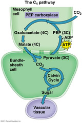
Spatial isolation.
1. CO2 fixed and produces four carbon oxaloacetate (4C) which is converted to malate in mesophyll cells
2. Malate transferred to bundle sheath cells which have less O2
3. Malate decarboxylated spatially isolating CO2. It is then fixed by RuBisCo and enters calvin cycle
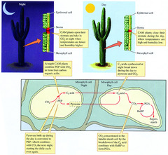
Crassulacean acid metabolism (CAM)
Temporal isolation
1. During day, stomata closed to prevent evaporation of water (transpiration)
2. During night stomata open to let CO2 in. CO2 is fixed producing oxaloacetate and afterwards malate, however malate stays in mesophyll cells.
3. During next day stomata are closed again and malate is decarboxylated. CO2 is temporally isolated from O2 which cannot enter during day.
Cell Division Key Terms
Genome: All DNA in a cell
Chromosomes- individual molecules of DNA that contain a portion of the entire genome
Homologous chromosome pairs- pair of chromosomes that contain the same genes in the same order with one inherited from each parent
Sister chromatid- identical attached copies of a single chromosome that form dyads
Dyads- replicated chromosomes containing two sister chromatids that look like an X
Centromere- region of DNA that connect sister chromatids in a dyad
Kinetochores- proteins on the sides of centromeres that help microtubules pull sister chromatids apart during cell division
Karyokinesis- division of nucleus
Cytokinesis- division of cytoplasm and cell membrane
Ploidy- describe number of chromosome set found in body. Diploid = 2 sets (46 in human), haploid = 1 set (23 in human)
Cell Division Terms continued
Sex chromosomes- One pair in human body, determine sex
Autosomes- 22 pairs in human body; nonsex chromosome
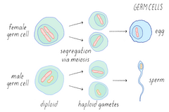
Gametes- haploid cells (sperm and egg)
Germ cells- diploid cells that divide and increase by mitosis and differentiate into gametocytes
Gametocytes- eukaryotic differentiated germ celsl that can undergo meiosis to produce gametes
Somatic- all body sells excluding gametes. Diploid in humans
The Cell Cycle
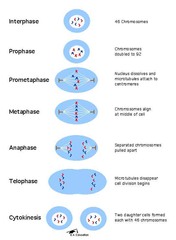
Divided into Interphase and M phase. Interphase proceeds mitosis and meiosis and 90% of the cell cycle happens during interphase. M phase is where karyokinesis and cytokinesis occur.
Components of Interphase
1. Gap Phase 1- cell grows in preparation for cell division. Checks for favorable conditions (sufficient cell size, nutrients, energy, and growth signals. Also check DNA integrity). If favorable, cell will enter S phase. If unfavorable cell will enter G0.
- G0- cell still carry out their functions but halt in cell cycle. Cells that do not divide are stuck here
2. Synthesis- Cell replicates its genome and moves to G2 phase when complete. centrosomes duplicate.
3. Gap phase 20 Cell continues to grow and prepare for cell division by checking DNA for any errors after replication. Also checks for mitosis promoting factor aka maturation promoting factor which needs to be present in adequate amounts for cell cycle continuation. Organelles replicated here
Cell Cycle Regulation
Surface to volume ratio- As cell grows, its volume increases more than its surface area. Cells rely on the surface area of their cell membrane for transport of material in and out of the cell. Cell division occurs if the S/V ratio decreases (surface area too small for volume)
- Cell specific checkpoints
- G1 = checks for favorable conditions to grow; enter G0 if unfavorable
- G2 = Checks accuracy of DNA replication and MPF levels
- M = check for chromosomal attachment to spindle fibers

Cyclin-dependent kinases
Phosphorylate certain substrates to signal cell cycle progress. Activated by cyclin, protein that cycles through stages of synthesis and degradation.
Growth factors
Bind to receptors in the plasma membrane to signal for cell division
Density dependent inhibition
Halting of cell division when density of cells is high
anchorage dependence
Cells divide only when attached to external surface
Microtubule Organizing Centers
Present in eukaryotic cells; organize extension of microtubules which are made of the protein tubulin. MTOCs are responsible for forming the spindle apparatus which guides chromosomes during karyokinesis.
Centrosomes- organelles found in animal cells that contain a pair of centrioles. Act as microtubule organizing centers.
Microtubules in spindle apparatus:
1. Kinetochore- extend from centrosomes and attach to kinetochores on chromosomes
2. Astral- extend from centrosomes to cell membrane to orient spindle apparatus
3. Polar- extend from two centrosomes and connect with one another. Push centrosomes to opposite end of cell.
Components of M Phase
Stage where karyokinesis and cytokinesis occur. Four stages:
1. Prophase- chromatin DNA condenses into chromosomes and nucleolus and nuclear envelope disappear. Spindle apparatus forms
2. Metaphase- chromosomes line up single file along metaphase plate guided by spindle apparatus
3. Anaphase- Kinetochore microtubules shorten to pull sister chromatids apart. Sister chromatids are now separate chromosomes, chromosome number doubles (in humans now have 92 chromosomes)
4. Telophase- chromosomes have separated and nuclear membranes reform. Nucleoli reappear and chromosomes decondense to chromatin
Cytokinesis- physical separation of cytoplasm and cell membrane into 2 daughter cells. In animal cells cytokinesis begins in late anaphase with formation of cleavage furrow (contractile ring of actin microfilaments and myosin motors that punch the cell in two)
In plants... cytokinesis begins in telophase with formation of cell plate. Cell plate is created by vesicles from golgi apparatus and ends up producing the middle lamella.
binary fission
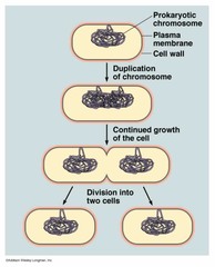
Used by archea, bacteria, and organelles (mitochondria, chloroplasts. During binary fission organisms replicate their genome while cell division is happening. Replicated DNA segregate to opposite ends of the cell and a septum forms to separate into 2 daughter cells.
Meiosis
Produces four haploid daughter cells from one diploid parent cell.
Meiosis I- reductional division. Produces two haploid daughter cells through separation of homologous chromosomes
1. Prophase I- chromatin condenses into chromosomes. Nucleolus and nuclear envelope disappear. Homologous chromosomes pair up, crossing over occurs
Synapsis- pairing of homologous chromosomes into tetrads ( pair of two homologous chromosome each with 2 sister chromatids.
Chiasmata- where 2 chromosomes of a homologous pair crossover during synapsis causing genetic recombination (exchange of DNA between chromosomes to produce genetically diverse offspring)
2. Metaphase I- tetrads randomly line up double file on metaphase plate. contributes to genetic diversity
3. Anaphase I- kinetochore microtubules shorten, separate homologous chromosomes from each other
4. Telophase and Cytokinesis I- After tetrads have been pulled to opposite poles, nuclear membrane reforms, nucleoli reform, chromosomes decondense into chromatin. A cleavage furrow forms in animal cells and cell plate in plants
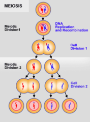
Meiosis II
Prophase II- chromatin condenses into chromosomes. Nucleolus and nuclear envelope disappear. Spindle apparatus forms.
Metaphase II- chromosomes line up single file at metaphase plate
Anaphase II- Kinetochore microtubules shorten and pull sister chromatids apart. Sister chromatids become separate chromosomes, chromosome number doubles
Telophase and cytokinesis- nuclear membrane reform, nucleoli reappear and chromosomes decondense to chromatin. Four haploid daughter cells produced
Genetic building Blocks
Nucleoside: Ribose sugar and nitrogenous base
Nucleotide: Ribose sugar, nitrogenous base, and phosphate
RNA- nucleotides have ribose sugar with hydroxyl on 2' and 3' carbon
DNA- nucleotides have deoxyribose sugar 3' has hydroxyl, 2' has hydrogen
Purines- double ringed nitrogenous bases = adenine and guanine
Pyrimidines- single ringed nitrogenous bases = cytosine and thymine
In DNA A binds T with 2 hydrogen bonds and G binds C with 3 hydrogen bonds
In RNA A binds U with 2 hydrogen bonds
DNA replication
Origin of replication is where DNA strands first separate to initiate DNA replication. Organisms with circular DNA (bacteria) have a single origin whereas linear DNA (humans) have multiple
DNA undergoes semiconservative replication where each new double helix produced by replication has one old and one new strand
Stages of DNA replication
1. Initiation- origins of replication form at A-T pairs since they have less hydrogen bonds and are easier to break
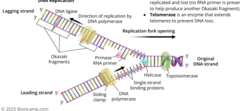
Helicase- unzips DNA by breaking hydrogen bonds between strands creating a replication fork. As it unzips the strands helicase leads to supercoiling
2. Elongation- producing new DNA strands using different types of enzymes
- Single-strand binding proteins- bind to uncoiled DNA strands preventing reattachment of the strands to each other
- Topoisomerase- Nicks DNA double helix ahead of helicase to relieve built up tension and supercoiling
- Primase- places RNA primers along the template strands to create 3' ends for nucleotide addition
Sliding clamp proteins- hold DNA polymerase to template strand
DNA polymerase- adds free nucleoside triphosphates to 3' ends. DNA polymerase can only add nucleotides onto preexisting 3' hydroxyl groups with primase.
3. Termination- Replication fork cannot continue, DNA replication ends
Telomeres- noncoding repeated nucleotide sequences at the ends of linear chromosomes. Necessary in eukaryotes because when the replication fork reaches the end of a chromosome, a small segment of DNA from the telomere is not replicated and lost.
Telomerase- enzyme that extends telomeres to prevent DNA loss
DNA strands
Leading strand- produced continuously because it has a 3' end that faces the replication fork (only needs one primer near origin of replication)
Lagging strand- produced discontinuously because its 3' end is facing away from origin of replication. Many RNA primers needed to produce short DNA fragments called Okazaki fragments.
Different DNA polymerase replaced RA primers with DNA
DNA ligase- glues separated fragments of DNA together
DNA organization
Nucleosomes are complexes of DNA wrapped around histone proteins
Histones positively charged, DNA negatively charged allowing proper binding
Chromatin: refers to the overall packaging of DNA and histones. Two types
1. Euchromatin- nucleosomes loosely packed so DNA is readily accessible for transcription
2. Heterochromatin- Nucleosomes are tightly packed so DNA is mostly inactive
Acetylation of histones removes positive charges, relaxing DNA-histone attractions and increases rate of transcription
Deacetylation of histones increases positive charge and decreases trasncription
Methylation of histones adds methyl groups either increasing or decreasing transcription.

Transcription
Genes- instructions within DNA that code for proteins. Must first be transcribed into RNA before being translated to proteins. In a gene the promoter region comes first then the gene operator then the gene.
DNA undergoes transcription to produce single stranded mRNA.
Steps of transcription
1. Initiation- promoter sequence next to gene attracts RNA polymerase to transcribe the gene.
2. Elongation- transcription bubble forms and RNA polymerase travels in 3' -> 5' direction on template strand, however it extends RNA in the 5' -> 3' direction
3. Termination: A termination sequence (terminator) signals to RNA polymerase to stop transcribing gene
Template/antisense strand- used to transcribe the mRNA
coding/sense strand- identical to mRNA except T replaced with U.

Eukaryotic transcriptional control
Unlike in prokaryotes, eukaryotic transcription occurs in the nucleus and uses RNA polymerase to transcribe most genes.
Transcription factors are needed in eukaryotes to help RNA polymerase to bind promoters. The TATA box is an AT-rich sequence in any promoters that transcription factors can recognize and bind to.
- Enhancers- DNA sites that activator proteins can bind to; help increase transcription of a gene.
- Silencers- DNA sites that repressor proteins can bind to; decrease transcription of a gene
Terminator sequence ends transcription. Includes poly-A signal in eukaryotes
DNA Binding motifs
Recognize and bind the major groove of regulatory sequences without unwinding the double helix. Four main types of binding motifs = zinc finger, helix-turn-helix, homeodomain, and leucine zipper