Vet Anatomy Test 1
 Call Kai
 Learn
Learn Practice Test
 Spaced Repetition
 Match
Match Flashcards
 Knowt Play
1/94

There's no tags or description
Looks like no tags are added yet.
Name | Mastery | Learn | Test | Matching | Spaced | Call with Kai | Chat |
|---|
No analytics yet
Send a link to your students to track their progress
95 Terms
Extrinsic muscles
One attachment to axial skeleton, one to appendicular skeleton (forelimb to trunk)
transfer weight to forelimb to stabilize scapula
propels limbs foreward
4 attach to scapula & 4 attach to humerus
Superior- Inferior on an animal is
Cranial- Caudal
Anterior-Posterior on an animal is
ventral- dorsal
The head is
an exception
for example: with the eyes we use superior, inferior, anterior, posterior
Sagittal plane
divides body into right/left parts
Median plane
midsagittal plane that divides body into left & right halves
Transverse plane
perpendicular to the long axis of the body
Dorsal plane
parallel to the back
Dorsal vs Ventral
dorsal directed toward the back, including manus and pes
ventral: directed toward the belly (NOT USED ON FEET)
Palmar vs plantar
palmar: surface of forepaw where pads are located distal to antebrachium
Plantar: surface of hindpaw where pads are located distal to crus
Cranial vs Caudal
used on legs & back
cranial: directed toward the head
Caudal: directed toward the tail
Rostral vs Caudal
used on head
rostral: directed toward the nose
caudal: directed toward the tail
Proximal vs Distal
proximal: toward main mass or origin
distal: away from main mass or origin
Axial vs Abaxial
used on digits
axial: directed toward the central axis of a limb
abaxial: directed away from the central axis of a limb
Extend versus flex
extend: increase flexor angle
flex: decrease flexor angle
Adduct vs Abduct
adduct: move limb toward body
abduct: move limb away from body
Pronation vs Supination
supinate: palm up
pronate: palm down
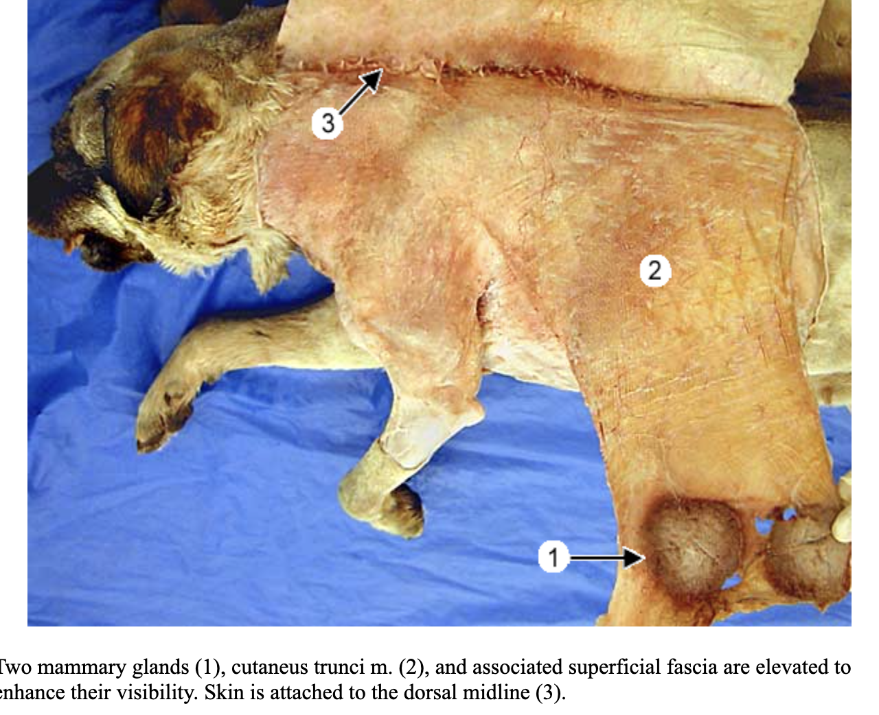
cutaneous trunci m.
1
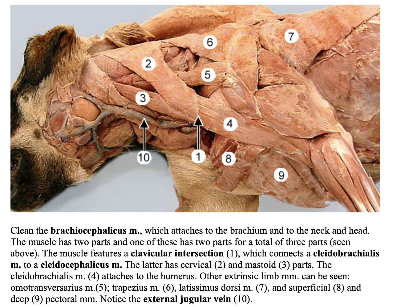
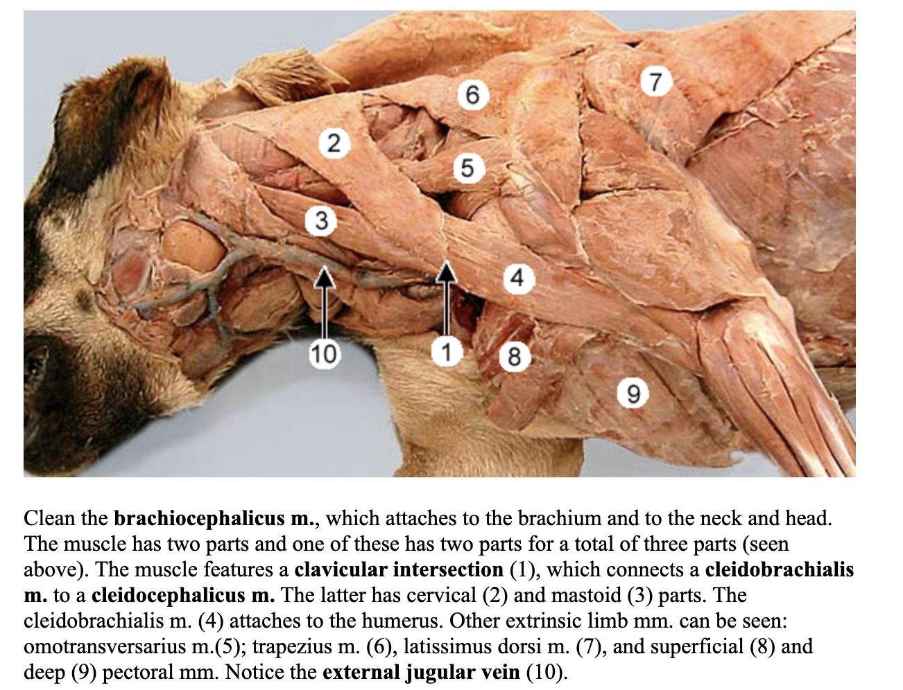
brachiocephalicus muscle
attach to arm & head

cleidobrachialis m.
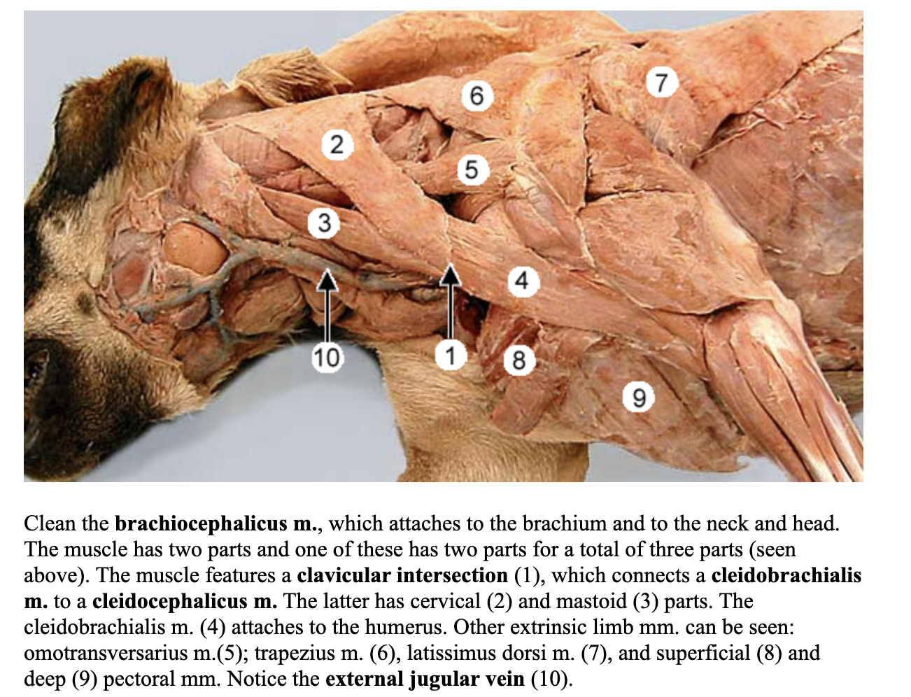
cleidocephalicus m.
separated into pars cervicalis & pars mastoidea
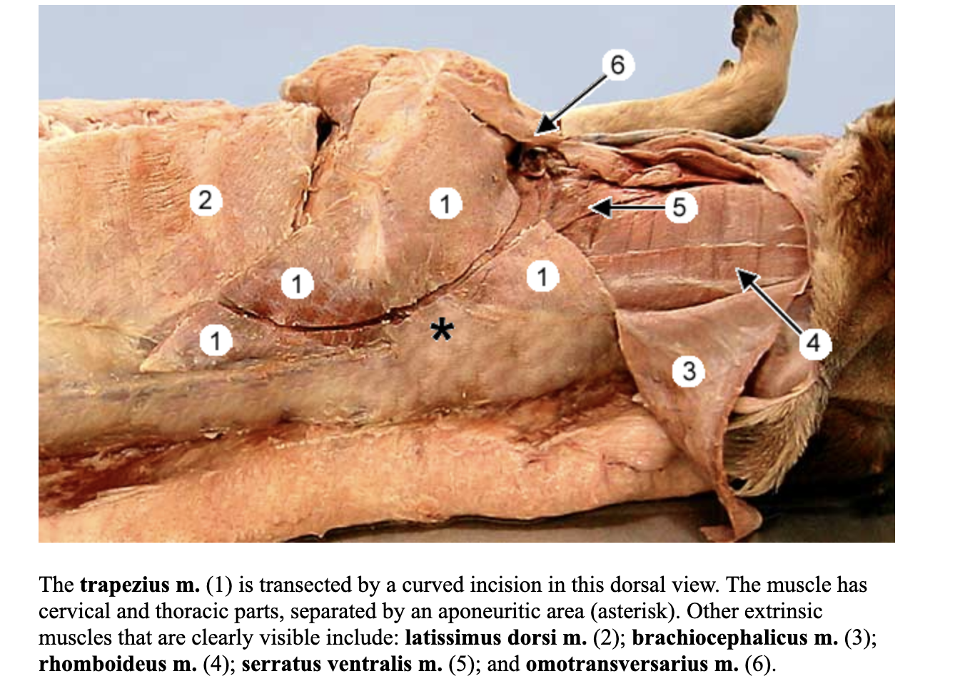
trapezius muscle
omotransversarius m.
on deep surface of this muscle is the superficial cervical lymph nodes
rhomboideus m.

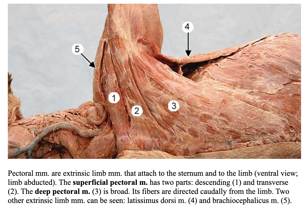
superficial pectoral m.
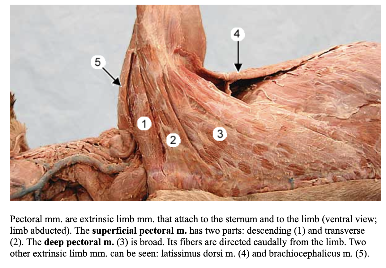
deep pectoral m.
latissimus dorsi m.

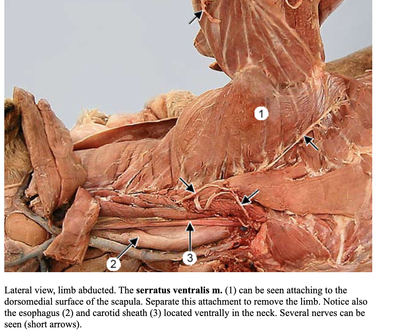
serratus ventralis m.
supraspinatus m.
attachment: supraspinous fossa of the scapula and greater tubercle of the humerus
action: extend and stabilize the shoulder
nerve: subscapular nerve
blood: subscapular artery
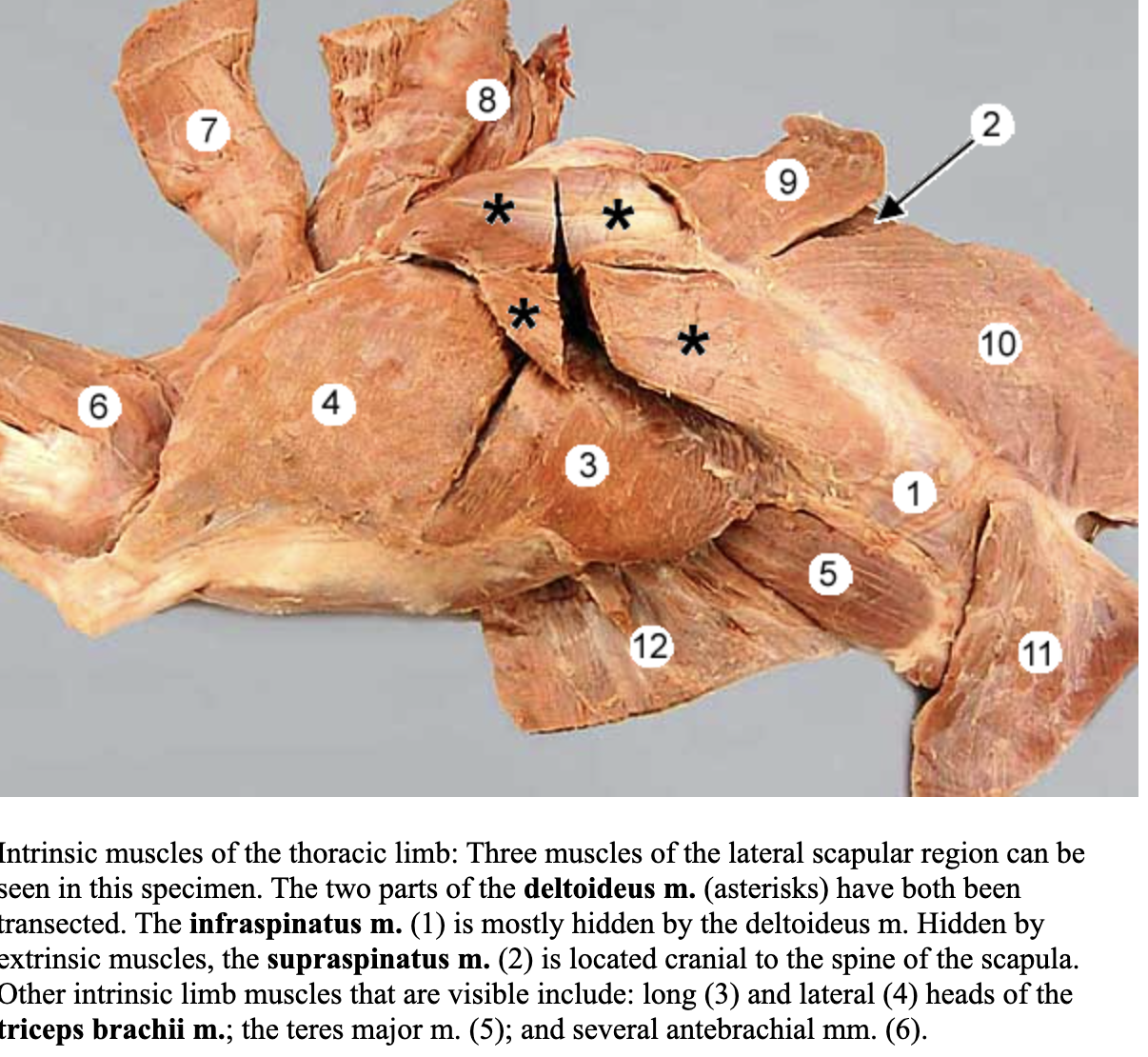
deltoideus m.
attachments: spine and acromion of the scapula and deltoid tuberosity of humerus
action: flex the shoulder joint
nerve: axillary nerve
blood: caudal circumflex humeral artery
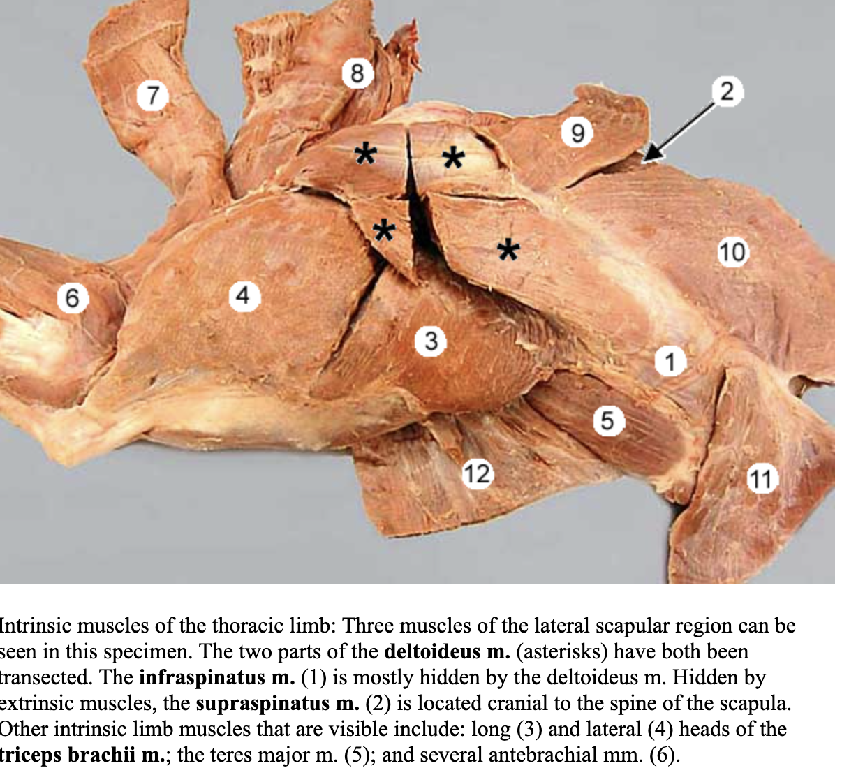
infraspinatus m.
attachments: infraspinous fossa of the scapula and greater tubercle of the humerus
action: flex and stabilize the shoulder; adbuct the limb at the shoulder joint
nerve: suprascapular nerve
blood: suprascapular artery
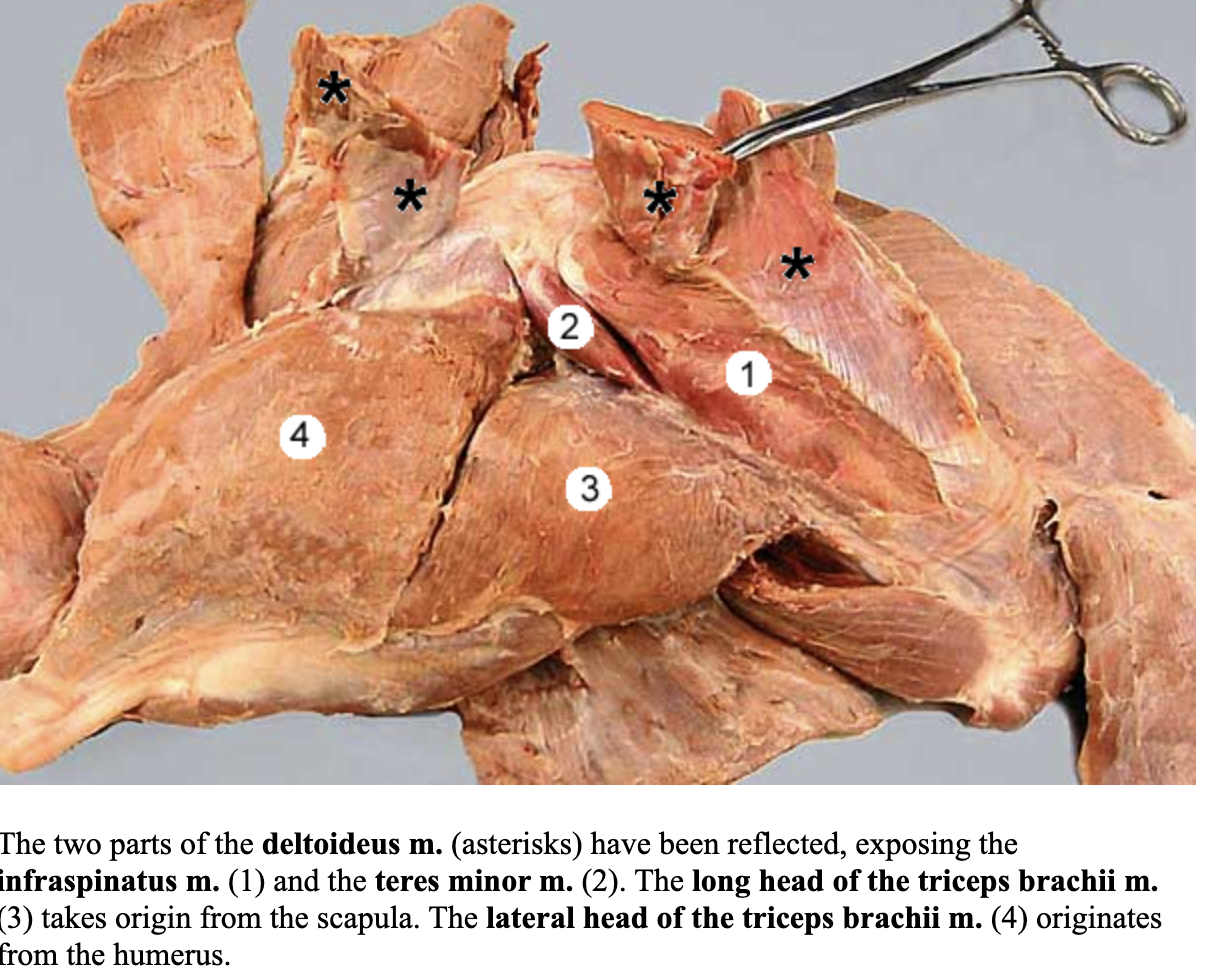
teres minor m.
attachments: caudal aspect of scapula and teres minor tuberosity of humerus
action: flex the shoulder joint
nerve: axillary nerve
blood: caudal circumflex humeral artery

subscapularis m.
attachments: subscapularis fossa of the scapula and lesser tubercle of the humerus
action: act as a shoulder stabilizer; adduct the limb at the shoulder joint
nerve: subscapular nerve
blood: subscapular artery
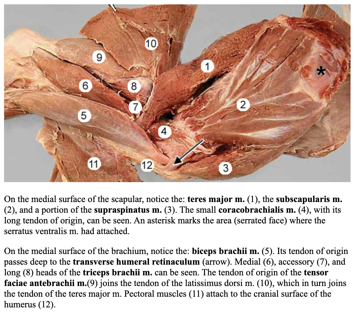
teres major m.
attachments: caudal border of the scapula and teres major tuberosity of the humerus
action: flex the shoulder joint
nerve: axillary nerve
blood: caudal circumflex humeral artery

coracobrachialis m.
attachments: coracoid process of scapula and lesser tubercle and teres major tuberosity of the humerus
action: stabilize the shoulder joint
nerve: musculocutaneous nerve
blood: cranial circumflex humeral artery
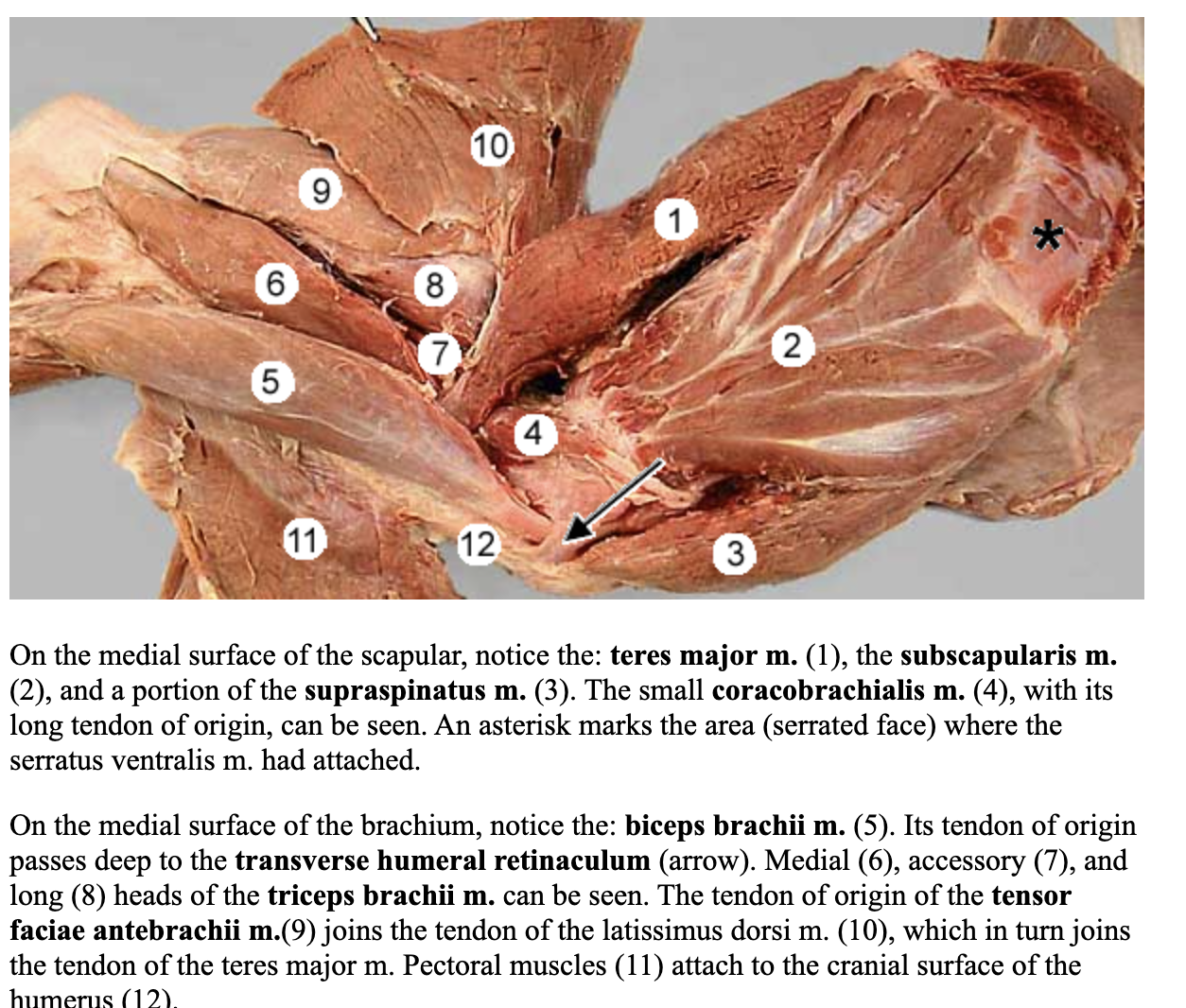
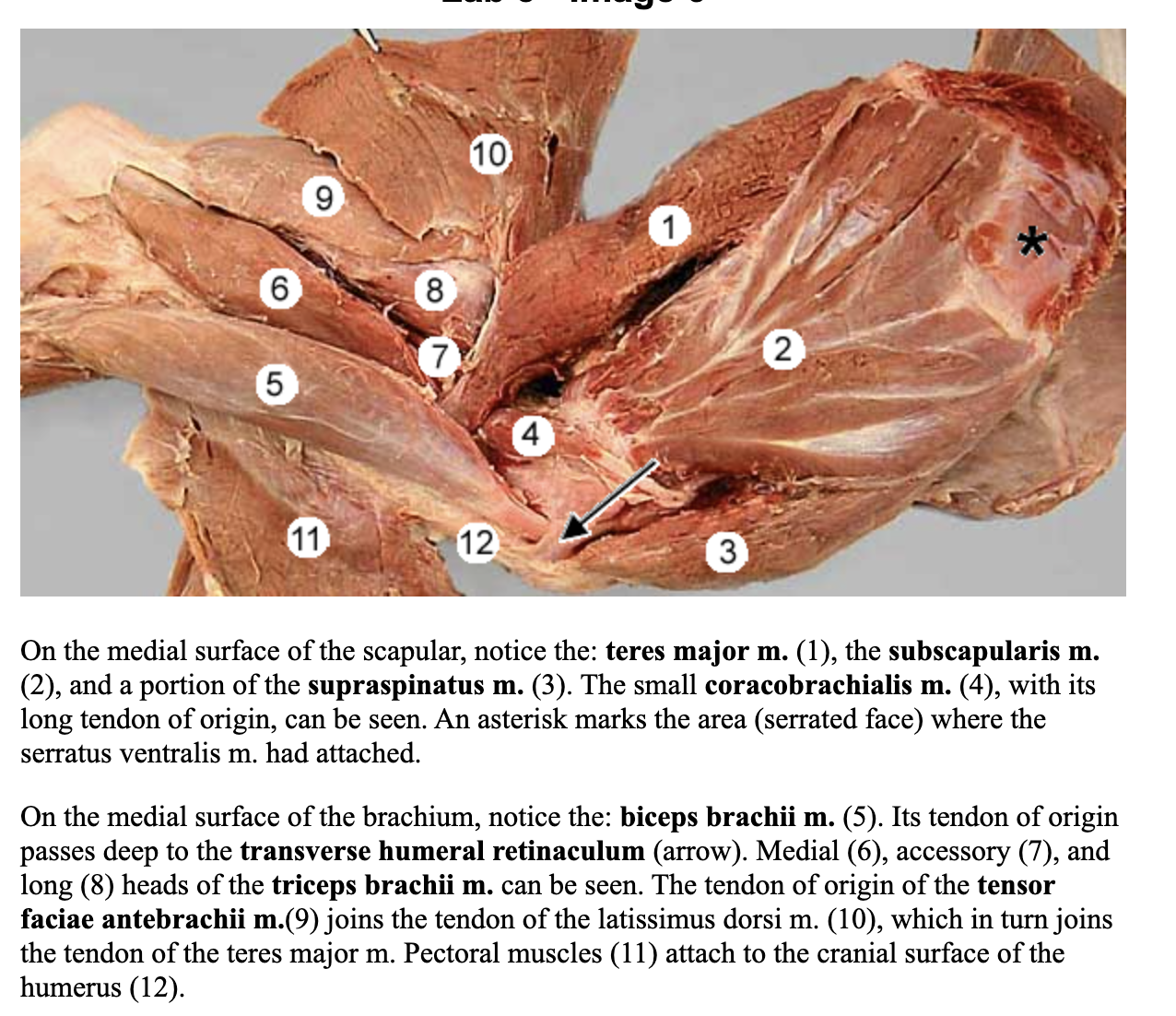
biceps brachii m.
attachments: supraglenoid tubercle of the scapula and proximal radius and ulna in combination with the tendon of the brachialis muscle
held in place by transverse humeral retinaculum (bridge looking foramen)
action: flex the elbow joint; extend the shoulder joint
nerve: musculocutaneous nerve
blood: brachial artery
brachialis m.
attachments: proximal humerus and medial aspect proximal radius and ulna
action: flex the elbow joint
nerve: musculocutaneous nerve
blood: brachial circumflex humeral branches

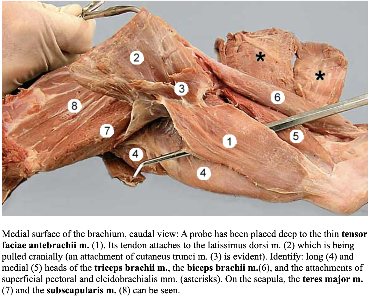
tensor fascia antebrachii m.
attachments: fascia on the lateral surface of the latissimus dorsi and olecranon
action: extend the elbow joint
nerve: radial nerve
blood: deep brachial artery
triceps brachii m.
attachments: long head: caudal border of the scapula to tuber olecrani
lateral head: tricipital line of humerus to tuber olecrani
medial head: crest of the lesser tubercle to tuber olecrani
accessory head: neck of the humerus to tuber olecranni
action: all heads extend the elbow joing; long head also flexes shoulder joint
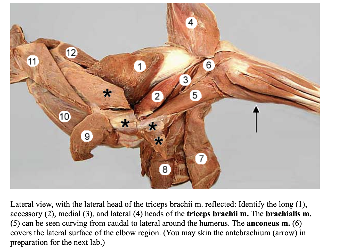
anconeus m.
attachments; lateral supracondylar crest and epicondyle of the humerus to the olecranon
action: extend the elbow joint
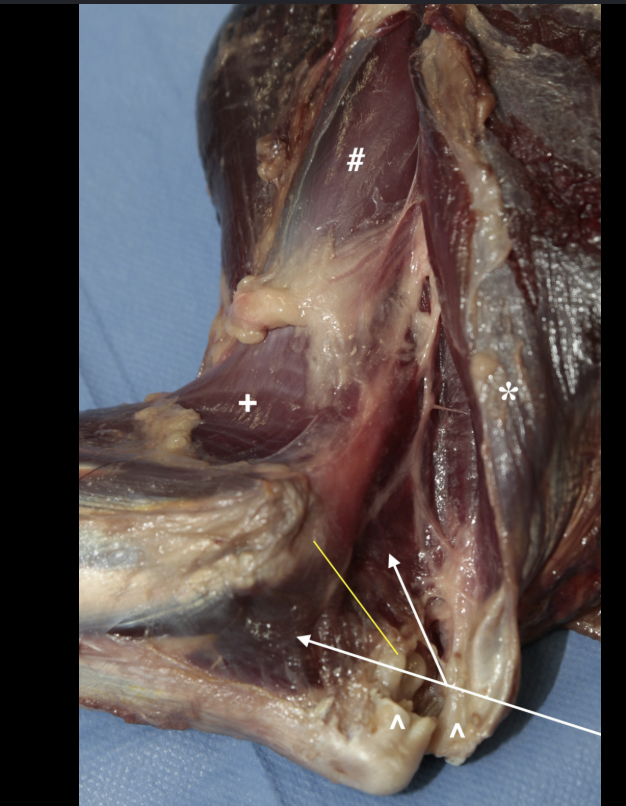
continue on page 16 with antebrachium muscles
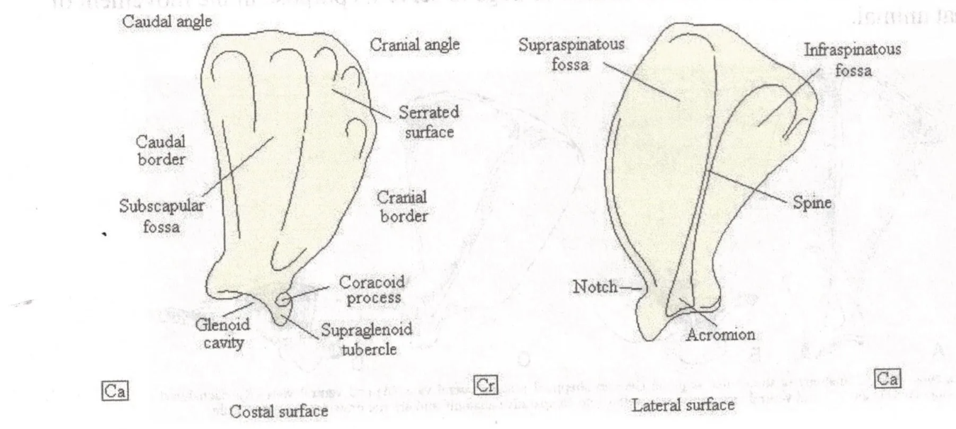
dog scapula glenoid cavity
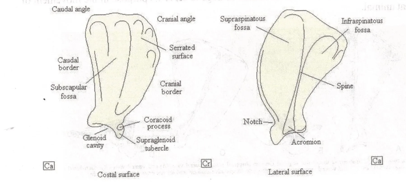
dog scapula supraglenoid tubercle
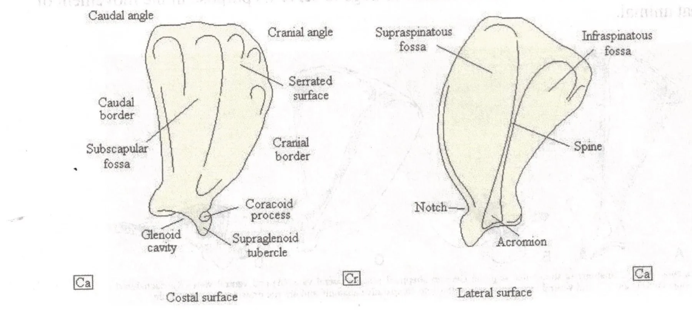
dog scapula coracoid process
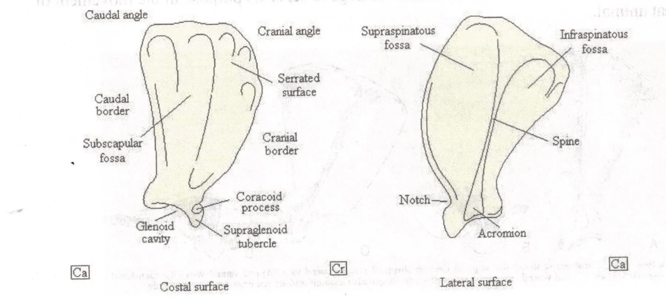
dog scapula spine

dog scapula acromion
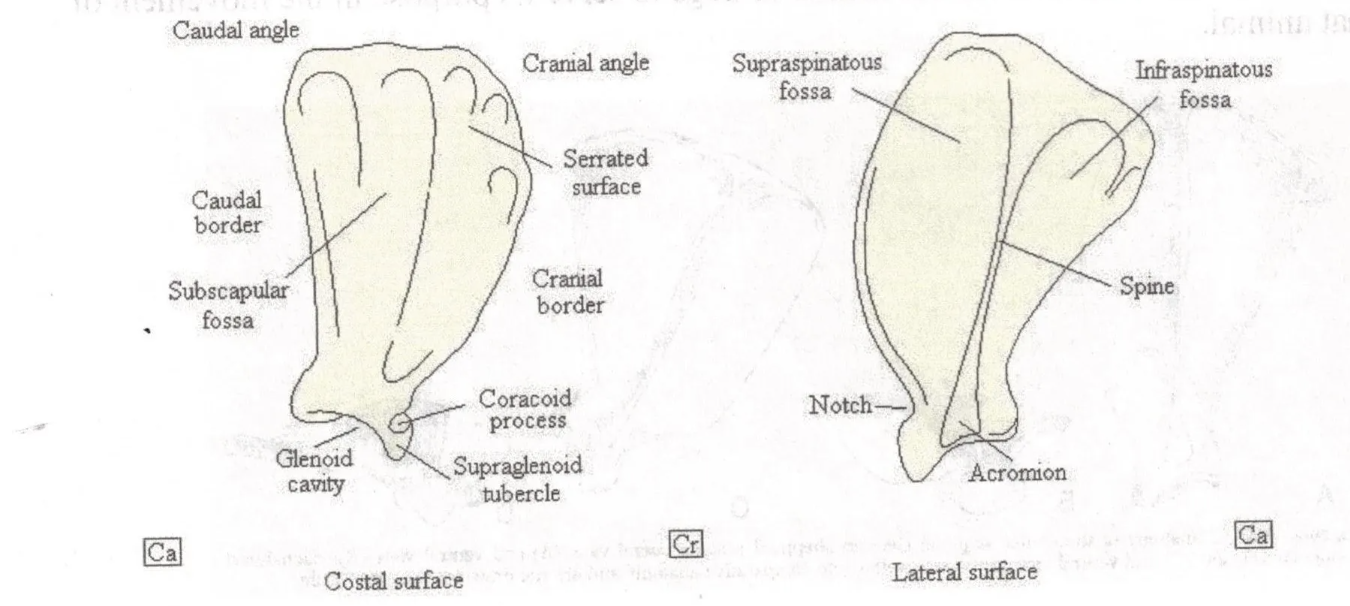
dog scapula supraspinous fossa and infraspinous fossa
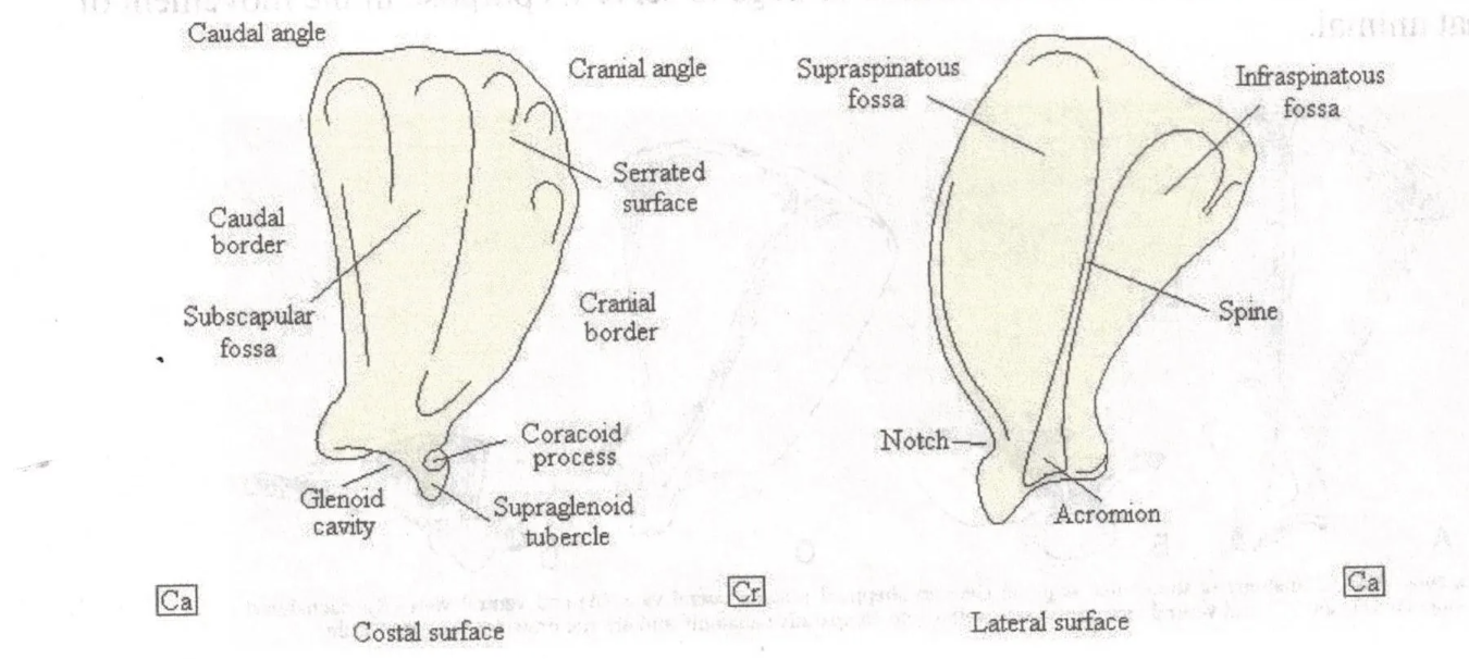
dog scapula serrated surface
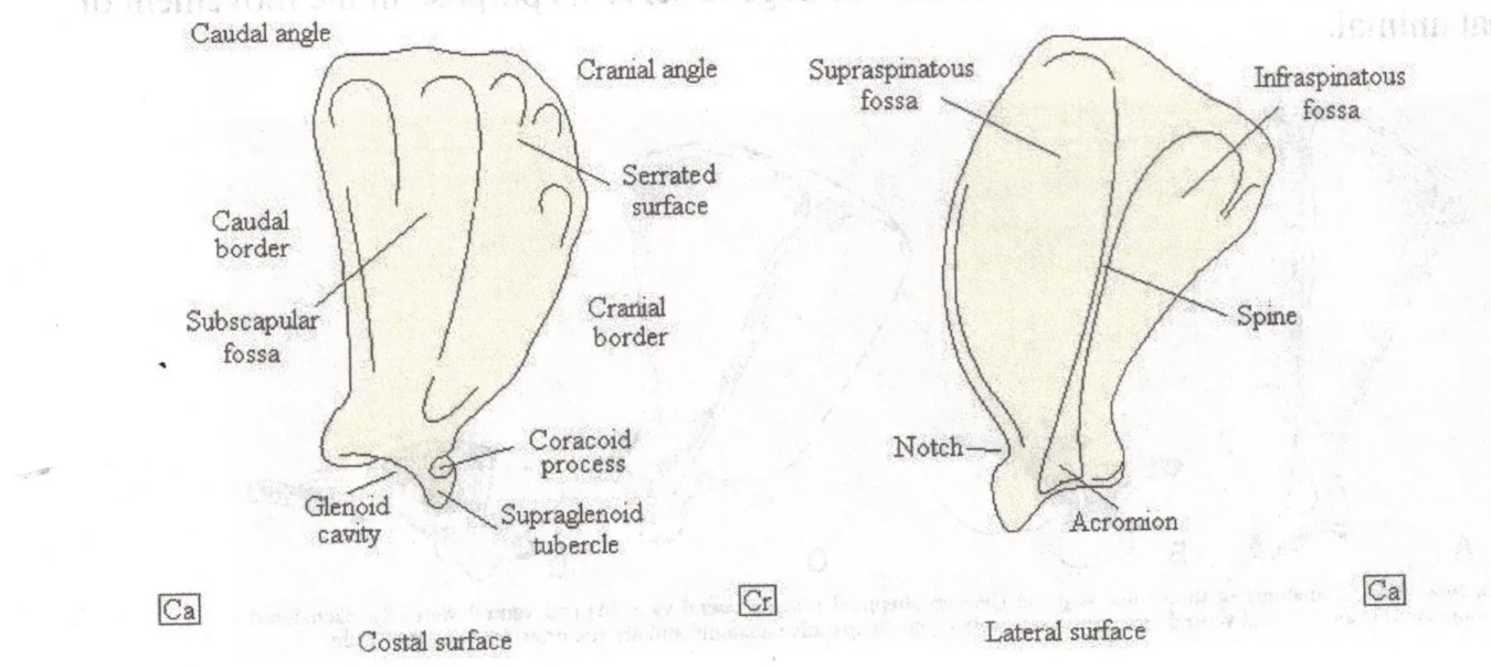
dog scapula subscapular fossa
name and describe the two joints of the humerus
shoulder joint: scapula and humerus
elbow joint: humerus and radius & ulna
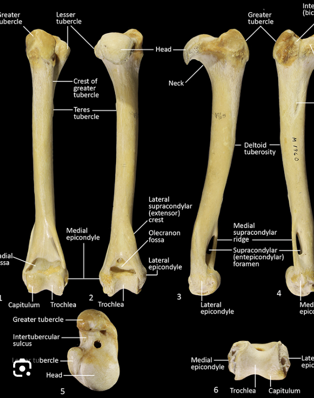
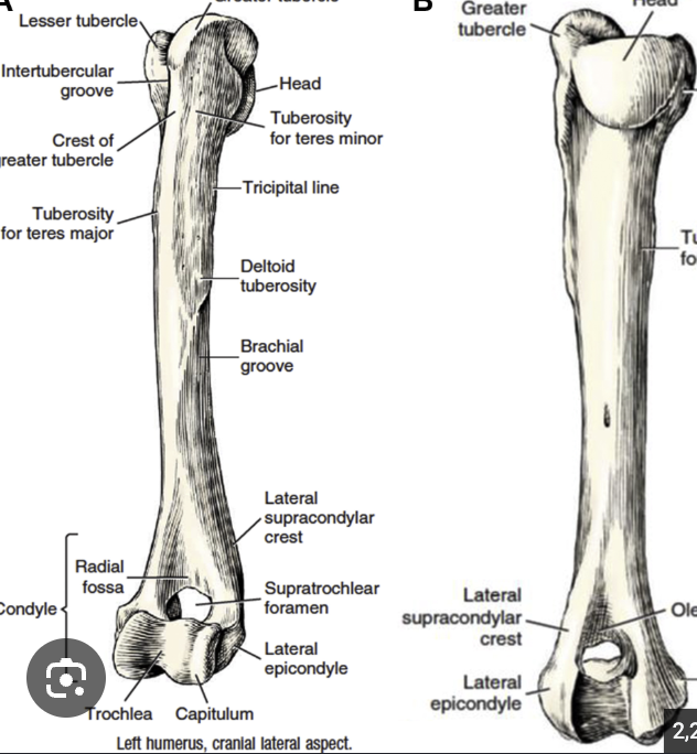
canine head of humerus
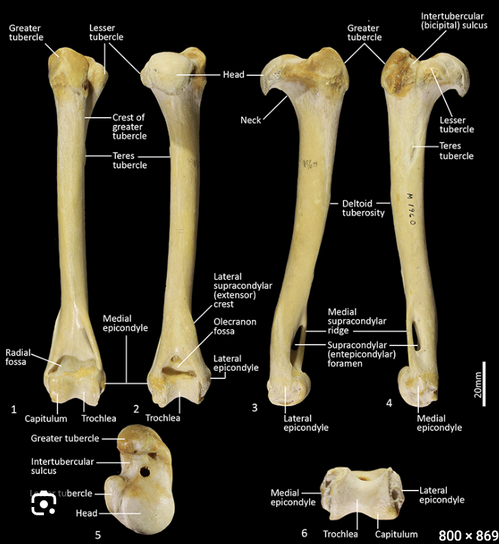
canine greater tubercle of humerus

canine lesser tubercle of humerus
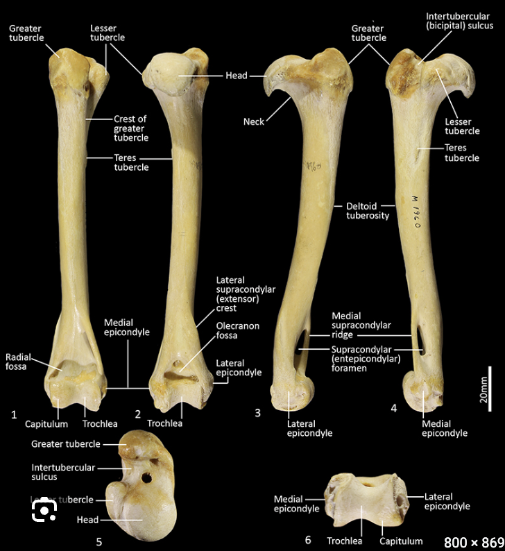
canine intertubercular groove of humerus
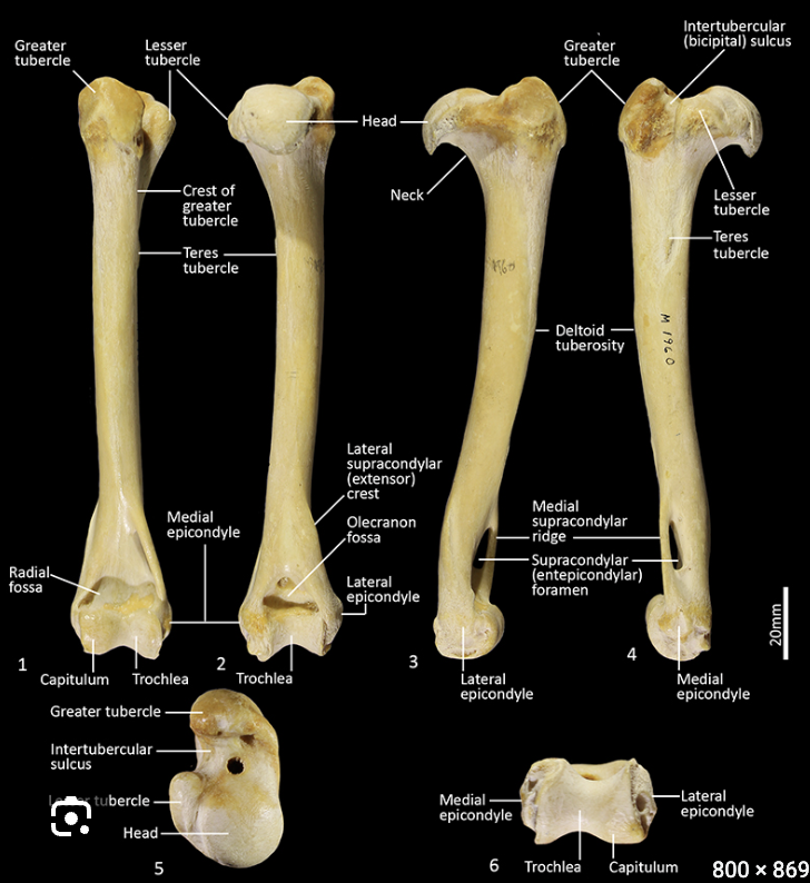
canine tricipital line
on same side as greater tubercle

canine deltoid tuberosity
right under tricipital line, on same side as greater tubercle
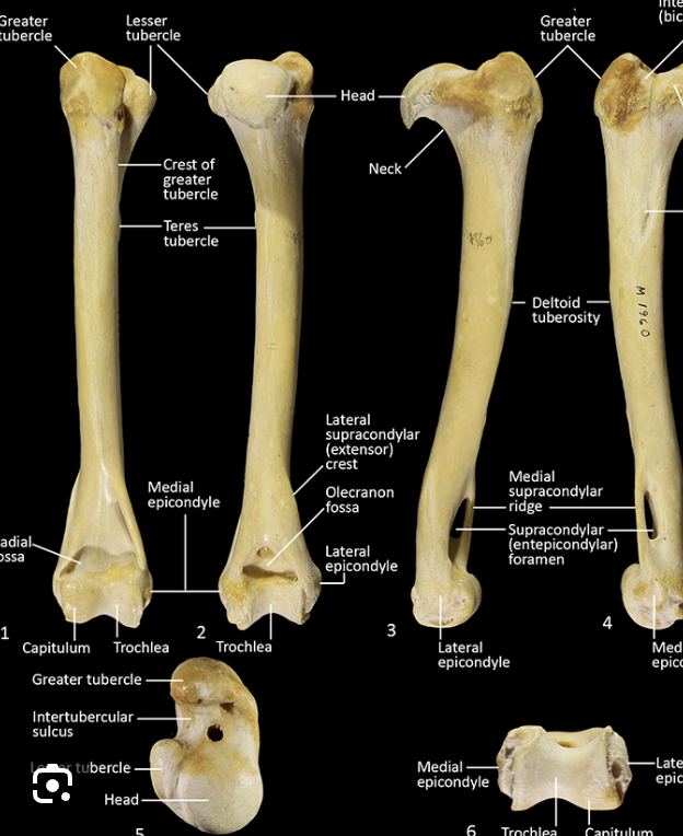
canine humeral condyle
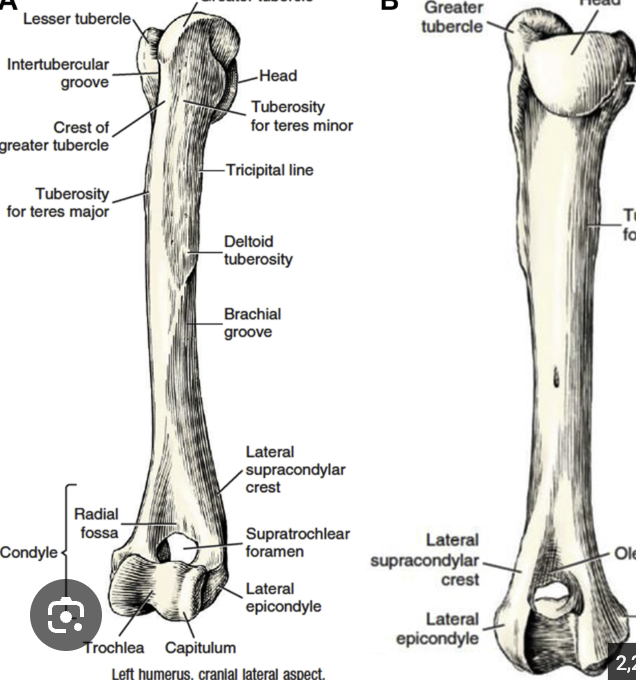
canine trochlea of humerus
same side as head

canine capitulum of humerus
same side as greater tubercle
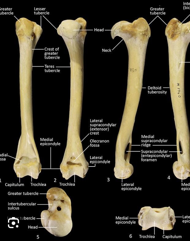
canine lateral and medial epicondyles
medial- on head side
lateral- on greater tubercle side
canine brachial groove of humerus
right under deltoid tuberosity
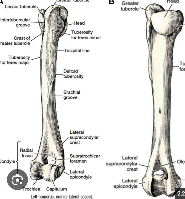
canine lateral supracondylar crest of humerus
same side as greater tubercle
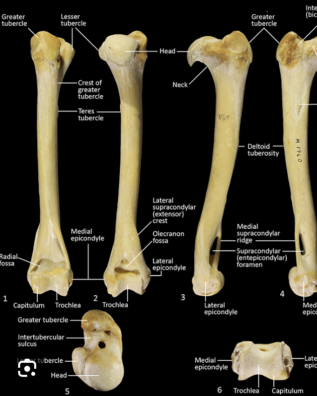
canine supratrochlear foramen of humerus
feline supracondylar foramen of humerus

canine head of radius
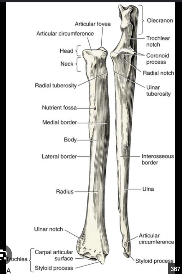
canine neck of radius
canine styloid process of radius
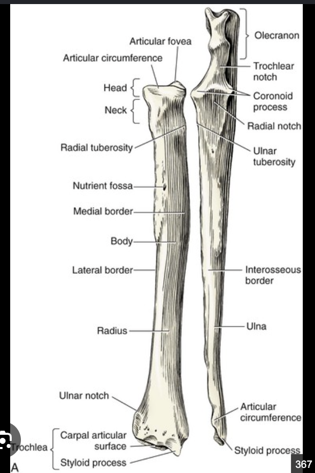
canine ulnar notch of radius
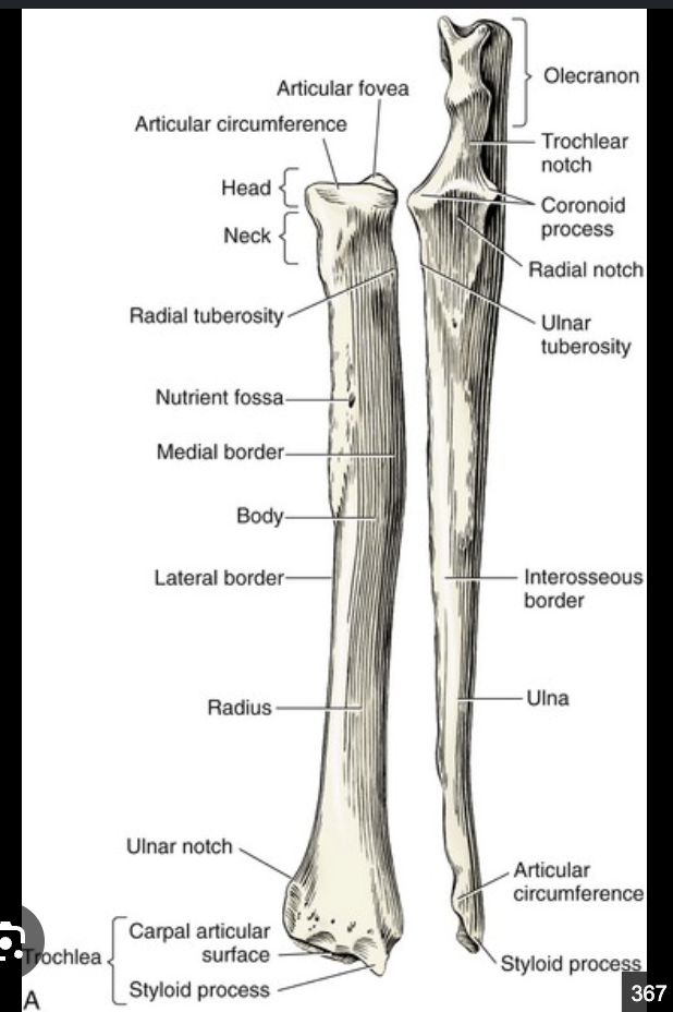
canine olecranon of ulna
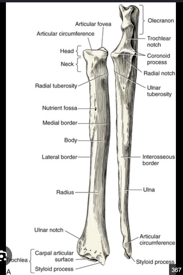
canine tuber olecrani of ulna
very top tip of olecranon

canine trochlear notch of ulna
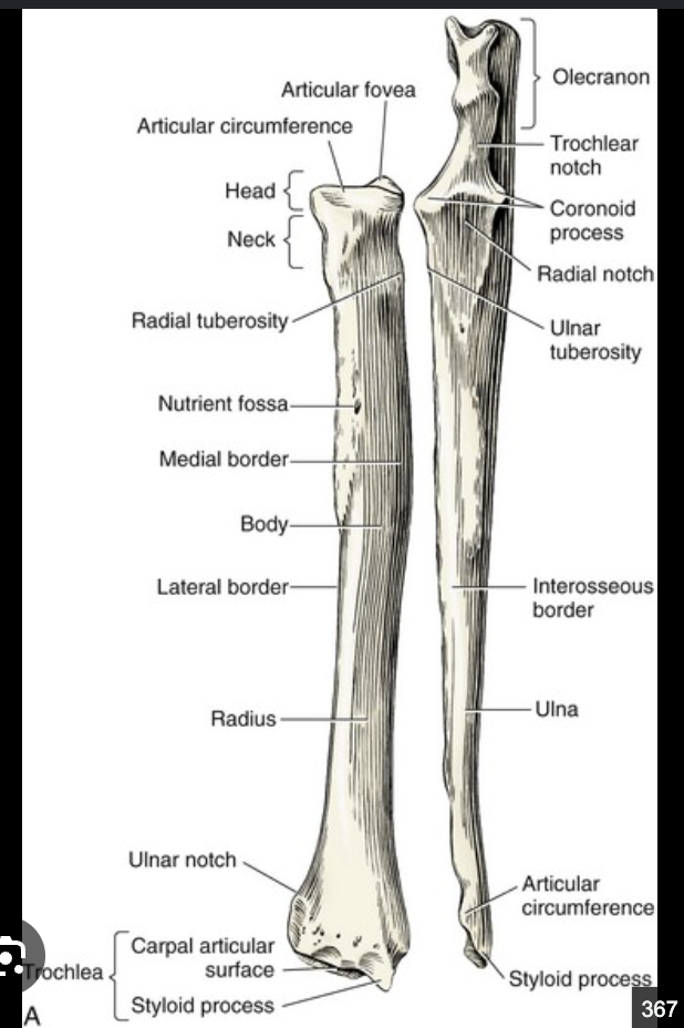
canine anconeal process of ulna
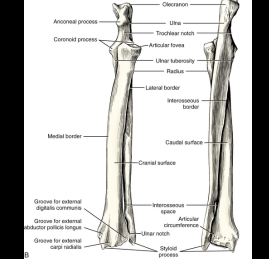
canine medial and lateral coronoid processes of ulna
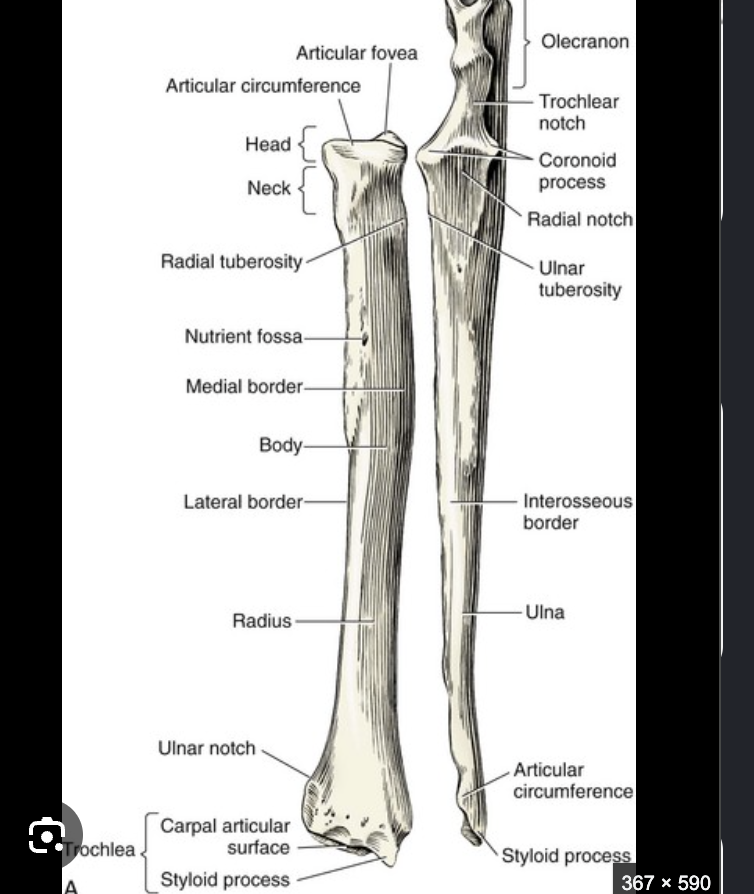
canine radial notch of ulna
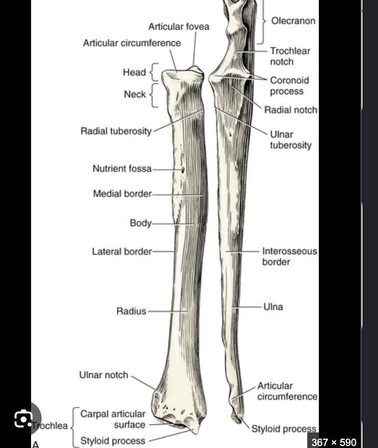
canine styloid process of ulna

what is the joint called between the antebrachium and the carpus
antebrachiocarpal joint
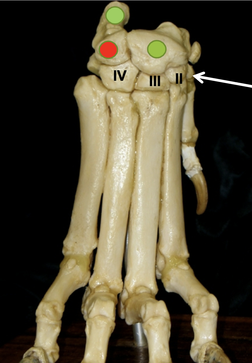
Name the bones of the carpus
Top left green: Accessory (always lateral)
Red: Ulnar
Right green: Intermedioradial
from white arrow left (medial to lateral): Carpal bone I, II, III, IV
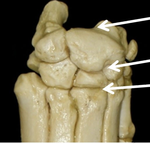
Name the joints pointed by the arrows
Top: Antebarchiocarpal joint
Middle: Carpal joint
Bottom: Carpometacarpal joint
Metacarpal bones are numbered from _
Medial to lateral
I-V
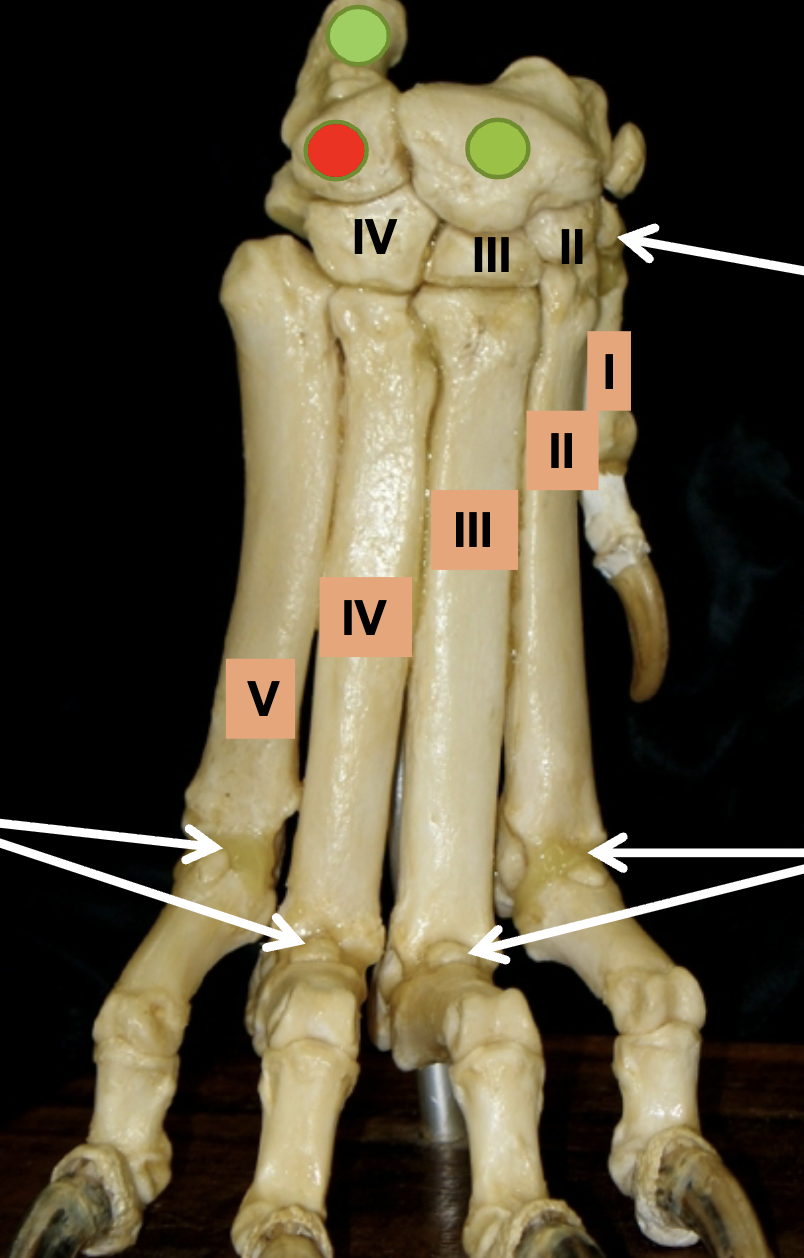
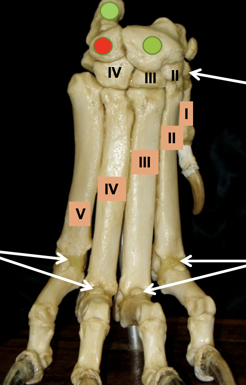
Name the joint of the metacarpals (bottom arrows)
Metacarpophalangeal joints
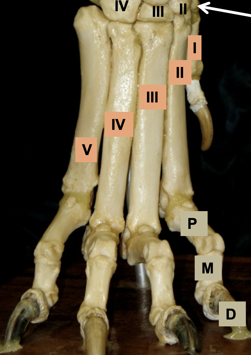
Name the joints of the phalanges
digit one has no middle
P: proximal interphalangeal
M: middle interphalangeal
D: distal interphalangeal
Feline dewclaw
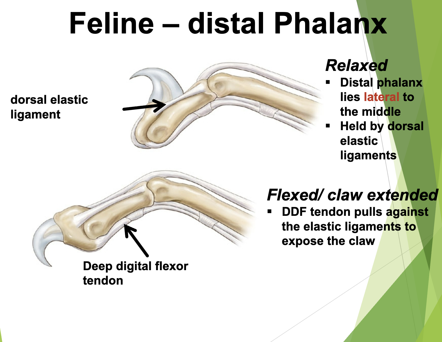
Flexor angles

Ionizing vs Nonionizing radiation
Ionizing: short wavelength, higher energy (DNA damage) ex: XRAY
Nonionizing: longer wavelength, lower energy
What happens when X-Ray strikes tissue
absorbed, scattered, or pass through (dark)
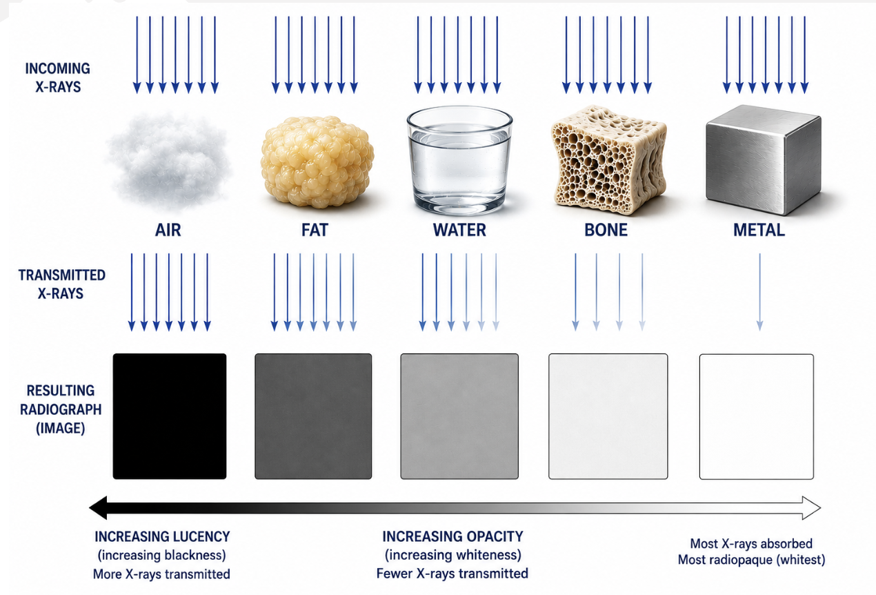
what are the different radiographic opacities
Air, fat, water, bone, metal
can only see margins if beside different opacity
What is radiographic summation
when images are superimposed they add together resulting in a mixed color
What is radiographic border effacement
when two structures of the same radiographic opacity are in contact, the margins cannot be distinguished
How are radiographic views named
for the direction the xray beam is traveling through the patient
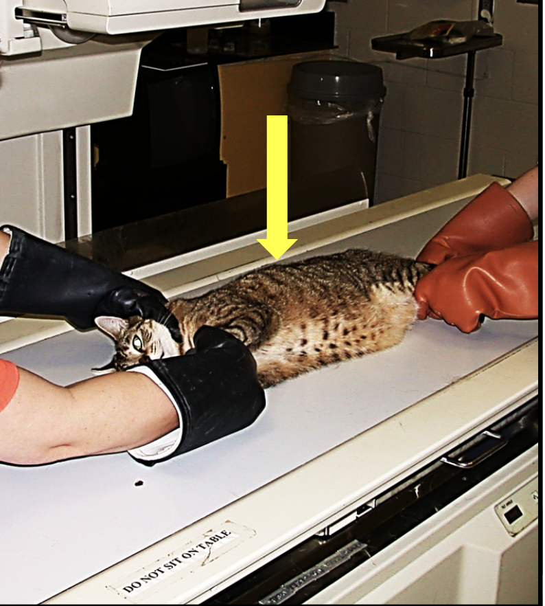
what is the name of this xray
right lateral
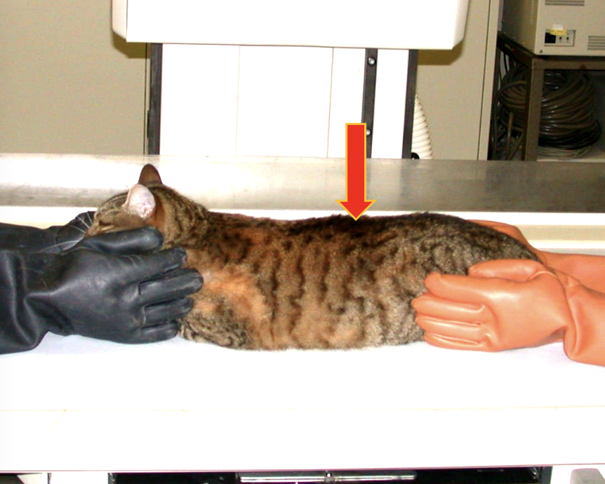
what is the name of this xray
dorsoventral
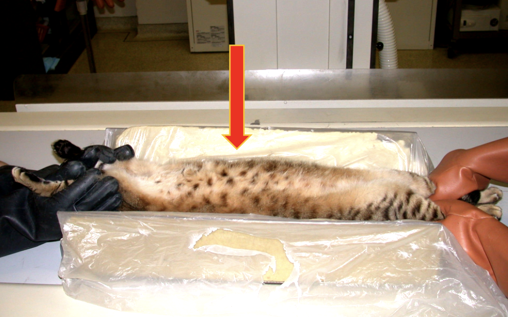
what is the name of this xray
ventrodorsal
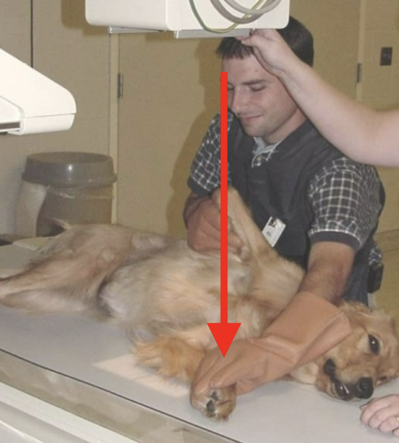
what is the name of this xray
lateral
what are the Roentgen signs
location, margination, number, opacity, shape, size
add xray flashcards