Oral radiology: Radiographic Properties
 Call Kai
 Learn
Learn Practice Test
 Spaced Repetition
 Match
Match Flashcards
 Knowt Play
1/29

There's no tags or description
Looks like no tags are added yet.
Name | Mastery | Learn | Test | Matching | Spaced | Call with Kai | Chat |
|---|
No analytics yet
Send a link to your students to track their progress
30 Terms
what are photographic proproperties
density
control
influence
contrast
control
influence
Factors affecting photographic properties
What are geometric properties
Factors affecting geometric properties
What does density mean and its types
Density = degree of darkinging on readiograph
Radiolucent = dark are on image
created by more exposure of film/sensor ← radiaton passing through less dense anatomy
Radiopaque = light are on image
radion absorbed by dense anatomy → film is less exposed
How well does radioation pass through: Metal, Air, Muslce, Bone?
Darkest/Most exposure/ less dense anatomy → Lightest/least exposure/less dense anatomy
Air (Radiolucent))→ Muscle → Bone →Metal (Radioopauqe)
Factors that can effect image density
Quantity of photons that interact with image
Controlled by milliamperage-seconds (mAs)
more mA→ more photons → more density
xray beam intensity is proportional to mA = more mA→ stronger beam
more time photon hit → more density
Influenced by kVp, collimation, FFD, filtration, patient factors
Denisty relationship to kVp, mA, exposure time, collimation
more kVP = more density
more mA = more Density
more time = more Density
shape beam => reduce fog => decrease density
What happens as kVp increases to its peak voltage value
xray intensity increases
slowly at first then it speeds up as it reaches its peak
Inverse square law
xray beam relationship is inverse to the square of distace betweeb target
How does Filtration and patient factors influence density
Filtraton= decrese density by removing low energy photons → less quantity of photons
Patient factors
thicker the person → more beam attenuated (beam loose intensity as it moves through matter) →lighter image (more white)
denser person = dense structure in subject → greater attenuation of xray beam → lighter image
Patient factors to note for xrays
Anatomic location
Anterior, posterior, maxillary molar
Age
>50 years or <10 years
size
Large or small
Oral Status
Edentulous or dentate
for these things adjust mA, leace kVp alone
What is constrast? types of contrast?
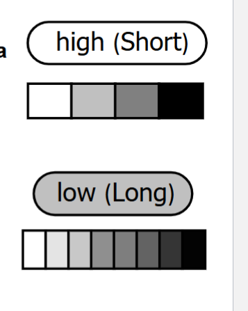
range or diffrence of density between light and dark regions on radiograph
High (short gray scale)
reduced range of grays between black and white on image and major diffrences
black and white image
it will be WHIIIITE AND BLAAAACK image
Low (long gray scale)
vast range of grays between black and white
gray image with slight diffences
what effects contrast
Controled by kVp
influenced by
Collinmation
Filtration
Patient Factors
contrast relationship between kVp, Collimation, filtration, patient factors
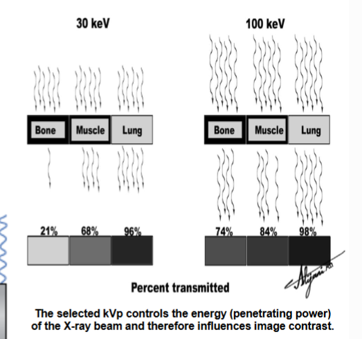
More kVp => stronger beam=> more penetrating power => less contrast
Has inverse relationship
Collimation shapes xray beam → reduces fog→ increases contrast
filtration → remove longer weaker waves → stronger beam→ decreases contrast
Patient Factors that effect contrast
variation of Thickness across body parts
Variation of tissue density(mass)
Effective atomic number: higher atomic # => Higher Contrast
ex: amalgam vs tooth structure
the subject contrast is proportional to amount of rays leaving adjacent section of subject]- what
What effect if any does the following (when increased) have on density?: mA, kVp,FFD, Collimation,Filtration
mA: increases
kVp: increases
FFD: decreases
Collimation: decreases
Filtration: decresses
What effect if any does the following (when increased) have on contrast?: mA, kVp,FFD, Collimation,Filtration
mA: n/a
kVp: decreases
FFD: n/a
Collimation: increases
Filtration: decresses
Image Sharpness
how defined (or good) is a boundary between areas of diffrent densities
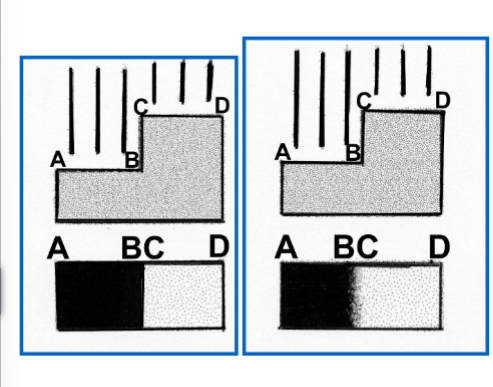
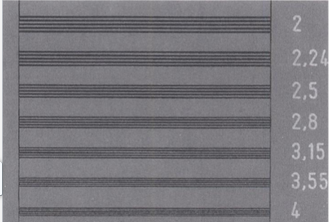
image Spatial Resolution
How well will small objects that are close tgr be revealed
Penumbra
Photons coming from different spots on the focal spot → zone of unsharpness on radiograph
smaller penumbra = sharper image
usally are created when hit edge of object
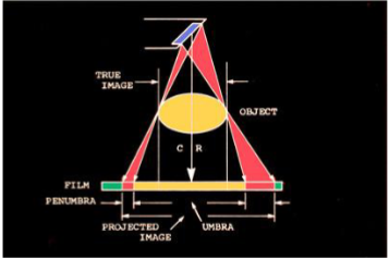
Componets of Image definition
image sharpness
image resolution
Factors that effect image definition
Controlled by focal spot size, focal spot to object distance, object to film distance
influenced by ;
motion
kVp & mA
screens
patients
image receptor
technique
3 ways to manipulate focal spot to control Penumbra
Spot size
use effective small focal spot and small angle to generate a small penumbra
Focal spot to object distance
Increase destance between focal spot and object with a long open ended cylinder; longer distance = smaller pneumbra
object to film distance
decrease distance between object and /film/sensor; decrese distace =
*These are actually the first 3 principles in order
Main influence of Image definition
Motion (tube, object, sensor)
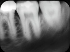
Types of Distortion
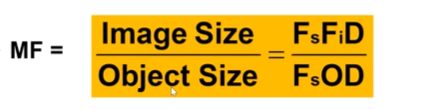
Size: magnification of object (image object bigger than actual object)
controlled by FFD (focal to film distance) and object to film distance (OFD)
when calculating top gonna always be a larger number than the bottom
Shape: magnification of different parts of objects unequally
so make sure to line the object up carefully
Types of shape distortion:
Dimensinal: 3D object to 2D media
Elongation: object looks longer on fim then irl
Foreshortening: object looks shorter on film than irl
*principle 4 : film needs to be parallel to long axis of object
*Principle 5: Central ray needs to be perpendicular to object AND film
Elongation and foreshortening caused by what
Elongation: ray is perpendicular to object but not to film
Foreshortening: film perpendicular to film but not to object
Shape techniques that effect distortion
Bisecting Angel:
put film as close to teeth as possible
perpendicularly angle cenral ray bisects the angle between the teeth and the film/sensor.
Paralleling Technique
place film paraelle to long axis of tooth
will lesses distortion
usual method
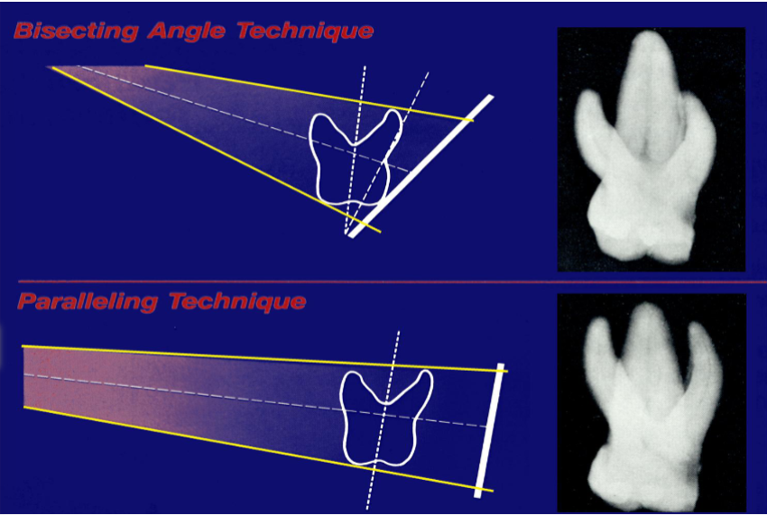
What are the 5 Principles
Use as small an effective focal spot as practical
Increase the distance between the focal spot and the object by using a long, open-ended cylinder
Minimize the distance between the object and the film
Position the film/sensor parallel to the long axis of the
object
Orient the central ray perpendicular to the object and
film/sensor.
Properties of radiographic image
Density: mAs, kVp
Contrst: kVp
Magnification/Unsharpness: Focal Spot, FFD,OFD
Shape distortion: Alignment