Anatomy Test 1 Review
 Call Kai
 Learn
Learn Practice Test
 Spaced Repetition
 Match
Match Flashcards
 Knowt Play
1/157

There's no tags or description
Looks like no tags are added yet.
Name | Mastery | Learn | Test | Matching | Spaced | Call with Kai | Chat |
|---|
No analytics yet
Send a link to your students to track their progress
158 Terms
Organ Level
The __________ of organization that includes a discrete structure made of more than one tissue type acting as a functional center.
A. Chemical Level
B. Organ Level
C. Cellular Level
D. Tissue Level
Spinal Cord
Choose all that apply. The vertebral cavity contains of which of the following organs?
A. Brain
B. Heart
C. Lungs
D. Inferior organs, such as bladder, rectum, reproductive organs, etc.
E. Great vessels (aorta and vena cavae), esophagus, and trachea
F. Spinal cord
G. Superior organs, such as stomach, liver, kidneys, etc.
Chemical Level
The __________ of organization includes small molecules, such as carbon dioxide, water, carbohydrates, and proteins.
A. Tissue Level
B. Organ Level
C. Cellular Level
D. Chemical Level
Nervous
_________ tissue type conducts electrical impulses to communicate bodily control.
A. Connective
B. Muscle
C. Nervous
D. Epithelial
Muscle
_________ tissue type is responsible for movement and maintenance of posture
A. Muscle
B. Epithelial
C. Nervous
D. Connective
Abdominal cavity
Identify H.
A. Mediastinum cavity
B. Pelvic cavity
C. Pleural cavity
D. Thoracic cavity
E. Pericardial cavity
F. Cranial cavity
G. Vertebral cavity
H. Abdominopelvic cavity
I. Abdominal cavity

Cellular Level
The __________ of organization that includes cells and their functional organelles.
A. Tissue Level
B. Chemical Level
C. Cellular Level
D. Organ Level
Metabolic rate is decreased
Which of the following is NOT a characteristic of neurons?
A. Mitosis or cell division does not occur
B. Longevity is increased
C. Metabolic rate is decreased
D. All of the above choices are characteristics of neurons
Stratified cuboidal epithelium
Identify the classification of epithelial tissue.
A. Stratified columnar epithelium
B. Simple columnar epithelium
C. Stratified squamous epithelium
D. Simple squamous epithelium
E. Stratified cuboidal epithelium
F. Pseudostratified columar epithelium
G. Stratified pseudostratified epithelium
H. Simple cuboidal epithelium

A. Areolar tissue
B. Adipose tissue
C. Reticular fibers
D. Dense regular connective tissue
E. Dense irregular tissue
F. Cartilage
Match the tissues below.
1. Dense irregular tissue
2. Reticular fibers
3. Cartilage
4. Dense regular connective tissue
5. Adipose tissue
6. Areolar tissue

Stratified columnar epithelium
Identify the classification of epithelial tissue.
A. Simple columnar epithelium
B. Simple cuboidal epithelium
C. Stratified cuboidal epithelium
D. Stratified squamous epithelium
E. Stratified columnar epithelium
F. Simple squamous epithelium
G. Pseudostratified columar epithelium
H. Stratified pseudostratified epithelium

Basal Lamina Layer - Superficial Layer
Reticular fiber layer - Deep Layer
The basment membrane of epithelial is composed of two main layers. Identify which layer is superficial and which layer is deep.
Basal Lamina Layer
Reticular fiber layer
A. Deep Layer
B. Superficial Layer
False
Adipose tissue is a highly metabolic tissue that has two different subtypes. White adipose tissue produces heat, and brown adipose tissue stores nutrients for energy.
A. True
B. False
Pseudostratified columnar epithelium
Identify the classification of epithelial tissue.
A. Simple squamous epithelium
B. Stratified squamous epithelium
C. Simple columnar epithelium
D. Pseudostratified columnar epithelium
E. Stratified pseudostratified epithelium
F. Stratified cuboidal epithelium
G. Stratified columnar epithelium
H. Simple cuboidal epithelium

True
The dermis layer is the second major layer of the integumentary system composed of vascularized connective tissue.
A. True
B. False
Acne
_______ occurs when a sebaceous gland becomes blocked by sebum.
A. Acne
B. Sebum
C. Holocrine secretion
D. Simple alveolar glands
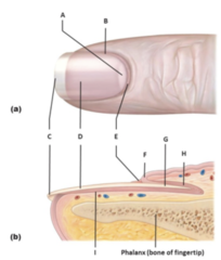
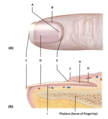
Nail Matrix
Identify the structure labeled H.
A. Eponychium (cuticle)
B. Free edge of nail
C. Nail bed
D. Lateral nail fold
E. Lunula
F. Body of nail
G. Nail matrix
H. Proximal nail fold
I. Root of nail

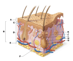
Papillary Layer
Identify the structure labeled C.
A. Dermis
B. Hair follicle
C. Epidermis
D. Hair root
E. Arrector pili muscle
F. Papillary layer
G. Hypodermis
H. Eccrine sweat gland
I. Reticular layer
J. Root nerve plexis
K. Sebaceous (oil) gland
Stratum basale
Identify epidermal layer E.
A. Stratum granulosum
B. Stratum basale
C. Stratum corneum
D. Stratum lucidum
E. Stratum spinosum

B. Keratinocytes produce antibodies and enzymes to protect the body against pathogens and harmful chemical exposures.
D. Keratinocytes are located in the stratum spinosum.
F. Keratinocytes are the most abundant cell type in the epidermis.
Choose all that apply. Which of the following statements are characteristics of keratinocytes?
A. Keratinocytes are begin to appear in the epidermis in the stratum basale.
B. Keratinocytes produce antibodies and enzymes to protect the body against pathogens and harmful chemical exposures.
C. Keratinocytes produce melanin pigments.
D. Keratinocytes are located in the stratum spinosum.
E. Keratinocytes are attached to disc-like sensory nerve endings.
F. Keratinocytes are the most abundant cell type in the epidermis.
Transverse
_______ body plane horizontally divides the body into inferior and superior halves.
A. Mid-sagittal
B. Frontal
C. Oblique
D. Transverse
Deep
Choose the correct anatomical directional term to fill in the blank.
Your stomach is ______ to your skin.
A. Superficial
B. Superior
C. Deep
D. Anterior
E. Inferior
F. Posterior
Medial
_______ means towards the middle of your body.
A. Ipsalateral
B. Medial
C. Contralateral
D. Lateral
Superior
Choose the correct anatomical directional term to fill in the blank.
Your head is ______ to your chest.
A. Proximal
B. Inferior
C. Deep
D. Superficial
E. Distal
F. Superior
B. Tissues can be defined as cells working together in functionally related communities.
C. Tissues can be defined as a combination of both living and non-living components.
D. Tissues can be defined as a collection of specialized cells and cell products.
Choose all that apply. Which of the following definition(s) of tissues is most accurate?
A. Tissues can be defined as a collection of cells operating independently of each other.
B. Tissues can be defined as cells working together in functionally related communities.
C. Tissues can be defined as a combination of both living and non-living components.
D. Tissues can be defined as a collection of specialized cells and cell products.
Not well vascularized
Which of the following is NOT a function of bone?
A. Not well vascularized
B. Supports hematopoiesis in the red bone marrow
C. Supports, moves, and protects the body
D. Highly resistant to compressive forces
True
Stratified epithelial tissues, like the epidermis, are named based on the cell shape of the apical layer. Therefore, the epidermis would be called stratified squamous epithelium.
A. True
B. False
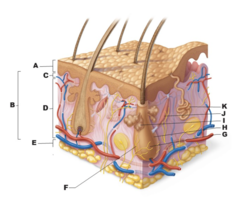
Hair root
Identify the structure labeled G.
A. Hair follicle
B. Sebaceous (oil) gland
C. Hypodermis
D. Arrector pili muscle
E. Reticular layer
F. Eccrine sweat gland
G. Epidermis
H. Root nerve plexis
I. Dermis
J. Papillary layer
K. Hair root
False
Melanin is the only pigment in a human's skin.
A. True
B. False
Hair follicle
Identify the structure labeled H.
A. Hypodermis
B. Sebaceous (oil) gland
C. Arrector pili muscle
D. Hair follicle
E. Papillary layer
F. Hair root
G. Reticular layer
H. Dermis
I. Root nerve plexis
J. Eccrine sweat gland
K. Epidermis

Eccrine sweat gland
Identify the structure labeled K.
A. Hair follicle
B. Dermis
C. Reticular layer
D. Epidermis
E. Sebaceous (oil) gland
F. Arrector pili muscle
G. Eccrine sweat gland
H. Papillary layer
I. Root nerve plexis
J. Hair root
K. Hypodermis

1. Tuberosity - D. Large rounded projection
2. Crest - B. Narrow prominent ridge of bone
3. Trochanter - E. Very large, blunt, and irregularly shaped process; only found on the femur
4. Line - C. Narrow but not prominent ridge of bone
5. Tubercle - A. Small rounded projection or process
Match the bone markings to their correct definitions.
1. Tuberosity
2. Crest
3. Trochanter
4. Line
5. Tubercle
A. Small rounded projection or process
B. Narrow prominent ridge of bone
C. Narrow but not prominent ridge of bone
D. Large rounded projection
E. Very large, blunt, and irregularly shaped process; only found on the femur
1. The region farthest from the marrow cavity that consists of resting hyaline cartilage. - E. Zone of reserve (resting)
2.The region of chondrocyte mitosis. - A. Zone of proliferation (multiplication)
3.The region where chondrocytes begin to get larger. - D. Zone of hypertrophy (enlargement)
4. The region where minerals are deposited in the matrix between lacunae - C. Zone of calcification
5. The region where chondrocyte death occurs leaving channels for vessel, osteoblast, and osteocyte invasion. - B. Zone of bone deposition
Match the following definitions to their correct zones of bone elogation.
1. The region farthest from the marrow cavity that consists of resting hyaline cartilage.
2.The region of chondrocyte mitosis.
3.The region where chondrocytes begin to get larger.
4. The region where minerals are deposited in the matrix between lacunae
5. The region where chondrocyte death occurs leaving channels for vessel, osteoblast, and osteocyte invasion.
A. Zone of proliferation (multiplication)
B. Zone of bone deposition
C. Zone of calcification
D. Zone of hypertrophy (enlargement)
E. Zone of reserve (resting)
Epicondyle - A. Raised area on or above a condyle
Spine - D. Sharp, slender, or pointed projection
Process - C. Any bony prominence
Head - E. Bony expansion carried on a narrow neck
Facet - B. Smooth, nearly flat articular surface
Match the bone markings to their correct definitions.
1. Epicondyle
2. Spine
3. Process
4. Head
5. Facet
A. Raised area on or above a condyle
B. Smooth, nearly flat articular surface
C. Any bony prominence
D. Sharp, slender, or pointed projection
E. Bony expansion carried on a narrow neck
Long bones
______ are longer than wide having a shaft and two ends. These bones can be found in all limb bones with exception of patellas, carpals, and tarsals.
A. Flat bones
B. Short bones
C. Irregular bones
D. Long bones
Inferior nasal concha
Identify the bone labeled K.
A. Temporal bone
B. Inferior nasal concha
C. Nasal bone
D. Lacrimal bone
E. Vomer bone
F. Maxilla bone
G. Parietal bone
H. Sphenoid bone
I. Mandible bone
J. Zygomatic bone
K. Ethmoid bone
L. Frontal bone

Vertebrochondral ribs
Identify the structure(s) labeled C.
A. False ribs
B. Vertebrochondral ribs
C. Xiphoid process
D. Sternum
E. Floating ribs (Vertebral ribs)
F. Manubrium
G. Body
H. True ribs (Vertebrosternal ribs)
I. Costal cartilage

Frontal bone
Identify the bone labeled B.
A. Lacrimal bone
B. Temporal bone
C. Maxilla bone
D. Parietal bone
E. Mandible bone
F. Ethmoid bone
G. Nasal bone
H. Inferior nasal concha
I. Frontal bone
J. Sphenoid bone
K. Vomer bone
L. Zygomatic bone

Connective
_____ tissue type forms the basis of organs and binds organs together.
A. Muscle
B. Connective
C. Nervous
D. Epithelial
Simple cuboidal epithelium
Identify the classification of epithelial tissue.
A. Stratified cuboidal epithelium
B. Simple cuboidal epithelium
C. Simple squamous epithelium
D. Stratified squamous epithelium
E. Stratified columnar epithelium
F. Simple columnar epithelium
G. Pseudostratified columnar epithelium
H. Stratified pseudostratified epithelium

True
Reticular tissue is similar to areolar tissue but contains reticular fibers that form caverns to hold maturing cells
A. True
B. False
Well vasularized
Which of the following is NOT a characteristic of dense regular connective tissue?
A. Wavy, regular appearance
B. There is no adipose tissue or immune cells
C. Well vascularized
D. Can be found in fascia, tendons, and ligaments
True
The irregular patterning of fibers in dense irregular connective tissue allows this tissue type to resist strong tensions coming from multiple different directions.
A. True
B. False
Simple squamous epithelium
Identify the classification of epithelial tissue.
A. Simple squamous epithelium
B. Stratified columnar epithelium
C. Stratified cuboidal epithelium
D. Stratified pseudostratified epithelium
E. Simple columnar epithelium
F. Pseudostratified columnar epithelium
G. Stratified squamous epithelium
H. Simple cuboidal epithelium

Stratum corneum
____ is the external epidermis layer and is composed of dead keratinocytes.
A. Stratum corneum
B. Stratum basale
C. Stratum spinosum
D. stratum lucidum
E. Stratum granulosum
True
A sudoriferous gland is commonly called a sweat gland and is widely distributed on the human body.
A. True
B. False
A. Thoracic cavity
B. Mediastinum cavity
C. Pleural cavity
D. Abdominal cavity
E. Peritoneal cavity
F. Pelvic cavity
I. Pericardial cavity
J. Abdominopelvic cavity
Choose all that apply. The ventral body cavity contains of which of the following cavities?
A. Thoracic cavity
B. Mediastinum cavity
C. Pleural cavity
D. Abdominal cavity
E. Peritoneal cavity
F. Pelvic cavity
G. Vertebral cavity
H. Cranial cavity
I. Pericardial cavity
J. Abdominopelvic cavity
Inferior
Choose the correct anatomical directional term to fill in the blank
Your pelvis is _____ to your shoulders
Lungs
Choose all that apply. The pleural cavities contains of which of the following organs?
A. Heart
B. Brain
C. Superior organs, such as stomach, liver, kidneys, etc.
D. Great vessels (aorta and vena cavae), esophagus, and trachea
E. Spinal cord
F. inferior organs, such as bladder, rectum, reproductive organs, etc.
G. Lungs
Distal
Choose the correct anatomical directional term to fill in the blank.
Your knee is _____ to your thigh.
A. Deep
B. Distal
C. Proximal
D. Inferior
E. Superior
F. Superficial
Anterior
Choose the correct anatomical directional term to fill in the blank.
Your navel (belly button is found on the ______ part of your body.
A. Posterior
B. Superior
C. Superficial
D. Deep
E. Inferior
F. Anterior
A. Anus
B. Urethra
C. Vagina
D. Mouth
E. Esophagus
Choose all that apply. Where would one find non-keratinized stratified squamous epithelium?
A. Anus
B. Urethra
C. Vagina
D. Mouth
E. Esophagus
F. Arm
A. Cells have a mesenchymal (embryonic) origin
C. Well vascularized tissue
D. Responsible for secreting the extracellular matrix
E. Very few cells in tissue
Choose all that apply. Which of the following are characteristics of connective tissue?
A. Cells have a mesenchymal (embryonic) origin
B. Large number of cells in tissue
C. Well vascularized tissue
D. Responsible for secreting the extracellular matrix
E. Very few cells in tissue
F. Cells have an apical-basal surface polarity
G. Highly innervated tissue
A. Store ions, like calcium, and organic salts, like phosphate
D. Form structural elements of the body such as the skeleton
E. Hold body fluid within the ground substance
F. Mount immune responses
G. Connect tissues and organs together
Choose all that apply. Which of the following are functions of connective tissue?
A. Store ions, like calcium, and organic salts, like phosphate
B. Act as endocrine glands to secrete hormones
C. Serve as a protective interface between differing environments
D. Form structural elements of the body such as the skeleton
E. Hold body fluid within the ground substance
F. Mount immune responses
G. Connect tissues and organs together
H. Maintain core body temperatures by shivering
B. Very mitotic
E. Possessing cellular polarity
F. Having cellularity
G. Avascular
H. Highly innervated
Choose all that apply. Which of the following characteristics define epithelial tissue?
A. Mainly composed of extracellular matrix
B. Very mitotic
C. Lacking of specialized contacts
D. Highly vascularized
E. Possessing cellular polarity
F. Having cellularity
G. Avascular
H. Highly innervated
I. Acellular
Body of nail
Identify the structure labeled D.
A. Lateral nail fold
B. Lunula
C. Free edge of nail
D. Nail matrix
E. Proximal nail fold
F. Eponychhium (cuticle)
G. Nail bed
H. Body of nail
I. Root of nail

D. Stratum corneum
Identify epidermal layer A.
A. Stratum spinosum
B. Stratum basale
C. Stratum lucidum
D. Stratum corneum
E. Stratum granulosum

Free edge of nail
Identify the structure labeled C.
A. Lateral nail fold
B. Nail bed
C. Lunula
D. Root of nail
E. Proximal nail fold
F. Body of nail
G. Free edge of nail
H. Eponuchiu (cuticle)
I. Nail matrix

Lateral
______ means away from the middle of your body
A. Lateral
B. Ipsilateral
C. Medial
D. Contralateral
Vertebral cavity
Cranial cavity
Choose all that apply. The dorsal body cavity contains of which of the following cavities.
A. Abdominal cavity
B. Pericardial cavity
C. Pleural cavity
D. Thoracic cavity
E. Pelvic cavity
F. Peritoneal cavity
G. Abdominopelvic cavity
H. Vertebral cavity
I. Mediastinum cavity
J. Cranial cavity
Well vascularized
Which of the following is NOT a characteristic of cartilage?
A. Well vascularized
B. Firm, flexible, and resistant to compressions
C. Can be found in the ears, nose, and trachea of adults
D. Not innervated
High cellularity
Well vascularized
Choose all that apply. Blood has a number of characteristics that are different from those of connective tissue. Which of the following characteristics of blood are exceptions to the connective tissue characteristics?
A. High cellularity
B. Mesenchymal origins
C. Surrounded by a non-living plasma matrix
D. Well vascularized
Dermis
Identify the structure labeled B.
A. Hair root
B. Hair follicle
C. Dermis
D. Eccrine sweat gland
E. Hypodermis
F. Root nerve plexus
G. Reticular layer
H. Papillary layer
I. Epidermis
J. Arrector pilli muscle
K. Sebaceous (oil) gland

Nail bed
Identify the structure labeled I.
A. Lateral nail fold
B. Body of nail
C. Nail bed
D. Proximal nail fold
E. Lunula
F. Eponuchium (cuticle)
G. Free edge of nail
H. Nail matrix
I. Root of nail
Stratum spinosum
____ is the epidermal layer containing maturing keratinocytes, Langerhans cells, and melanocytes.
A. Stratum spinosum
B. Stratum granulosum
C. Stratum corneum
D. Stratum basale
E. Stratum lucidum
Stretch tension
_____ is the pulling forces applied to the exterior side of bone.
A. Stretch tension
B. Compression
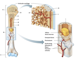
Distal epiphysis
Identify the structure labeled C.
A. Endosteum
B. Proximal epiphysis
C. Spongy bone
D. Distal epiphysis
E. Epiphyseal line
F. Diaphysis
G. Compact bone
H. Periosteum
I. Medullary cavity
Foramen- round or oval opening through a bone
Fissure- narrow slit-like opening in the bone
Epicondyle- raised area on or above a condyle
Condyle- round articular projection
Fossa- shallow basin-like depression in a bone
Match the bone markings to their correct definitions.
1. Foramen
2. Fissure
3. Epicondyle
4. Condyle
5. Fossa
A. Narrow slit-like opening in the bone
B. Shallow basin-like depression in a bone
C. Round articular projection
D. Round or oval opening through a bone
E. raised area on or above a condyle
Compact bone
______ is the dense, solid outer layer of bone.
A. Spongy (Trabecular) bone
B. Compact Bone
Lamina
Identify the vertebral structure labeled H.
A. Vertebral foramen
B. Lamina
C. Transverse process
D. Articular process and facet
E. Body (centrum)
F. Pedicle
G. Spinous process
H. Vertebral arch

Vomer bone
Identify the bone labeled E.
A. Maxilla bone
B. Sphenoid bone
C. Foramen magnum
D. Zygomatic bone
E. Palatine bone
F. Temporal bone
G. Vomer bone
H. Petrous region of temporal bone
I. Maxilla bone
J. Parietal bone

Parietal bone
Identify the structure labeled B.
A. Mandible
B. Temporal bone
C. Lambdoid suture
D. Nasal bone
E. Frontal bone
F. Squamous suture
G. Occipital bone
H. Parietal bone
I. Sphenoid bone
J. Ethmoid bone
K. Coronal suture
L. Maxilla
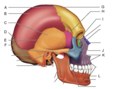
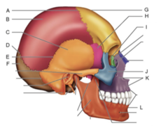
Ethmoid bone
Identify the structure labeled I.
A. Nasal bone
B. Ethmoid bone
C. Frontal bone
D. Temporal bone
E. Squamous suture
F. Sphenoid bone
G. Lambdoid suture
H. Mandible
I. Parietal bone
J. Occipital bone
K.Maxilla
L. Coronal suture
Brain
The cranial cavity contains which of the following organs?
A. Lungs
B. Brain
C. Inferior organs, such as bladder, rectum, reproductive organs, etc.
D. Spinal cord
E. Superior organs, such as stomach, liver, kidneys, etc.
F. Heart
G. Great vessels, esophagus, and trachea
Heart
Choose all that apply, The pericardial cavity contains which of the following organs?
A. Brain
B. Spinal cord
C. Heart
D. Inferior organs
E. Lungs
F. Great vessels, esophagus, and trachea
G. Superior organs
Abdominopelvic cavity
Identify B
A. Thoracic cavity
B. Pericardial cavity
C. Abdominopelvic cavity
D. Cranial cavity
E. Pelvic cavity
F. Pleural cavity
G. Abdominal cavity
H. Mediastinum cavity
I. Vertebral cavity

Mediastinum cavity
Identify E
A. Thoracic cavity
B. Pericardial cavity
C. Abdominopelvic cavity
D. Cranial cavity
E. Pelvic cavity
F. Pleural cavity
G. Abdominal cavity
H. Mediastinum cavity
I. Vertebral cavity

Neuron
Which of the following is NOT a neuroglial/glial cell type?
A. Astrocytes
B. Neuron
C. Oligodendrocytes
D. Schwann cells
False
Lacuna are sites were immature osteoblasts or chondroblasts are found.
A. True
B. False
True
Areolar tissue is one of the most widespread tissues and can be found underlying the epithelium, surrounding small blood vessels and nerves, and borders of most tissues in the body.
A. True
B. False
1. Areolar connective tissue - Loose connective tissue
2. Adipose tissue - Loose connective tissue
3. Reticular fibers - Loose connective tissue
4. Dense irregular tissue - Dense connective tissue
5. Dense regular - Dense connective tissue
6. Elastic fibers - Dense connective tissue
Place the following types of connective tissue proper into their correct subclasses.
1. Areolar connective tissue
2. Adipose tissue
3. Reticular fibers
4. Dense irregular tissue
5. Dense regular
6. Elastic fibers
A. Dense connective tissue
B. Loose connective tissue
ABC- skeletal
DEF- cardiac
GHI- smooth
Match the following muscle characteristics to the correct type of muscle.
A. Composed of long, cylindrical bundles of fibers that are multinucleated
B. Striated and voluntarily controlled
C. Attached to bones, fascia, and skin
D. Composed of branching chains of cells
E. Cells are connected by intercalated discs
F. Striated and under involuntary control
G. Composed of uninucleated cells arranged in layers with opposing orientation
H. Can be found in the lining of hollow organs and blood vessels
I. Not striated and under involuntary control
1. skeletal
2. cardiac
3. smooth
A. The hypodermis anchors the integumentary system to the underlying structures
B. The hypodermis stores fat and helps insulate the body
C. The hypodermis expands with weight gain
E. The hypodermis contains areolar and adipose connective tissue
Which of the following statements are functional characteristics of the hypodermis.
A. The hypodermis anchors the integumentary system to the underlying structures
B. The hypodermis stores fat and helps insulate the body
C. The hypodermis expands with weight gain
D. The hypodermis is superficial to the skin
E. The hypodermis contains areolar and adipose connective tissue
F. The hypodermis is composed of squamous epithelium and adipose
Root of nail
Identify the structure labeled G
A. Root of nail
B. Lateral nail fold
C. Nail matrix
D. Free edge of nail
E. Proximal nail fold
F. Eponychium (cuticle)
G. Nail bed
H. Body of nail
I. Lunula
Sebaceous gland
Identify the structure labeled I
A. Epidermis
B. Root nerve plexus
C. Sebaceous gland
D. Hypodermis
E. Papillary layer
F. Hair follicle
G. Reticular layer
H. Dermis
I. Hair root
J. Eccrine sweat gland
K. Arrector pili muscle

Gap junctions
____ are located all along the lateral surface of cells and are made of hollow cylinders of proteins and allow small molecules to pass through.
A. Gap junctions
B. Integrins
C. Tight junctions
D. Desmosomes
E. Connexins
Arrector pili muscle
____ is the name of the muscle that when stimulated will cause hair to stand erect.
A. Arrector pili muscle
B. Goosebump flexor muscle
C. Horripilatory muscle
D. Erector muscle
Thick superficial epithelial layer - epidermis
Deep fibrous connective tissue layer - dermis
Fatty layer deep to the dermis - hypo
Match the following integumentary layers to their definition
A. Thick superficial epithelial layer
B. Deep fibrous connective tissue layer
C. Fatty layer deep to the dermis
1. Epidermis
2. Dermis
3. Hypodermis
Fibrocartilage, EF
Elastic cartilage, CD
Hyaline cartilage, AB
Match the following characteristics to the type of cartilage.
A. Most abundant cartilage that looks like frosted glass
B. Composed of collagen fibrils and provides support and flexibility
C. Composed of elastic fibers and tolerates repeated bending
D. Found in epiglottis and outer ear
E. A hybrid cartilage composed alternating collagen fibrils and rows of chondrocytes
F. Occurs in ligaments, intervertebral discs, and knee meniscus and resist strong compression and tension
1. Fibrocartilage
2. Elastic cartilage
3. Hyaline cartilage
Osteoclasts, GH
Osteoprogenitor cells, EF
Osteocytes, AB
Osteoblasts, CD
Match the following to the correct cell type (14)
A. Maintains and monitors proteins and mineral content of one matrix
B. Occupies lacunae located between calcified layers of bone matrix
C. Actively producing and secreting the bone matrix especially during osteogenesis
D. Located on the inner and outer surfaces of bone
E. Stem cells that differentiate to heal bond
F. Randomly dispersed throughout bone, but most concentrated at periosteum and endosteum
G. Responsible for breaking down and resorption of bone
H. Leukocyte derived cell capable of secreting hydrochloric acid and lysosomal enzymes
1. Osteoclasts
2. Osteoprogenitor cells
3. Osteocytes
4. Osteoblasts
A. Intervertebral disc
C. Ends of moveable bone
D. Trachea and bronchi
E. Ribs
F. External ear
G. Nose
Which of the following locations contains cartilage?
A. Intervertebral disc
B. Spongy bone trabeculae are too small to contain vessels
C. Ends of moveable bone
D. Trachea and bronchi
E. Ribs
F. External ear
G. Nose
Optic canal
____ and superior orbital fissure are structures on the sphenoid bone responsible for cranial eye nerves.
A. Foramen rotundum and foramen ovale
B. Foramen spinosum
C. Greater wings and lesser wings
D. Optic canal
True
The cervical vertebrae are the smallest, lightest, most flexible triangle vertebrae and are forked with the exception of C7 or the vertebra prominens.
A. True
B. Fasle
Foramen spinosum
____ are holes in the sphenoid bone where the meningeal artery for parietal and temporal bones pass through.
A. Optic canal
B. Foramen rotundum and foramen ovale
C. Greater wings and lesser wings
D. Foramen spinosum
Pleural cavity
Identify F.
A. Pelvic Cavity
B. Pericardial Cavity
C. Mediastinum Cavity
D. Thoracic Cavity
E. Cranial Cavity
F. Abdominopelvic Cavity
G. Abdominal Cavity
H. Vertebral Cavity
I. Pleural Cavity

Middle cranium
Identify the bone labeled B.
A. Middle cranium
B. Anterior cranium
C. Posterior cranium

Eccrine (Merocrine) sweat glands- ABC
Apocrine sweat glands- DEF
Match the following characteristics to either eccrine (merocrine) or apocrine sweat glands.
A. Called a true sweat gland and is the most numerous of sweat glands.
B. Most abundantly found on palms, soles of feet, and forehead.
C. Will most likely cause a sweaty handshake in a nervous situation.
D. Sweat glands confined to axillary, anal, and genital areas.
E. Larger sweat glands that open onto hair follicles to release a viscous substance,
F. Cause you to smell really funky!
1. Eccrine (Merocrine) sweat glands
2. Apocrine sweat glands
Root nerve plexus
Identify the structure labeled F
A. Epidermis
B. Root nerve plexus
C. Sebaceous gland
D. Hypodermis
E. Papillary layer
F. Hair follicle
G. Reticular layer
H. Dermis
I. Hair root
J. Eccrine sweat gland
K. Arrector pili muscle

Serous fluid: A
Parietal serosa: C
Visceral serosa: B
Match the following serous cavity components to their definition.
Answer Choices:
A. Watery Substance that fills the serous cavity
B. Inside Layer of the serous cavity
C. Outside layer of the serous cavity
1. Serous fluid
2. Parietal serosa
3. Visceral serosa
Pelvic cavity
Identify I
A. Pericardial cavity
B. Pelvic cavity
C. Abdominal cavity
D. Thoracic cavity
E. Pleural cavity
F. Abdominopelvic cavity
G. Vertebral cavity
