A&P Lab Practical 2
 Call Kai
 Learn
Learn Practice Test
 Spaced Repetition
 Match
Match Flashcards
 Knowt Play
1/200

Earn XP
Description and Tags
Structures → identify/define,; Muscles → identify + actions + innervation; Starred muscles → identify + actions + innervation + origins + insertions; Joint structures → identify/define; Nerves → identify + what they innervate; Actions → definition Directional terms → definition
Name | Mastery | Learn | Test | Matching | Spaced | Call with Kai | Chat |
|---|
No analytics yet
Send a link to your students to track their progress
201 Terms
Tibia – lateral condyle
The proximal end of the tibia is greatly expanded. The two sides of this expansion form the medial and lateral condyles of the tibia.
The superior surface of each condyle is smooth and flattened. These areas articulate with the medial and lateral condyles of the femur to form the knee joint.
anterior superior iliac spine
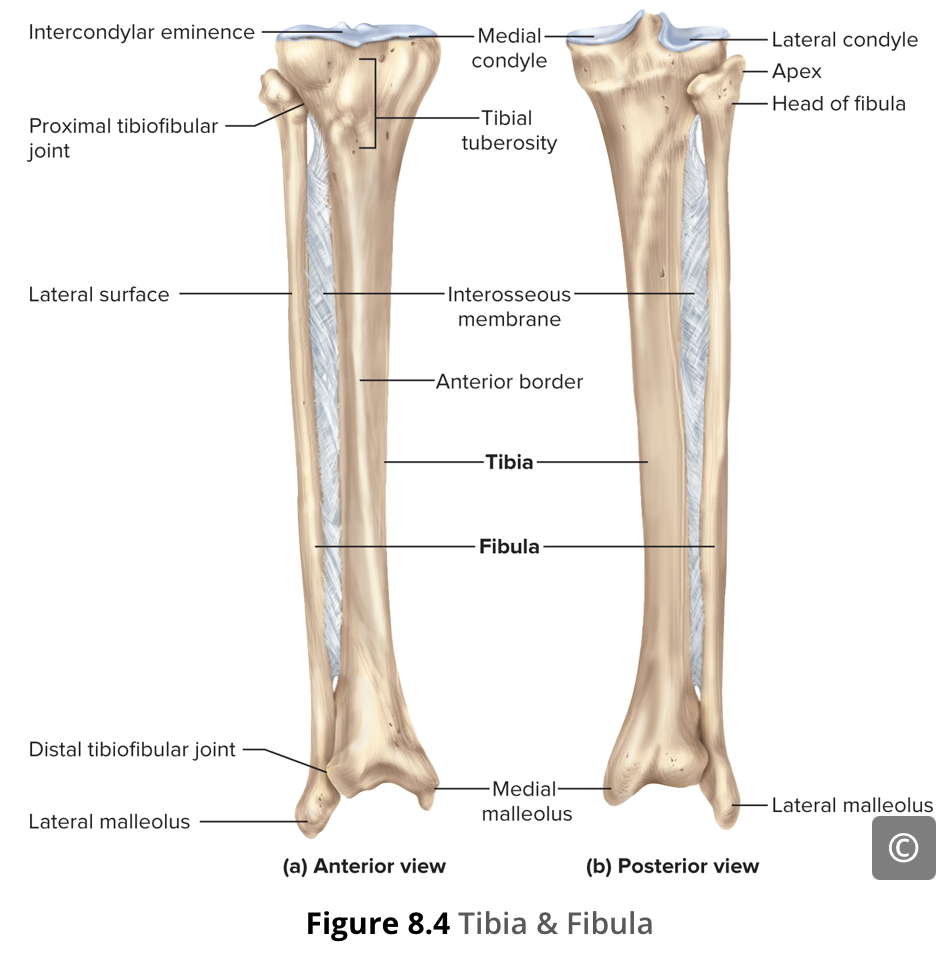
The rounded, anterior termination of the iliac crest, serve as attachment points for muscles of the thigh.

anterior inferior iliac spine
Inferior to the anterior superior iliac spine is a rounded protuberance, serve as attachment points for muscles of the thigh.

Coccyx
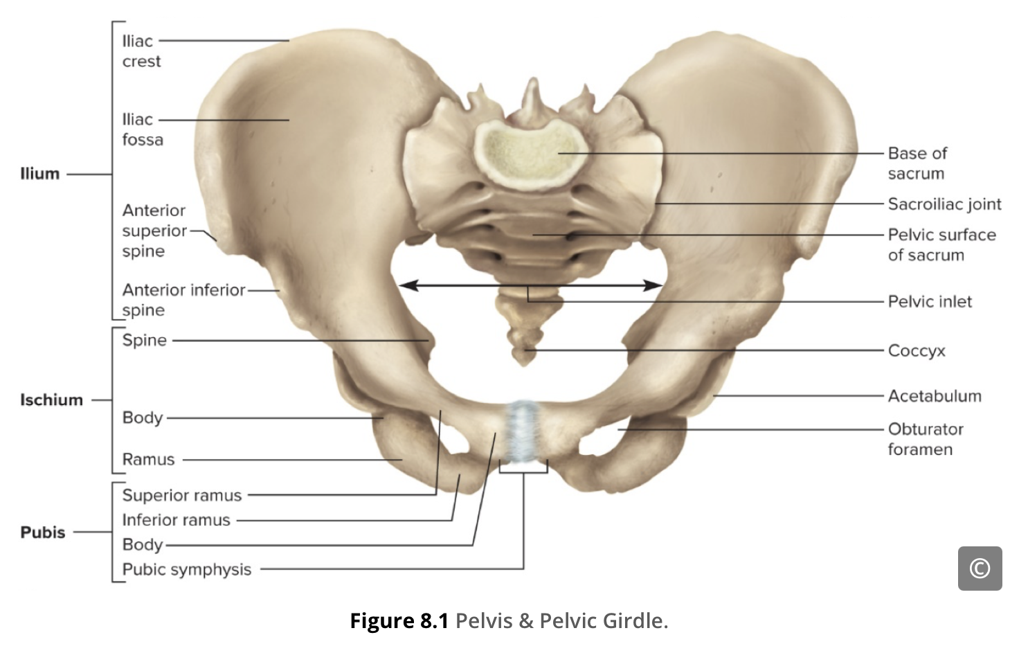
The bony pelvis is the entire structure formed by the two hip bones, the sacrum, and the coccyx. The coccyx, also known as the tail bone, is attached inferiorly to the sacrum (Figure 8.1).
Greater sciatic notch
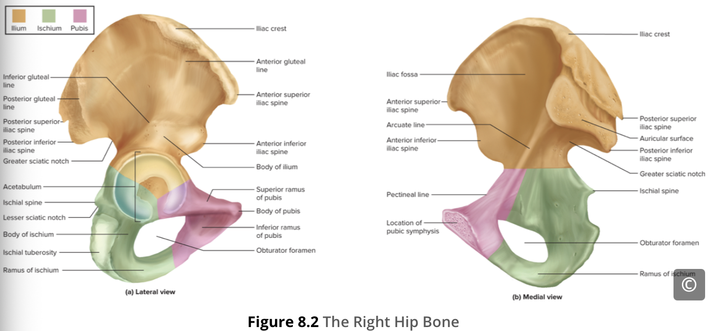
The large, inverted U-shaped indentation located on the posterior margin of the lower ilium
Iliac crest
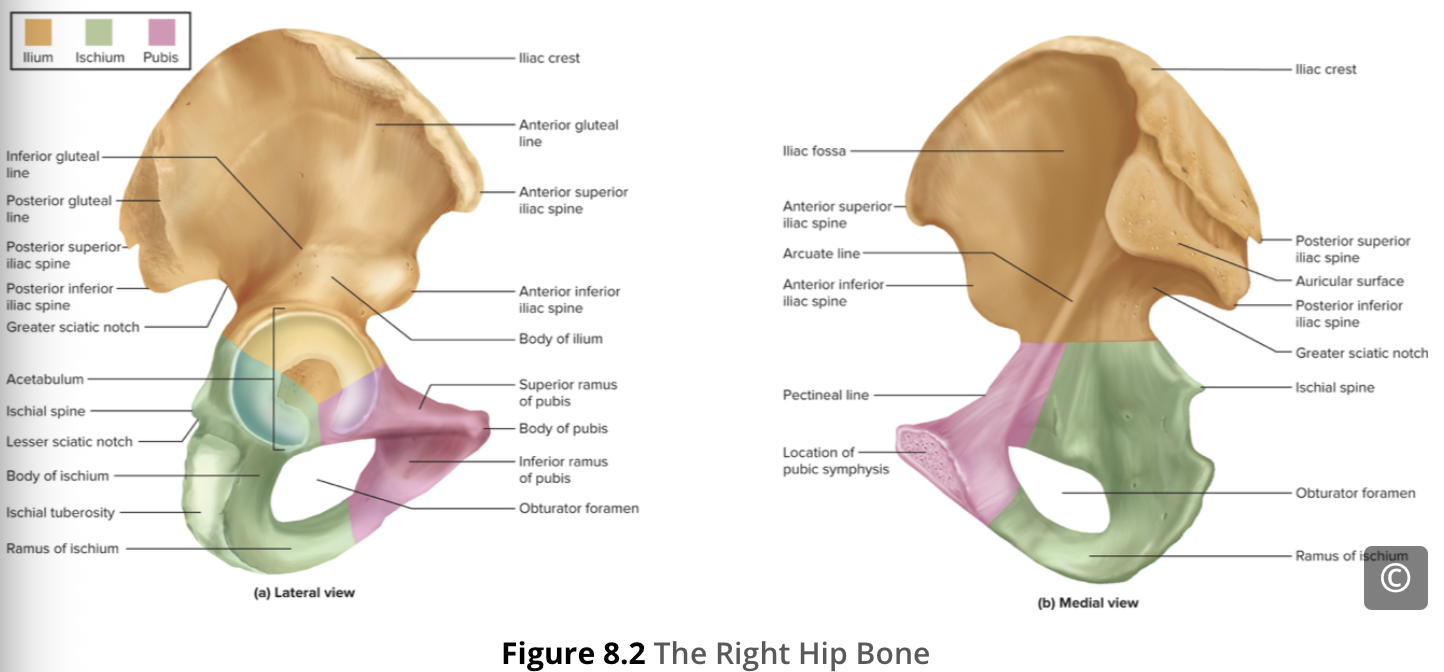
When you place your hands on your waist, you can feel the arching, superior margin of the ilium along your waistline (Figure 8.2). This curved, superior margin of the ilium is the iliac crest.

Iliac fossa
The shallow depression located on the anteromedial (internal) surface of the upper ilium

Ischial ramus
Projecting superiorly and anteriorly from the ischial tuberosity is a narrow segment of bone called the ischial ramus.
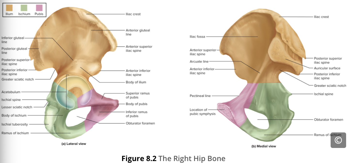
Ischial spine
The bony projection separating the lesser sciatic notch and greater sciatic notch

Ischial tuberosity
The large, roughened area of the inferior ischium. This serves as the attachment for the posterior thigh muscles and also carries the weight of the body when sitting.

Obturator foramen
a large, oval or triangular opening in the hip bone formed by the pubis and ischium bones

Os coxa – acetabulum
is the socket portion of the hip joint. This space is deep and has a large articulation area for the femoral head, thus giving stability and weight bearing ability to the joint.

Os coxa – ilium
The ilium is the fan-like, superior region of the hip bone forming the largest part of the hip bone. The ilium is firmly connected to the sacrum at the largely immobile sacroiliac joint (see Figure 8.1).

Os coxa – ischium
The ischium forms the posteroinferior region of each hip bone. It supports the body when sitting.

Os coxa – pubis
The pubis forms the anterior portion of the hip bone. The pubis curves medially, where it joins to the pubis of the opposite hip bone at a specialized joint called the pubic symphysis.
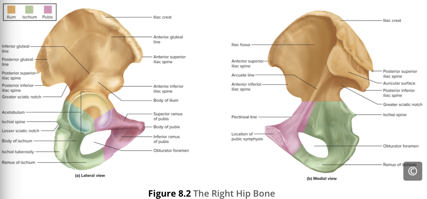
Pubic symphysis
joins the 2 hip bone pubic body
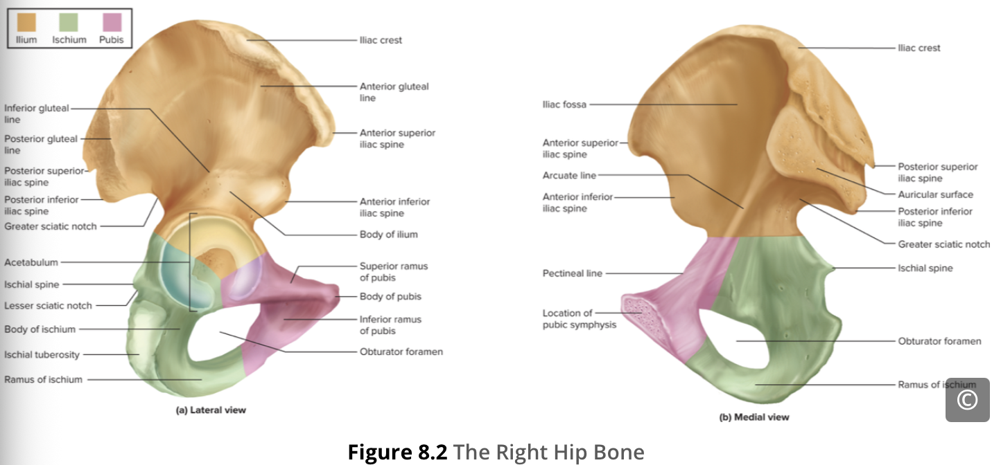
Pubis – body
The enlarged medial portion of the pubis is the pubic body
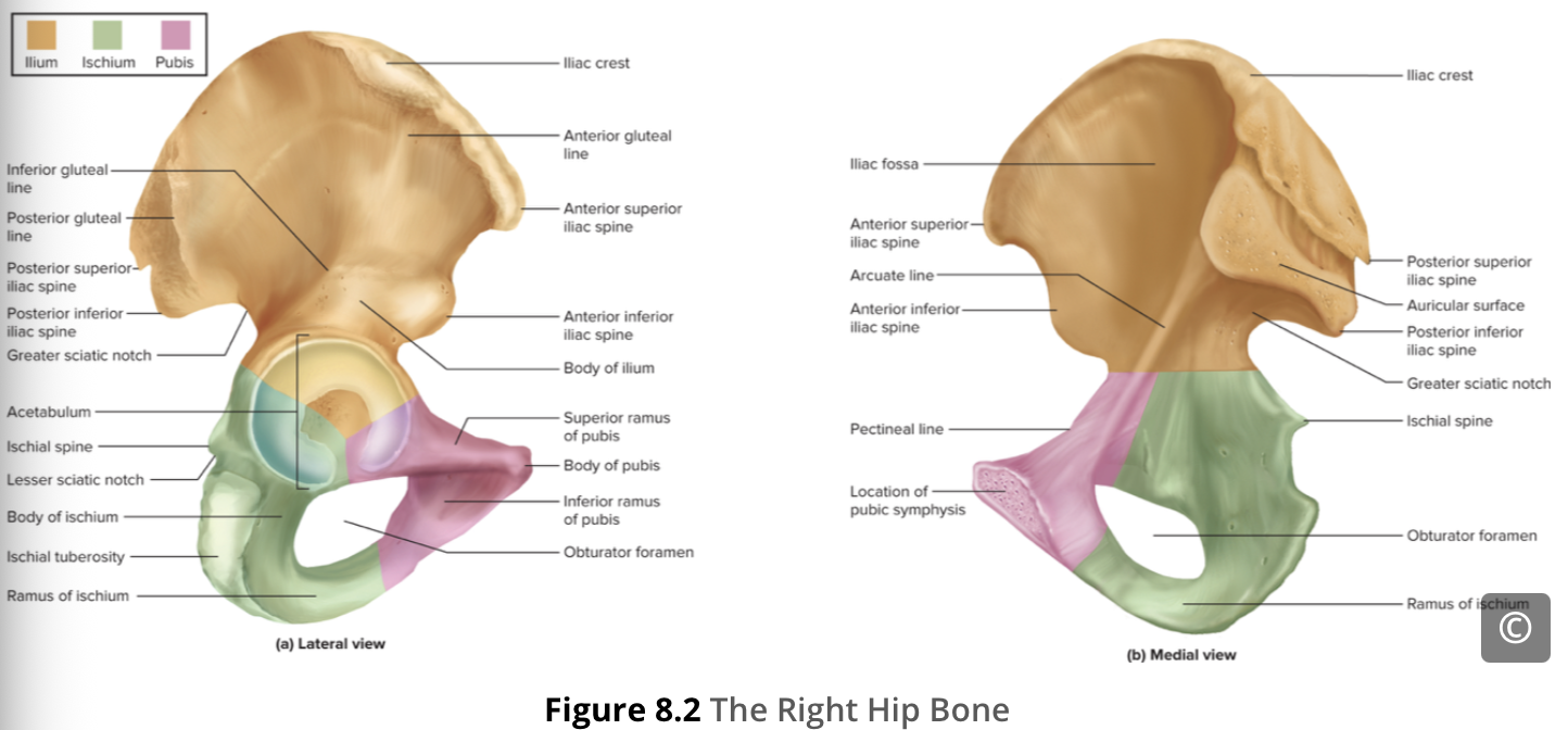
Pubis – inferior ramus
Extending downward and laterally from the pubic body

Pubis – superior ramus
the segment of bone that passes laterally from the pubic body to join the ilium.

Sacral plexus
pass across and through the openings (sacral foramina) of the bone known as the sacrum.
Sacrum
The bony pelvis is the entire structure formed by the two hip bones, the sacrum, and the coccyx. The coccyx, also known as the tail bone, is attached inferiorly to the sacrum (Figure 8.1).

Sacroiliac joint
connects the lower spine (sacrum) to the pelvis (ilium) and acts as a shock absorber to transfer upper body weight to the legs

Femur
the single bone of the thigh, It is the longest and strongest bone of the body, and accounts for approximately one-quarter of a person’s total height.

Femur – fovea capitis
a minor indentation on the medial side of the femoral head that serves as the site of attachment for the ligament of the head of the femur.
This ligament (extra info) spans the femur and acetabulum but is weak and provides little support for the hip joint. It does, however, carry an important artery that supplies the head of the femur.

Femur – gluteal tuberosity
a roughened area extending inferiorly from the greater trochanter

Femur – greater trochanter
the large, upward, bony projection located above the base of the neck.
Multiple muscles that act across the hip joint attach to the greater trochanter, which, because of its projection from the femur, gives additional leverage to these muscles.

Femur – head
The rounded, proximal (close to point of attachment) end of the femur, articulates with the acetabulum of the hip bone to form the hip joint

Femur – lateral condyle
the smooth portion that covers the distal and posterior aspects of the lateral expansion
articulate with the tibia to form the knee joint
Femur – lateral epicondyle
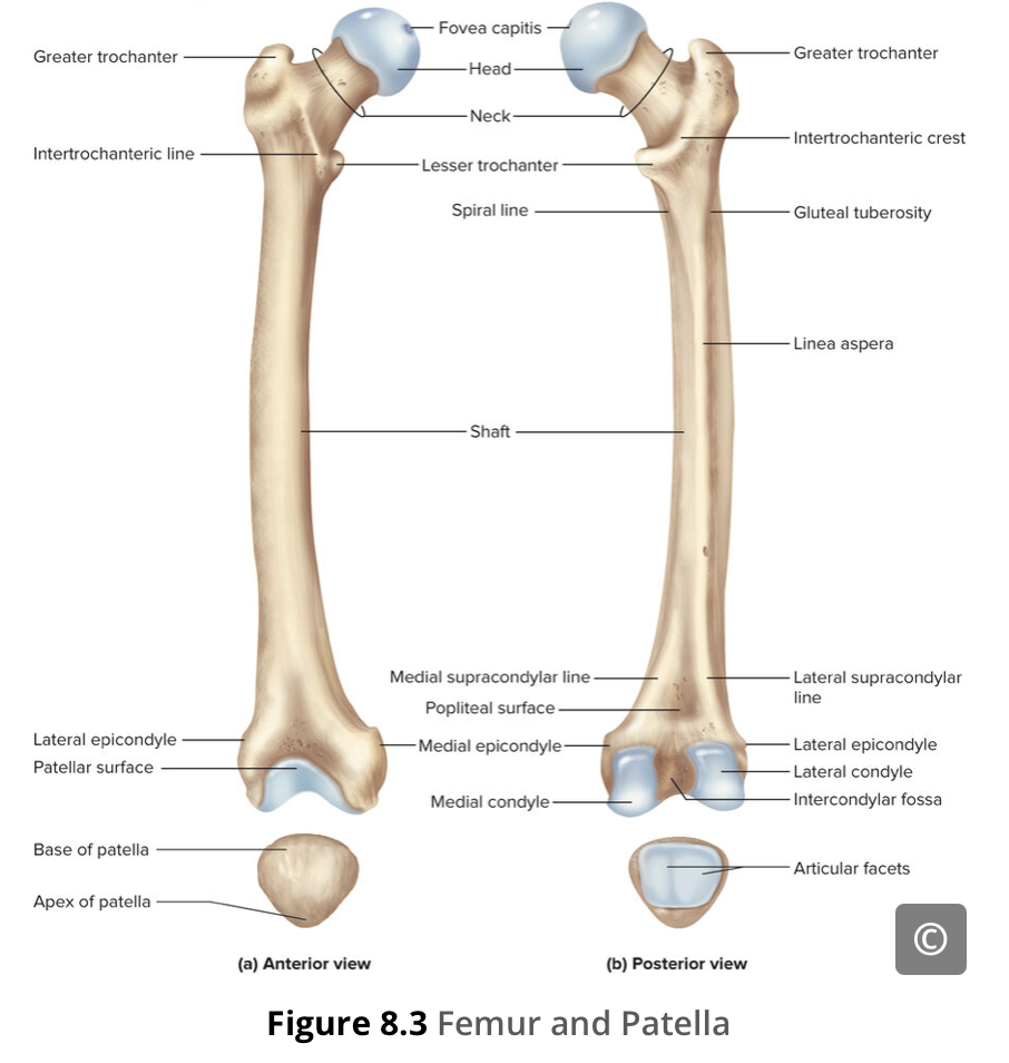
The roughened area on the outer, lateral side of the condyle
provide attachment points for muscles and supporting ligaments of the knee

Femur – lesser trochanter
a small, bony prominence that lies on the medial aspect of the femur, just below the neck.

Femur – linea aspera
the roughened ridge that passes distally along the posterior side of the mid-femur
Multiple muscles of the hip and thigh regions make long, thin attachments to the femur along the linea aspera
Femur – medial condyle
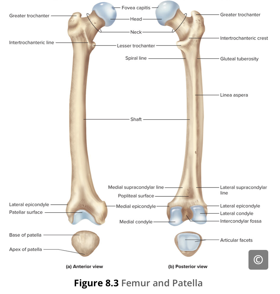
the smooth region of the distal and posterior medial femur
articulate with the tibia to form the knee joint

Femur – medial epicondyle
the irregular outer, medial side of the medial condyle
provide attachment points for muscles and supporting ligaments of the knee

Femur – neck
The narrowed region below the head. This is a common area for fractures of the femur.

Femur – shaft
The elongated shaft of the femur has a slight anterior bowing or curvature.
Patella
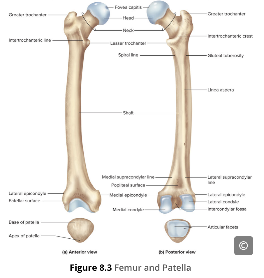
the kneecap, largest sesamoid bone of the body, found in the tendon of the quadriceps femoris muscle, articulates with the patellar surface of the femur and thus prevents rubbing of the muscle tendon against the distal femur, lifts the tendon away from the knee joint, which increases the leverage power of the quadriceps femoris muscle as it acts across the knee, does not articulate with the tibia

Anterior cruciate ligament
Posterior cruciate ligament
Transverse ligament
Fibular collateral ligament
Tibial collateral ligament
Medial meniscus
Tibia – articular cartilage
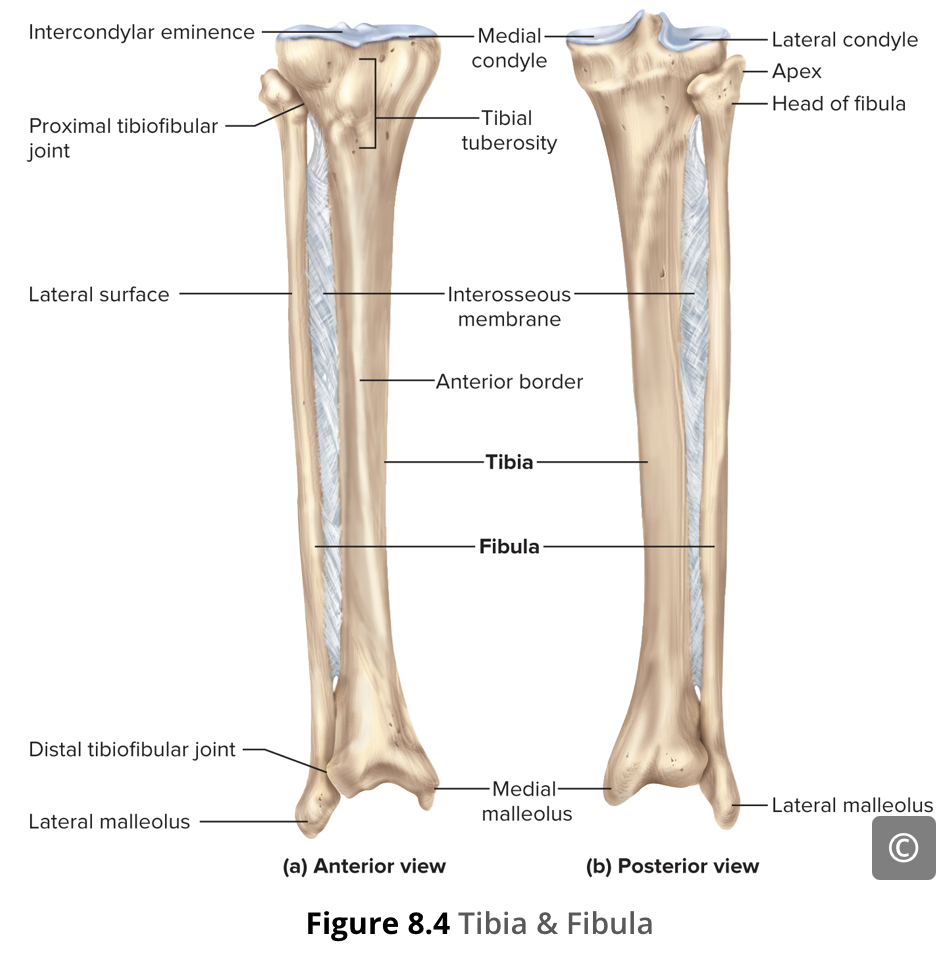
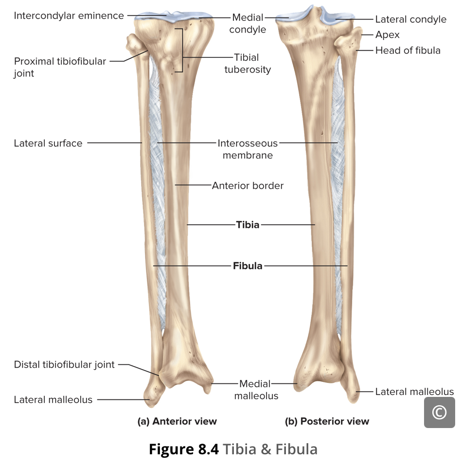
Tibia
the larger, weight-bearing bone located on the medial side of the lower leg, the second longest bone of the body, after the femur

Tibia – anterior border
begins at the tibial tuberosity and runs inferiorly along the length of the tibia.
Both the anterior border and the medial side of the triangular shaft are located immediately under the skin and can be easily palpated along the entire length of the tibia

Tibia – intercondylar eminence
an irregular, elevated area that serves as the inferior attachment point for two supporting ligaments of the knee

Tibia – medial condyle
The proximal end of the tibia is greatly expanded. The two sides of this expansion form the medial and lateral condyles of the tibia.
The superior surface of each condyle is smooth and flattened. These areas articulate with the medial and lateral condyles of the femur to form the knee joint.

Tibia – medial malleolus
The large expansion found on the medial side of the distal tibia

Tibia – shaft
the long, middle, prism-shaped section of the shinbone located between the knee and ankle

Tibia – tibial tuberosity
an elevated area on the anterior side of the tibia, near its proximal end. It is the final site of attachment for the muscle tendon associated with the patella
Fibula
the thin bone on the lateral side of the lower leg, The fibula does not bear weight. It serves primarily for muscle attachments and thus is largely surrounded by muscles. Only the proximal and distal ends of the fibula can be palpated.

Fibula – head
the small, knob-like, proximal end of the fibula.

Fibula – lateral malleolus
forms the easily palpated bony bump on the lateral side of the ankle.
Foot
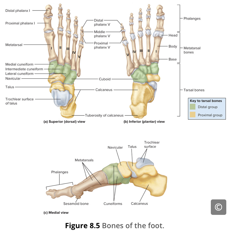
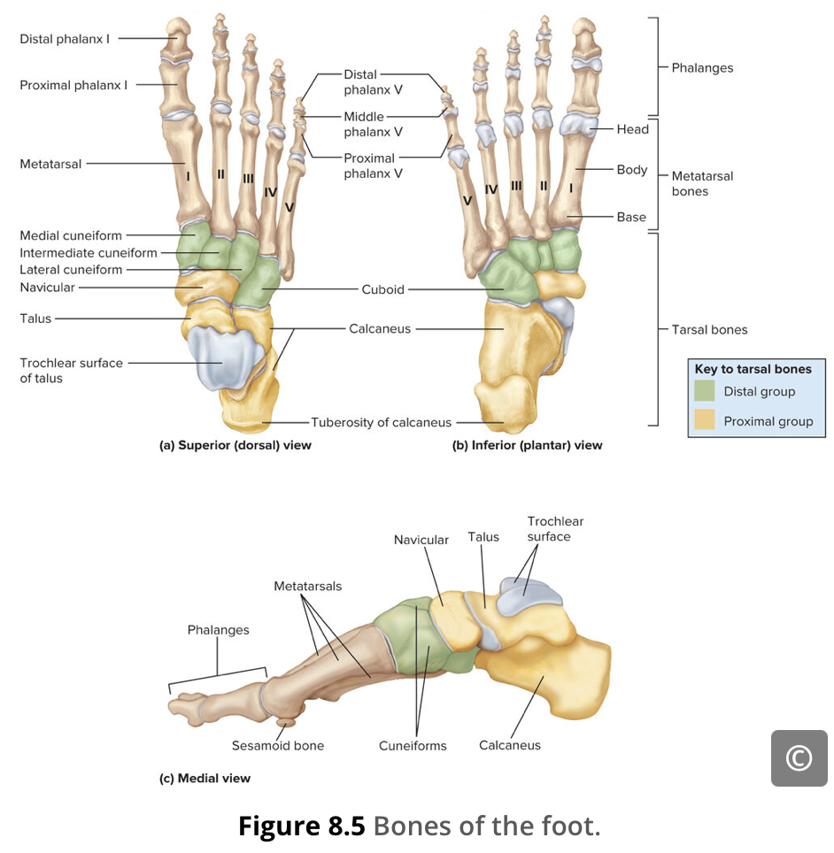
distal to the ankle
Talus
relatively square-shaped, upper surface that articulates with the tibia and fibula to form the ankle joint.
Three areas of articulation form the ankle joint: The superomedial surface of the talus articulates with the medial malleolus of the tibia, the top of the talus articulates with the distal end of the tibia, and the lateral side of the talus articulates with the lateral malleolus of the fibula.
Calcaneus
the largest bone of the foot, which forms the heel
Navicular
The talus bone articulates anteriorly with the navicular bone, which in turn articulates anteriorly with the three cuneiform (“wedge-shaped”) bones.
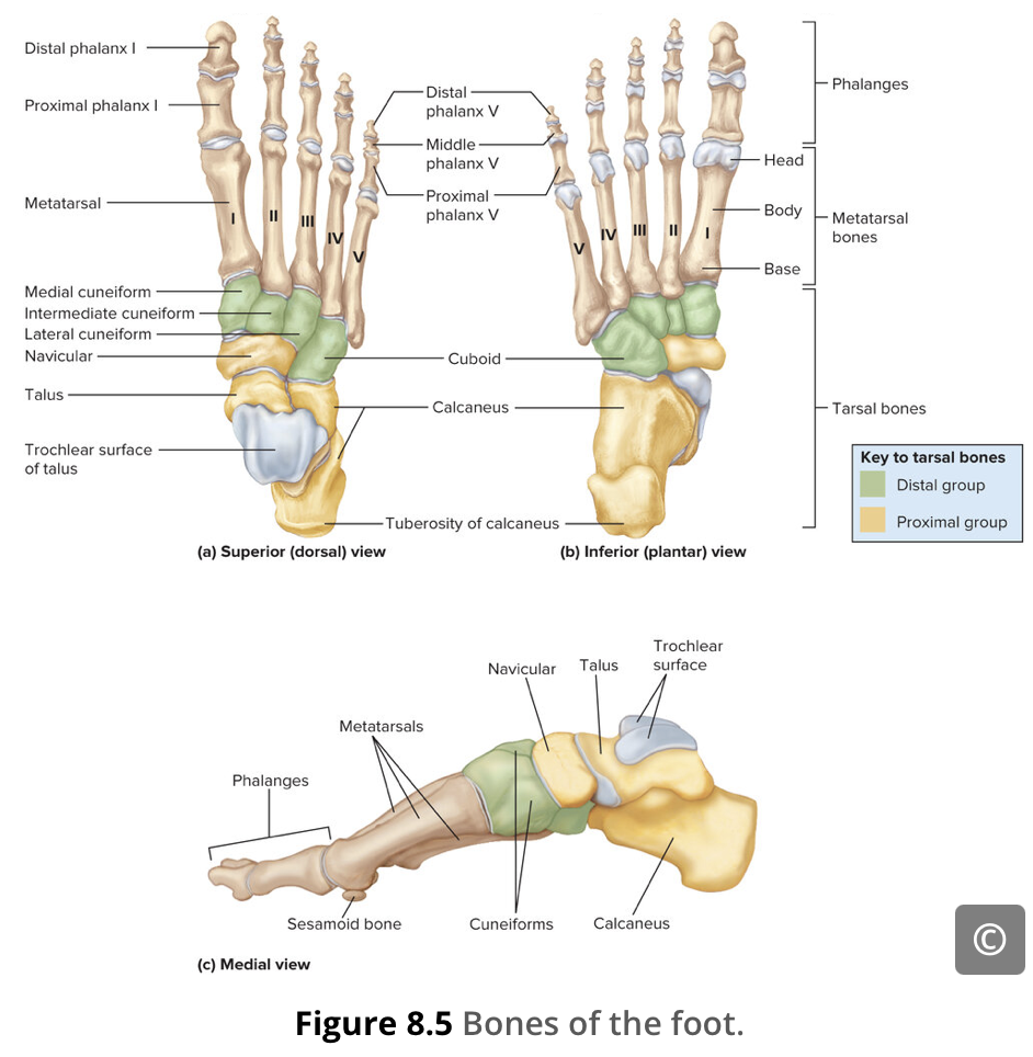
Cuboid
has a deep groove running across its inferior surface, which provides passage for a muscle tendon.

Medial cuneiform
These bones are the medial cuneiform, the intermediate cuneiform, and the lateral cuneiform. Each of these bones has a broad superior surface and a narrow inferior surface, which together produce the transverse (medial-lateral) curvature of the foot
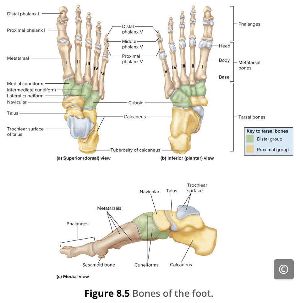
Intermediate cuneiform
These bones are the medial cuneiform, the intermediate cuneiform, and the lateral cuneiform. Each of these bones has a broad superior surface and a narrow inferior surface, which together produce the transverse (medial-lateral) curvature of the foot
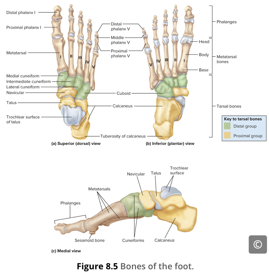
Lateral cuneiform
These bones are the medial cuneiform, the intermediate cuneiform, and the lateral cuneiform. Each of these bones has a broad superior surface and a narrow inferior surface, which together produce the transverse (medial-lateral) curvature of the foot
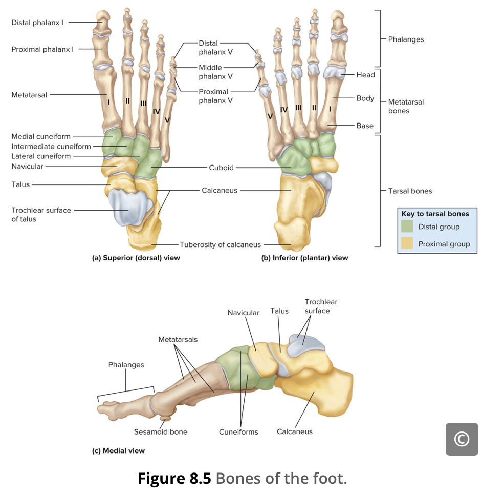
Tarsal bones
seven bones found in the posterior foot.

Metatarsals
five bones found in the mid-foot
The first metatarsal bone is shorter and thicker than the others. The second metatarsal is the longest. The base of the metatarsal bone is the proximal end of each metatarsal bone. These articulate with the cuboid or cuneiform bones. The base of the fifth metatarsal has a large, lateral expansion that provides for muscle attachments.

Phalanges
fourteen phalanx bones in the distal foot
The toes are numbered 1–5, starting with the big toe (hallux) on the medial side of the foot. The big toe has two phalanx bones, the proximal and distal phalanges. The remaining toes all have proximal, middle, and distal phalanges. A joint between adjacent phalanx bones is called an interphalangeal joint.

Phalanx
14 small digital bones that form your toes

Proximal phalanges
Each metatarsal bone articulates with the proximal phalanx of a toe to form a metatarsophalangeal joint

Middle phalanx / phalanges
The toes are numbered 1–5, starting with the big toe (hallux) on the medial side of the foot. The big toe has two phalanx bones, the proximal and distal phalanges. The remaining toes all have proximal, middle, and distal phalanges.
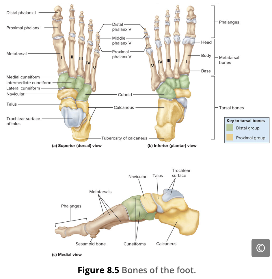
Distal phalanges
The toes are numbered 1–5, starting with the big toe (hallux) on the medial side of the foot. The big toe has two phalanx bones, the proximal and distal phalanges. The remaining toes all have proximal, middle, and distal phalanges.
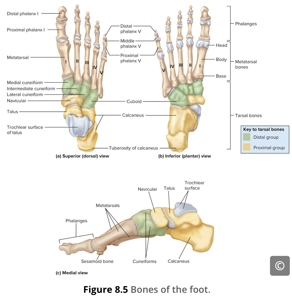
Toes
The anterior half of the foot is formed by the five metatarsal bones, which are located between the tarsal bones of the posterior foot and the phalanges of the toes (Figure 8.5).
The toes contain a total of 14 phalanx bones (phalanges), arranged in a similar manner as the phalanges of the fingers (Figure 8.5). The toes are numbered 1–5, starting with the big toe (hallux) on the medial side of the foot. The big toe has two phalanx bones, the proximal and distal phalanges. The remaining toes all have proximal, middle, and distal phalanges. A joint between adjacent phalanx bones is called an interphalangeal joint.
Hallux
the big toe

Hip Joint
a large ball-and-socket joint where the head of your thigh bone (femur) connects to the cup-shaped socket (acetabulum) of your pelvis
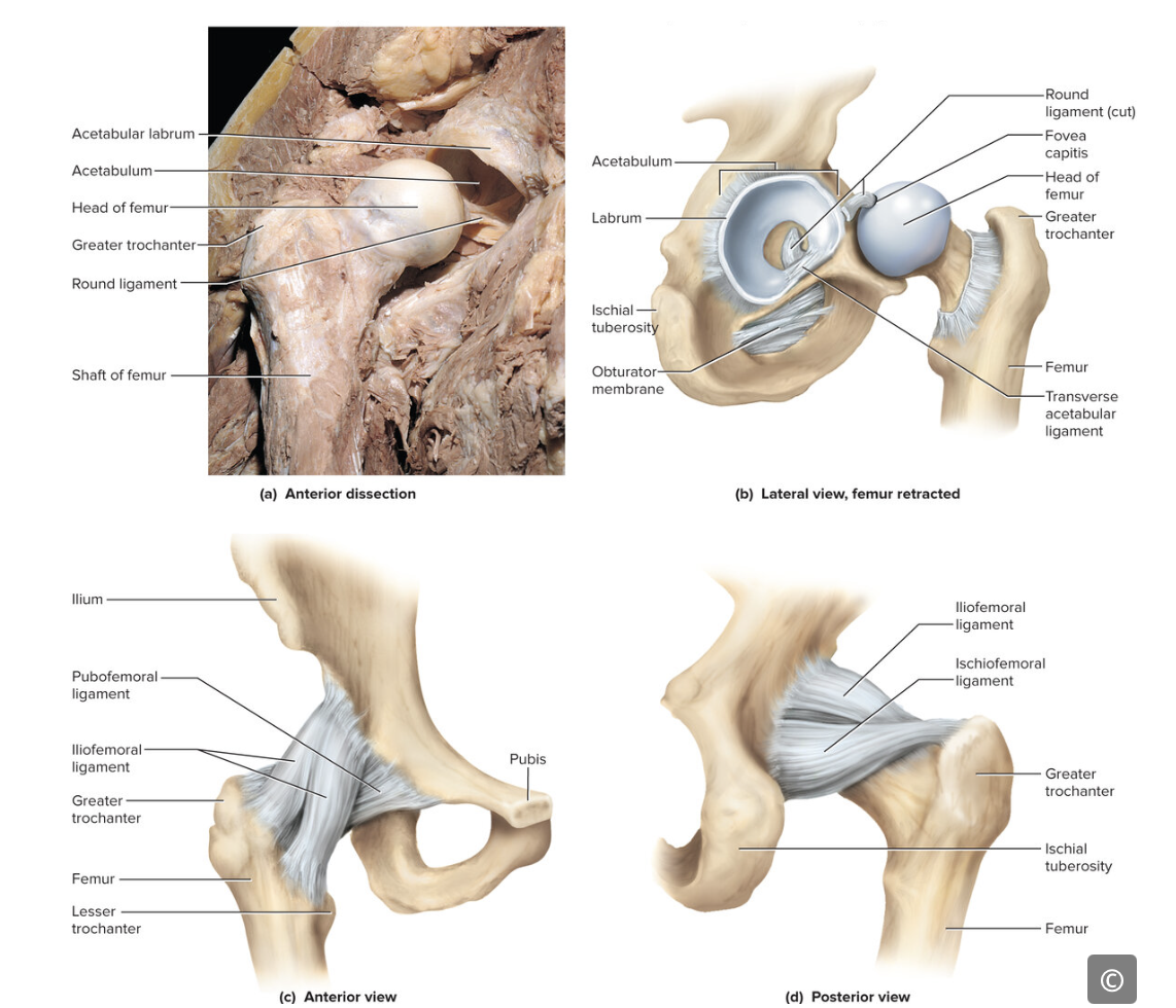
Round ligament
Transverse acetabular ligament
Labrum
fibrocartilage lip attached to the outer margin of the acetabulum.

Pelvic Girdle / Hip
Formed by a pair of hip bones (coxae bones) that are joined to the axial skeleton by the sacrum. These left and right hip bones are attached by the pubic symphysis. The bony pelvis is the entire structure formed by the two hip bones, the sacrum, and the coccyx.
Immobile and stable structure

Lateral meniscus
Interosseous membrane
the sheet of dense connective tissue that connects the tibia and fibula bones.

Ankle
Medial ligament
Anterior talofibular ligament
Posterior talofibular ligament
Calcaneofibular ligament
Anterior tibiofibular ligament
Posterior tibiofibular ligament
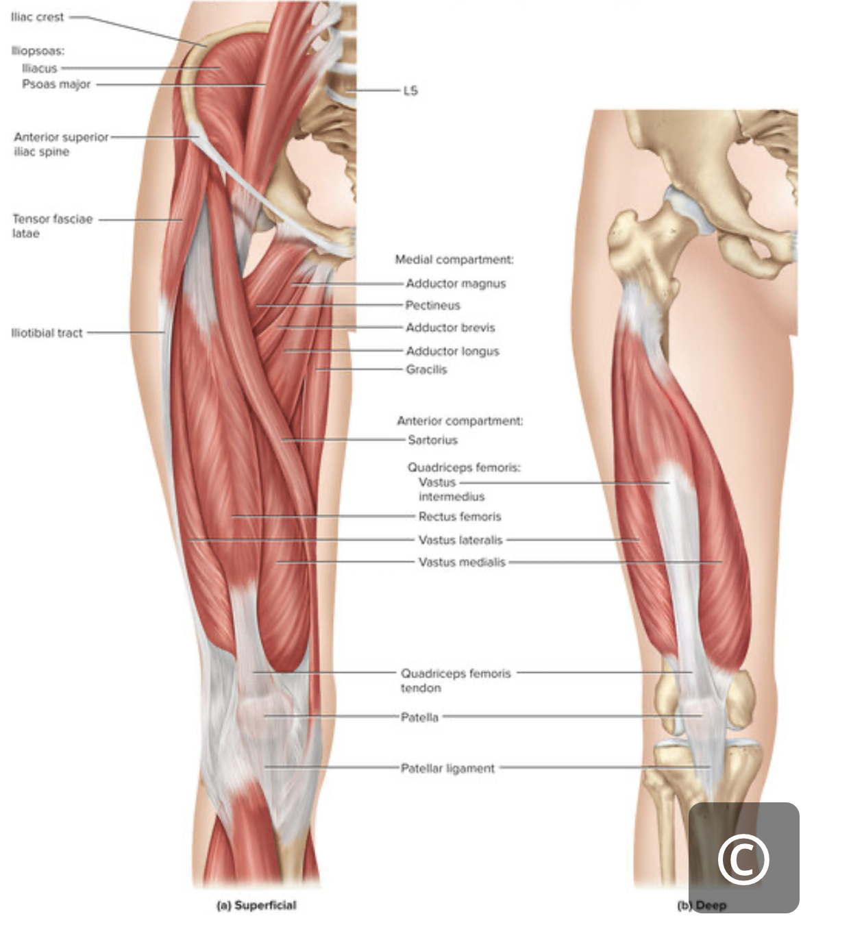
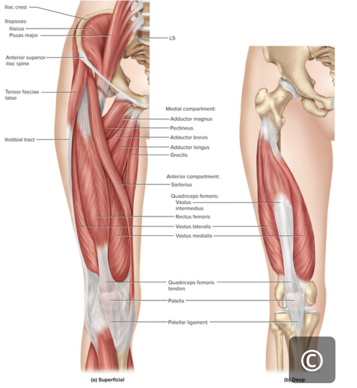
Iliopsoas
Flex hip and laterally rotate thigh; flex torso | (Psoas major) Lumbar vertebrae (L1-L5); thoracic vertebra (T12) (Iliacus) Iliac fossa; iliac crest; lateral sacrum | Lesser trochanter | Femoral nerve |
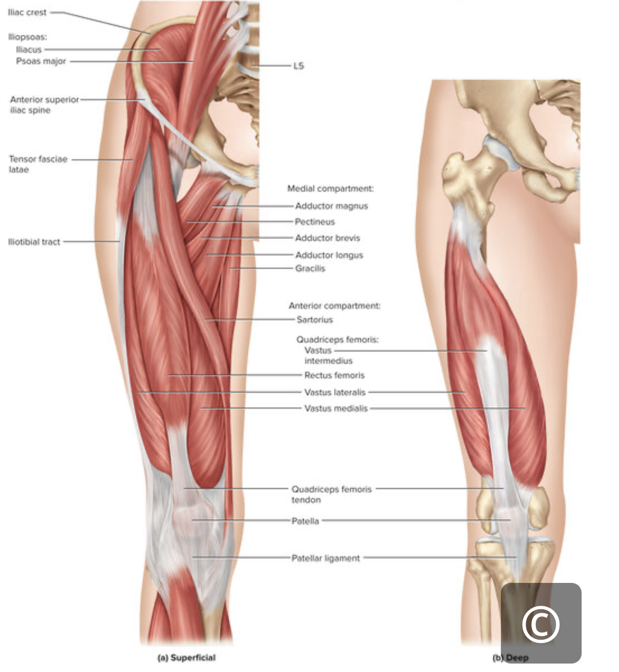
Sartorius
longest muscle in body
Flex & abduct hip; laterally rotate thigh; flex knee | Anterior superior iliac spine | Medial aspect of proximal tibia | Femoral nerve |

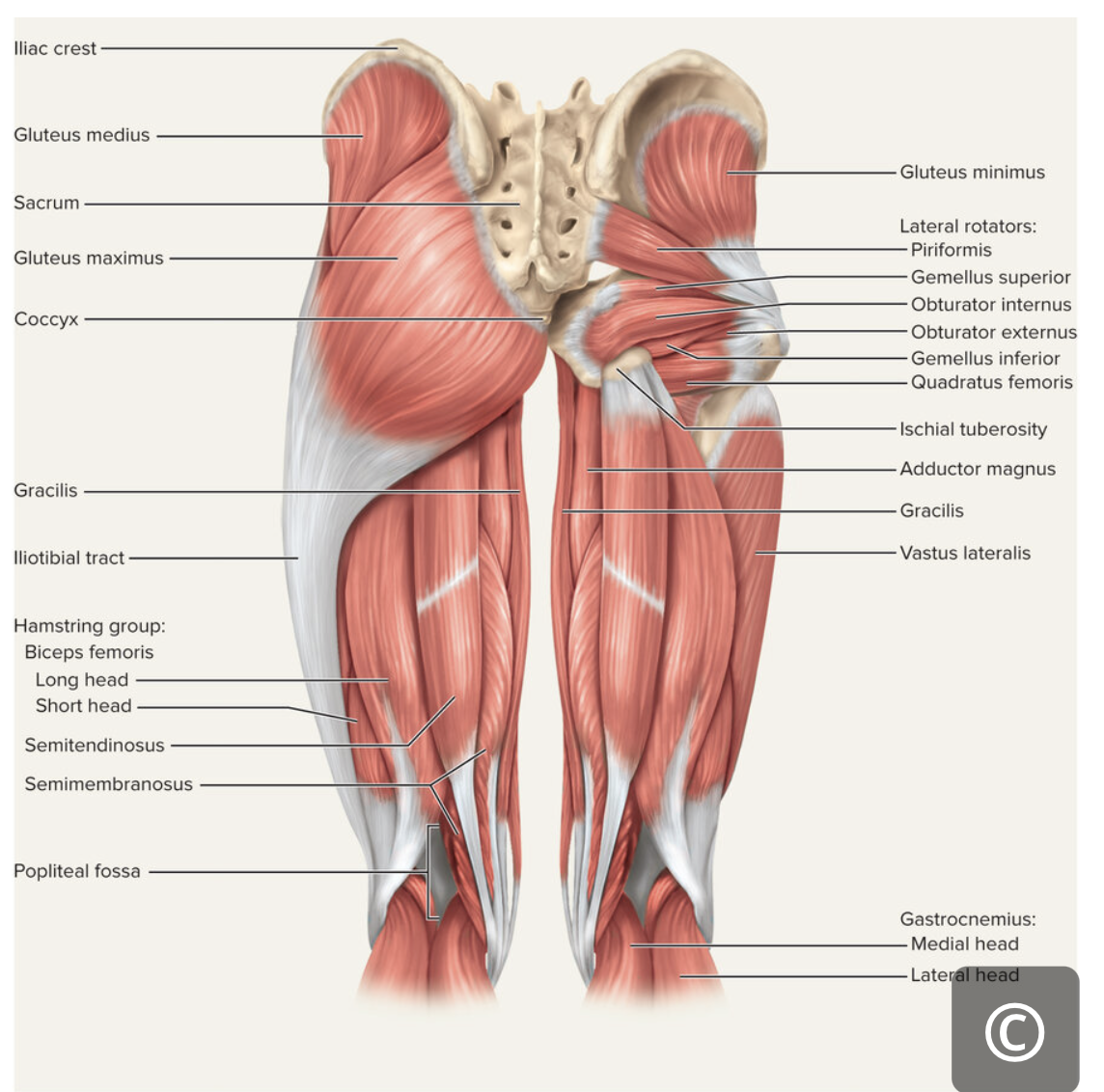
Gracilis
Adduct hip and flex knee | Inferior ramus and body of pubis; ischial ramus | Medial surface of tibia | Obturator nerve |

Pectineus
Pectineus | Adduct & flex hip; medially rotate thigh | N/A | N/A | Femoral nerve |

Piriformis
Laterally rotates thigh; abducts hip | N/A | N/A | Spinal nerves L5-S2 |
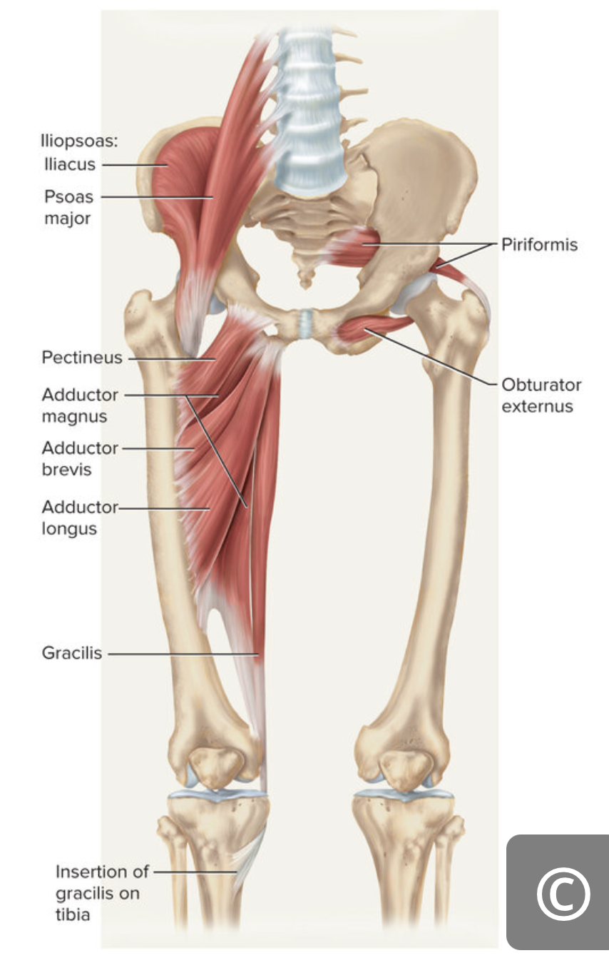
Adductor brevis
Adduct and flex hip | N/A | N/A | Obturator nerve |
Adductor longus
Adduct hip and medially rotate thigh; flex hip | N/A | N/A | Obturator nerve |
Adductor magnus
Adductor magnus | Adduct hip and medially rotate thigh; extend hip | N/A | N/A | Obturator nerve |
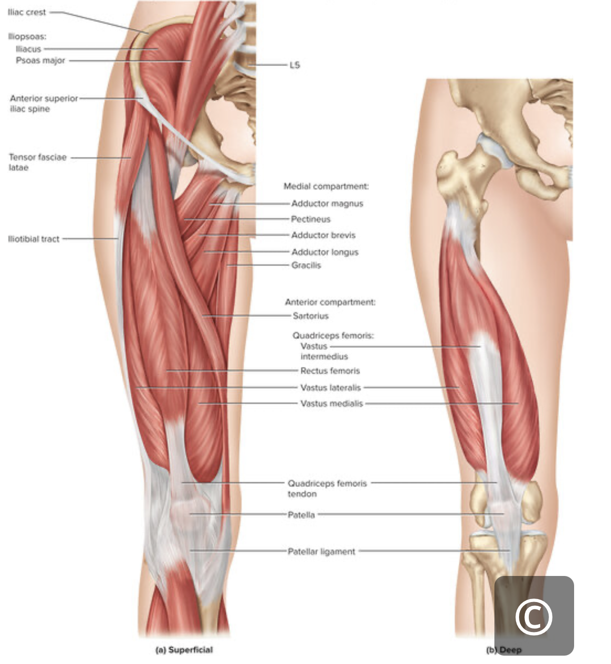
Rectus femoris
Extend knee; flex hip | Anterior inferior iliac spine; superior margin of acetabulum | Patella; tibial tuberosity | Femoral nerve |

Vastus lateralis
Extend knee | Greater trochanter; linea aspera | Patella; tibial tuberosity | Femoral nerve |

Vastus intermedius
Extend knee | Proximal femur shaft | Patella; tibial tuberosity | Femoral nerve |

Vastus medialis
Extend knee | Linea aspera | Patella; tibial tuberosity | Femoral nerve |

Tensor fasciae latae
Tensor fascia latae | Flex and abduct hip | N/A | N/A | Superior gluteal nerve |

Gluteus maximus
Extend hip | Dorsal ilium; sacrum; coccyx | Gluteal tuberosity of femur; iliotibial tract | Inferior gluteal nerve |
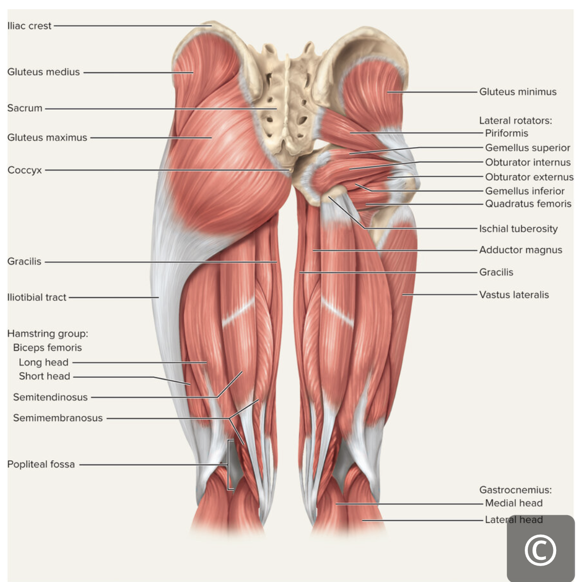
Gluteus medius
Abduct hip | Superior lateral surface of ilium | Greater trochanter of femur | Superior gluteal nerve |

Gluteus minimus
Abduct hip | Inferior lateral surface of ilium | Greater trochanter of femur | Superior gluteal nerve |