MB 251 Exam 1
 Call Kai
 Learn
Learn Practice Test
 Spaced Repetition
 Match
Match Flashcards
 Knowt Play
1/84

There's no tags or description
Looks like no tags are added yet.
Name | Mastery | Learn | Test | Matching | Spaced | Call with Kai | Chat |
|---|
No analytics yet
Send a link to your students to track their progress
85 Terms
Magnification
Ratio of size of image to size of object
Series of lenses is used to achieve magnification of specimens
Definition: The ability of a lens to enlarge the apparent size of an object relative to its actual size.
Mechanism: Microscopes use a series of lenses in combination to systematically bend light and amplify the image.
The Magnification Equation:
Magnification (M)= Image/Actual
Note: The "Image Size" is what you see in the microscope or micrograph; the "Actual Size" is the real-world size of the microbe.
Rearranging the Equation:
To find Image Size:
To find Actual Size: Magnification x Image
Scale Bar
Visual reference for the actual size of organisms/structures in a microscopy image (micrograph)
Used to measure the size and dimensions of organisms and to determine the magnification power used to visualize the image
Purpose: A visual reference for the actual real-world size of structures in an image.
Functions:
Allows for quick measurement of organism dimensions.
Used to mathematically determine the exact magnification power of an image.
Serves as a comparison tool
Unit Conversions
Matching Units: Units must match before performing calculations (e.g., both must be in micrometers).
Converting Larger to Smaller Units: Move the decimal point to the right.
Example: To convert 3.5 centimeters (cm) to micrometers (\(\mu\)m):
Move decimal one place right for millimeters (mm) \(\rightarrow\) 35 mm.
Move decimal three more places for micrometers (\(\mu\)m) \(\rightarrow\) 35,000 \(\mu\)m.
Magnification Units: Typically expressed as "X" (e.g., 3500x) because the actual units cancel out during division.
Resolution
The ability of a microscope to distinguish two points as separate entities
Image clarity
Resolving power = /2NA
= wavelength of light (shorter is better)
NA = numerical aperture of lens (measure of light refraction)
Definition: The clarity of an image; the ability to distinguish two close points as separate, distinct entities.
Importance: Magnification without adequate resolution results in a giant, blurry image (similar to blurry pixels in a zoomed-in digital photo).
Mathematical Relationship: \(\text{Resolving Power} = \text{Wavelength} / \text{Numerical Aperture}\).
Factors Affecting Resolution:
Wavelength: Shorter wavelengths are better for achieving finer resolution.
Numerical Aperture: A fixed physical property of a lens representing its ability to gather light and manage refraction.
Resolving Power Threshold: If two objects are closer together than the microscope’s resolving power, they will appear as one blurry object rather than two distinct ones.
Light Microscopy
Relies on key properties of light:
They are electromagnetic waves with different frequencies (number of wavelengths in a specific timeframe)
They interact with materials by being reflected, absorbed, or transmitted
The waves can change direction (refract) when traveling into a new medium.
Light waves (400-700 nm) pass through a specimen then a series of magnifiying lenses
Maximum magnification
<1500x (most microscopes are 1000x)
Maximum resolution
200 nm
Can view live or dead cells depending on type of microscopy
Properties of light in microscopy
Nature of Light: Light is an electromagnetic wave and a form of kinetic energy.
Wavelength and Frequency:
Wavelength is the distance between the peak of one wave and the next.
Longer wavelengths (e.g., red light) have low frequencies.
Shorter wavelengths (e.g., purple light) have high frequencies.
Light Interactions:
Reflection: Light bounces off a material.
Absorption: Material captures the energy of the light.
Transmission: Light travels through a substance.
Refraction: Light changes direction when moving between different media (e.g., air to glass). This bending allows lenses to focus light on a focal point to magnify an image.
Working Distance and refraction
Working distance= Distance between the front surface of cover glass or specimen when it is in sharp focus
Oil immersion is used to improve resolution when working distance is too small to collect refracted light
Compound Light Microscope Fundamentals
Operating Range: Standard light microscopes use light waves between 400 to 700 nanometers.
Technical Limits:
Maximum Magnification: Limited to under 1,500x (most lab microscopes top out at 1,000x).
Maximum Resolution: Roughly 200 nanometers.
Anatomy of the Microscope:
Light Source: Located at the base.
Condenser Lens: Focuses the broad beam of light into a narrow point to pass through the specimen.
Stage: Holds the slide and specimen.
Objective Lenses: Rotatable lenses (typically 10x, 40x, 100x) that provide the initial magnification.
Ocular Lenses: Eyepiece lenses, typically set at 10x magnification.
Calculating Total Magnification: Multiply the power of the objective lens by the power of the ocular lens (e.g., 100x objective × 10x ocular = 1,000x total magnification).
Key Operational Concepts
Working Distance: The physical distance between the specimen's surface and the objective lens. To see an image in sharp focus, the stage must be moved so this distance matches the lens's focal length.
Oil Immersion:
The Problem: At high magnification (100x), the lens is so close to the slide that light refracts away into the air and misses the lens.
The Solution: A drop of immersion oil is placed on the slide. Because oil has similar refractive properties to glass, light does not bend away and instead enters the objective lens.
Use Cases: Standard for viewing small prokaryotes (bacteria and archaea), whereas eukaryotic cells are often viewed at 40x without oil.
Types of light microscopy
Technique | Description & Key Features | Best Used For... |
Brightfield | Light passes directly through the specimen; usually requires staining for contrast. | Standard bacterial morphology; external structures (cells are usually killed). |
Darkfield | An opaque disc blocks direct light; light hits the specimen at an angle and scatters into the lens, creating a light image on a dark background. | Live organisms; delicate bacteria like spirochetes (e.g., syphilis). |
Phase Contrast / DIC | Uses refraction and interference to create high contrast without stains. DIC uses two beams for a 3D texture. | Delicate internal structures in live eukaryotic cells. |
Fluorescence | Uses fluorophores (dyes) that absorb an excitation wavelength and emit a specific emission wavelength (glowing light). | Pinpointing specific proteins, pathogens, or cell components with high precision. |
Direct Fluorescent Antibody (DFA) | Uses antibodies tagged with fluorescent dyes that bind to specific pathogens. | Highly sensitive clinical diagnosis (e.g., identifying rabies virus in tissue). |
Confocal | Uses a laser to perform "optical sectioning," scanning one layer of a thick specimen at a time. | Constructing 3D models of complex communities like biofilms. |
Bright-Field Microscopy
Light is passed through a specimen that has been stained to improve contrast
Staining typically requires cells to be killed
Simple to use
Often used to examine bacterial morphology or external structures
Dark-field microscopy
Light enters at an angle, so it is scattered off the specimen and reflected into the objective lens
Image appears light against a dark background
No staining required
Used on live organisms
Used to identify spirochete bacteria such as Treponema pallidum, the causative agent of syphilis
Used for morphology of eukaryotic microbes
Phase contrast and DIC
Use refraction and interference of a specimen to create high contrast, high-resolution images without staining
Allows better images of live cells without need for staining
Alters the wavelength of light that pass through the specimen
Requires a more complex light microscope
Often used to view internal structures in live eukaryotic cells
Fluorescence microscopy
Used for the examination of naturally fluorescent specimens or specimens with fluorescent tags/stains
Tags specific proteins, pathogens, cell components
Cells can be alive or dead
Requires fluorescent microscope
Direct Fluorescence antibody assay
Uses fluorescent tagged, pathogen-specific antibodies
Can reveal presence of viruses and bacteria too small to visualize
Confocal Microscopy
Performs optical sectioning with thick objects
Overcomes some limitations of light emission from fluorescent microscopes
Numerous applications including study of biofilms
Electron Microscopy
Uses an electron beam and magnetic lenses
Electron beams have a wavelength of ~0.004 nm
Magnification= 100,000X - 1,000,000X
Resolution= 0.1 nm
Two main types
Transmission electron microscopy
Scanning electron microscopy
Purpose: Used to visualize structures smaller than one micrometer, such as viruses or fine internal details of prokaryotic cells, which are beyond the capabilities of light microscopy.
Mechanism: Uses a beam of electrons instead of photons of light.
Lenses: Employs magnetic lenses to bend and focus negatively charged electrons, whereas light microscopes use glass lenses to refract light.
Wavelength: Electrons have an incredibly short wavelength of approximately 0.004 nanometers, which directly contributes to higher resolving power.
Magnification: Ranges from 100,000x to 1,000,000x.
Resolution: Achieves a limit of 0.1 nanometers (compared to the 200 nm limit of light microscopy).
Transmission Electron Microscopy (TEM)
Primary Use: Observing fine internal details inside a cell or virus particles.
How it Works:
An electron beam is fired directly through an ultra-thin section of a specimen.
Image Formation: Electrons scatter based on the density of the sample. Denser regions scatter more electrons, resulting in darker areas on the detector, while lighter regions indicate where more electrons passed through.
Specimen Preparation (Highly Intensive):
Thickness: Sections must be less than 100 nm thick (ideally 10 to 50 nm).
Process: Fixation (with formaldehyde) \(\rightarrow\) Dehydration (with solvents like ethanol) \(\rightarrow\) Infiltration (with liquid resin/epoxy) \(\rightarrow\) Curing into a hard block \(\rightarrow\) Slicing into ultra-thin sections.
Alternative Methods: Specialized structural clearing like freeze fracturing or freeze etching.
Scanning Electron Microscopy (SEM)
Primary Use: Observing surface detail and topography; produces detailed 3D images.
How it Works:
A beam of electrons scans back and forth across the surface of the specimen rather than passing through it.
Secondary Electrons: The beam knocks loose "secondary electrons" from the specimen, which are gathered by a detector to build a topographical map.
Specimen Preparation:
The specimen must be dried and coated with a thin layer of metal, typically gold, to allow the electron beam to knock electrons loose.
Visual Hallmark: Highly detailed surface texture is the classic characteristic of an SEM micrograph.
Light vs Electron Microscopy

Purpose and Classification of Staining
Need for Staining: Because microbial cells are primarily composed of water and are transparent, they are difficult to see without intervention.
Benefits: Staining increases contrast, which allows for better observation of a microbe's size, shape, and arrangement. It also helps identify structural properties like flagella or endospores.
Two Main Categories of Dyes:
Visible Colored Dyes: These work based on electrical charges.
Basic Dyes: Carry a positive charge. Since bacterial surfaces are negatively charged, these dyes are attracted to and bond directly to the cell (positive stains).
Acidic Dyes: Carry a negative charge. They are repelled by the cell and instead stain the background, leaving the cell clear (negative stains).
Fluorescent Dyes: These bind to specific structures and emit light at a distinct wavelength when activated by a specific excitation wavelength.
Staining Procedures
Simple Staining: Employs a single dye to highlight the overall shape and size of an organism.
Differential Staining: Uses multiple dyes sequentially to distinguish between different types of microbes based on their structural properties.
The Gram Stain
The Gram stain is the most widely used differential stain in clinical microbiology. It separates bacteria into two groups—Gram-positive and Gram-negative—based on their cell wall structure.
The Four-Step Procedure:
Primary Stain (Crystal Violet): Flooding the slide with crystal violet stains all cells purple.
Mordant (Iodine): Iodine acts as a mordant, forming large chemical complexes with the crystal violet to "lock" the dye into the cell wall. At this stage, all cells remain purple.
Decolorizer (Alcohol): This is the critical differentiation step.
Gram-positive cells: Their thick peptidoglycan layer dehydrates, trapping the large dye complexes inside; they remain purple.
Gram-negative cells: The alcohol removes the dye from their different cell wall structure, leaving them clear.
Counterstain (Safranin): Safranin (a red dye) is added to colorize the clear cells. Gram-positive cells stay purple, while Gram-negative cells turn red.
Mnemonic: Positive = Purple.
Specialized Differential Stains
Beyond the Gram stain, other specialized techniques are used to identify specific pathogens or structures:
Acid-fast Stain: Specifically used to identify the genus Mycobacterium (which includes pathogens for TB and leprosy). These bacteria have unique, thick mycolic acids in their cell walls that prevent standard Gram staining from working.
Endospore Stain: A specialized heat-driven process used to visualize endospores, which are indestructible survival structures found in genera like Bacillus and Clostridium.
Capsule Stain: Detects the slippery polysaccharide coating (capsule) surrounding some bacteria. Because capsules do not pick up charges, the procedure uses both a positive and a negative stain to leave the capsule as a clear, colorless halo.
Flagella Stain: Used to observe thin filaments used for locomotion. Because flagella are too thin for standard light microscopy, this stain uses a mordant to build up layers of dye until they are thick enough to see.
Explain the discoveries of key people in the field of microbiology (CO 2)
Hans Christian Gram: Invented the Gram stain. This clinical technique remains the most widely used staining procedure in clinical microbiology today, separating the vast majority of bacteria into Gram-positive and Gram-negative groups based on their cell wall structure.
Describe the relative sizes of various microbe classifications (CO 1)
Human Eye Limit: The human naked eye can typically only see objects that are 1 millimeter (mm) or larger. Microorganisms are classified as organisms that cannot be seen by the naked eye.
Bacteria (Prokaryotes): Individual bacteria, such as a single bacillus, can measure around 2.9 micrometers ((\mu\text{m})) in length. Prokaryotic structures and cells have fine details that are smaller than 1 micrometer.
Viruses: These particles are smaller than 1 micrometer and are too small to be resolved or visualized using standard light microscopes. They require electron microscopy to be seen.
Note: More general size classifications for eukaryotic microbes vs. prokaryotes are covered in the Week 2 lectures (which were excluded from this query).
Discuss the timeline of evolution for microorganisms (CO 4)
Determine what type of microscopy would be used in various microbiological scenarios, including the key differences between light and electron microscopy (CO 2)
Light Microscopy: Relies on light waves (400–700 nm) and glass lenses to refract light to a focal point. It is limited to a maximum magnification of under 1,500x (usually 1,000x in labs) and a resolution of roughly 200 nanometers. It can view both live and dead cells.
Electron Microscopy: Uses a beam of electrons (wavelength of ~0.004 nm) and magnetic lenses to focus negatively charged electrons. It achieves a magnification range of 100,000x to 1,000,000x and an incredible resolution limit of 0.1 nanometers. Because of intensive sample preparation (dehydration, curing, vacuum), it is exclusively used for dead/fixed specimens.
Microscopy Scenarios & Applications
Brightfield: Best for examining standard bacterial morphology or external structures. Since cells are transparent, they must typically be killed and chemically stained to provide contrast.
Darkfield: Forces light to hit the specimen at an angle, scattering light into the lens to create a bright image on a dark background. Best for studying live, delicate organisms that cannot be stained, such as Treponema pallidum (the spirochete that causes syphilis).
Phase Contrast / DIC: Manipulates light refraction and interference to add sharp contrast and edges. DIC uses two light beams to create a striking 3D texture. Best for viewing delicate internal structures of live eukaryotic cells in real time.
Fluorescence: Uses specific excitation and emission wavelengths of light to make naturally fluorescent or fluorescently tagged specimens glow. Best for pinpointing and tagging specific proteins, pathogens, or cell components.
Direct Fluorescent Antibody (DFA) Assay: Chemically links fluorescent tags to specific antibodies that bind to a target pathogen. Best for sensitive clinical diagnosis of pathogens (e.g., detecting rabies virus in tissue).
Confocal: Uses a laser to scan a thick specimen layer-by-layer (optical sectioning), which a computer then reconstructs into a sharp 3D model. Best for studying complex, thick microbial communities (biofilms) without background blur.
Transmission Electron Microscopy (TEM): Fires an electron beam through an ultra-thin (10–50 nm) specimen. Best for observing fine internal details inside a cell or virus particles.
Scanning Electron Microscopy (SEM): Scans a beam of electrons across a gold-coated specimen surface to detect secondary electrons. Best for high-contrast 3D surface detail and topography.
Calculate image size and magnification using a scale bar (CO 2)
The Formulas
Magnification ($M<strong>)</strong>=(Image Size (with ruler)/Actual Size (written on scale bar))2223</p></li><li><p><strong>ActualSize(</strong>A<strong>)</strong>=(Image Size/M)24</p></li><li><p><strong>ImageSize(</strong>I$) = M×Actual Size24
Example Calculation Steps
Match your units: If you measure the image with a ruler in centimeters (cm), convert it to micrometers ((\mu\text{m})) so it matches the scale bar's real-world unit23.
To convert a larger unit (cm) to a smaller unit ((\mu\text{m})), move the decimal point to the right25.
Example: 3.5 cm→35 mm→35,000μm25.
Find Magnification ($M$):
If the scale bar image measures 3.5 cm (35,000μm)2526 and its written actual size is 10μm27:
M=35,000μm/10μm=3500x28. (Units cancel out; magnification is written as "X")28.
Find Actual Microbe Size ($A$):
If a bacterium measures 1 cm on that same micrograph with a ruler2629:
A=1 cm/3500=0.00029 cm430.
Move the decimal point to convert back to micrometers: 0.00029 cm→2.9μm4.
Explain the properties of magnification and resolution (CO 2)
Magnification: The ability of a lens to enlarge the apparent size of an object relative to its actual size. It is achieved by combining a series of lenses to systematically bend (refract) light and amplify the image.
Resolution: The clarity of the image; specifically, the ability of a microscope to distinguish two close points as separate, distinct entities.
Wavelength Relationship: Resolving power is calculated as (\text{Wavelength} / \text{Numerical Aperture}). Because wavelength is the numerator, shorter wavelengths of light yield finer resolution.
Numerical Aperture: A physical, fixed property of the lens measuring its ability to gather light and manage refraction.
Resolving Power Threshold: If two microbes are closer together than the microscope's resolving power limit, they will merge and appear as one giant, blurry object.
Compare and contrast the purpose of different staining procedures (CO 2)
Simple Staining: Uses one single dye to highlight the overall shape and size of an organism.
Positive Stain: Uses a basic dye (positive charge). Since bacterial cell surfaces are negatively charged, the positive dye is electrostatically attracted to and bonds directly to the cell, staining the cell itself.
Negative Stain: Uses an acidic dye (negative charge). The negative dye is repelled by the negative bacterial surface, staining only the background and leaving the transparent cell clear.
Differential Staining: Uses multiple dyes sequentially to structurally differentiate one type of microbe from another.
Gram Stain: Differentiates bacteria based on cell wall structures (Gram-positive cells have a thick peptidoglycan layer; Gram-negative cells have a thin peptidoglycan layer and an outer lipid membrane).
Primary Stain (Crystal Violet): Stains all cells purple.
Mordant (Iodine): Locks the dye into place by forming large, insoluble chemical complexes.
Decolorizer (Alcohol): Dehydrates thick peptidoglycan in Gram-positive cells, trapping the purple dye. In Gram-negative cells, it degrades the outer membrane, allowing the purple dye to wash away completely and leaving them clear.
Counterstain (Safranin): Stains the clear Gram-negative cells red/pink, while Gram-positive cells remain purple.
Acid-fast Stain: Specifically identifies the genus Mycobacterium (pathogens causing TB and leprosy). These bacteria have super-thick, waxy mycolic acids in their cell walls that resist taking up standard Gram stains.
Endospore Stain: A specialized, heat-driven stain used to bypass the indestructible, resistant coats of endospores (dormant survival structures) to identify spore-formers like Bacillus or Clostridium.
Capsule Stain: Detects the slippery polysaccharide capsule. Because polysaccharides do not carry a charge, they repel dyes. This technique uses both a positive and negative stain; the background and cell are stained, leaving the capsule visible as a clear, colorless halo.
Flagella Stain: Uses a mordant to coat delicate, thin motor filaments (flagella) repeatedly. This builds up layers of dye until the filaments are thick enough to be resolved under a standard light microscope.
Taxonomy & Evolutionary Context of Prokaryotes
Domains of Life: Life is divided into three primary domains: Bacteria, Archaea, and Eukarya. Both Bacteria and Archaea consist of single-celled prokaryotic organisms.
Structural Complexity: Although prokaryotes are often pictured as simple blobs, they feature high levels of structural complexity, including external structures for attachment and mobility, alongside various internal storage structures.
Archaea Domain:
Although they look very similar to bacteria under a microscope, evolutionary analysis reveals they share a more recent common ancestor with Eukarya.
Discovered as a distinct domain in the 1970s when scientists were first first able to molecularly examine their genomes.
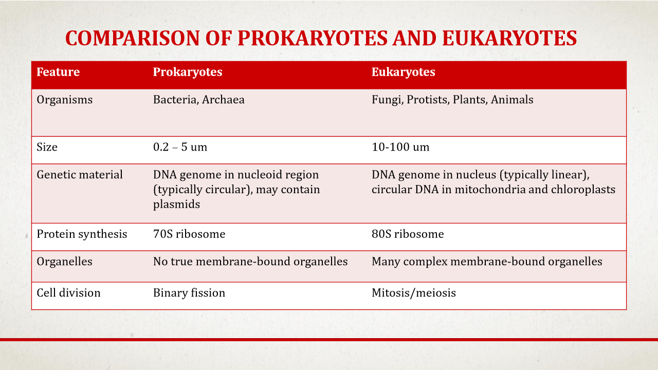
General Comparisons: Prokaryotes vs. Eukaryotes
ote: These are general guidelines; exceptions exist in microbiology (e.g., some bacteria are larger than 10 (\mu)m, and others have linear genomes rather than circular).
Feature | Prokaryotes | Eukaryotes |
Typical Size | Smaller: 0.2 to 5 micrometers ((\mu)m) | Larger: 10 to 100 micrometers ((\mu)m) |
Genomic DNA | Circular genome residing in the cytoplasm (nucleoid); often contains plasmids | Linear genome stored inside a membrane-bound nucleus |
Ribosome Size | 70S ribosomes | 80S ribosomes |
Membrane-bound Organelles | Absent (though some have useful internal membrane invaginations) | Present (true organelles) |
Cell Division | Binary fission (rapid division) | Mitosis and meiosis |
Cellular Shape, Arrangement, and Pathogens
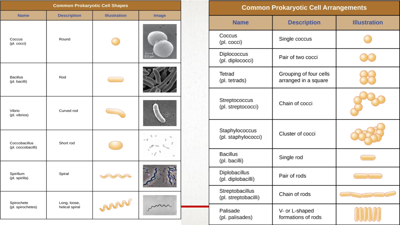
Identifying cell shape and arrangement is crucial for diagnostics and clinical identification.
Three Common Shapes:
Cocci: Spherical.
Bacilli: Rod-shaped.
Spirochetes: Corkscrew-shaped.
Cell Division & Arrangements:
Cocci arrangements:
Diplococci: Pairs.
Tetrads: Groups of four.
Streptococci: Long chains.
Staphylococci: Grape-like clusters.
Bacilli arrangements:
Diplobacilli: Pairs.
Streptobacilli: Long chains.
Palisades: Attached side-by-side along their long ends.
Taxonomic Correlation & Pathogens: Genus names often describe cell arrangement:
Streptococcus pyogenes (causes strep throat) is arranged in chains.
Staphylococcus aureus (causes staph infections) is identified by grape-like clusters of cocci in wound cultures.
Bacterial Cellular Architecture
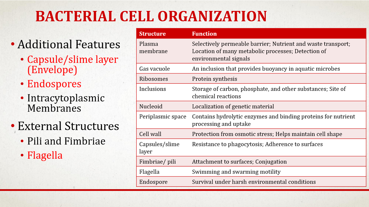
Bacterial structures are classified into three levels of prevalence:
Ubiquitous (found in virtually all bacteria):
Plasma membrane, DNA genome, ribosomes, and a bacterial cytoskeleton (gives structure to the cytoplasm).
Common (found in most bacteria):
Cell wall, plasmid DNA (extra-chromosomal pieces offering extra traits), and various inclusion bodies (used to store nutrients/minerals).
Additional (found in only some bacteria):
Capsules, endospores, and flagella.
Intracytoplasmic membranes: Some species contain invaginations of the cell membrane (tubes or vesicles) that increase surface area for metabolic processes like photosynthesis and respiration.
The Plasma Membrane & Environmental Adaptations
Structure: A selectively permeable lipid bilayer composed of phospholipids (glycerol molecule, phosphate head, and two hydrophobic fatty acid tails).
Thermal Adaptation: Because they are single-celled organisms exposed directly to their surroundings, bacteria actively adjust membrane lipid composition to maintain optimal fluidity:
High Temperature: The cell adds more saturated fatty acids (which contain single carbon-carbon bonds and make the membrane more solid) to prevent the membrane from becoming too fluid (similar to warmed butter).
Low Temperature: The cell increases unsaturated fatty acids (which contain carbon-carbon double bonds and keep the membrane fluid).
Hopanoids: Molecules analogous to cholesterol in eukaryotic cell membranes that help regulate stability and fluidity.
Membrane Transport Mechanism
Because bacteria frequently reside in nutrient-poor environments where nutrient concentrations are lower outside than inside, active transport is heavily favored over passive transport.
Active Transport: Required to pull molecules into the cell against a concentration gradient.
Primary Active Transport: Powered directly by ATP.
Secondary Active Transport: Powered by concentration gradients.
Group Translocation (Unique Active Transport):
Definition: A process where a molecule is chemically modified during its transport across the membrane.
Mechanism (Phosphotransferase System - PTS): Uses the high-energy compound phosphoenolpyruvate (PEP) to transport and phosphorylate sugars like glucose.
The Trap: Glucose is modified into glucose 6-phosphate (G6P) as it enters. G6P cannot easily exit the cell, effectively trapping it inside.
Driving Inward Flow: Because G6P is chemically distinct from free glucose, the intracellular concentration of free glucose remains extremely low, allowing more glucose to continue flowing inward passively.
System Redundancy:
Bacteria often utilize multiple transport systems for the same nutrient (e.g., bringing glucose in via both an ATP-powered ABC transporter and a PEP-powered PTS system).
These parallel systems have different affinities (some work better in high concentrations, others in low) and function optimally under different environmental conditions (such as varying pH). This redundancy is key to bacterial survival and adaptation.
Functions of the Cell Wall and Osmotic Protection
Primary Roles: The primary functions of the bacterial cell wall are to maintain cell shape and prevent osmotic lysis.
The Mechanism of Osmosis: Water naturally flows across the cell membrane from an area of high water concentration to low water concentration.
Hypotonic Environments: In hypotonic environments, water concentration is higher outside the cell than inside, causing water to rush into the cell. Without a rigid, protective cell wall, the resulting osmotic pressure would cause the cell to burst (lyse). Most bacteria cannot control the water concentration of their surroundings, making the cell wall essential for survival.
Isotonic Environments: Isotonic environments have equal water concentrations inside and outside the cell. Bacteria that lack a cell wall must live in these constant environments at all times and typically exist as intracellular parasites within the cytoplasm of eukaryotic cells to avoid osmotic damage.
Chemical Composition of Peptidoglycan
Alternating Sugars: All bacterial cell walls contain peptidoglycan, a mesh network composed of alternating sugar chains of N-acetyl glucosamine (NAG) and N-acetyl muramic acid (NAM).
Pentapeptide Side Chain: Attached to each NAM sugar is a short pentapeptide (a chain of five amino acids) containing both L and D amino acids.
Cross-linking: The long, repeating NAG-NAM sugar-peptide chains surround the entire outer surface of the cell membrane. These chains are cross-linked into a durable mesh by the enzyme DD-transpeptidase.
Gram-Negative Cross-linking: These bacteria possess direct cross-links between the peptides of adjacent sugar chains.
Gram-Positive Cross-linking: These bacteria use a peptide interbridge (an additional chain of amino acids) to connect the oligopeptides of neighboring chains rather than linking them directly.
Gram-Positive vs. Gram-Negative Architecture
Bacteria are categorized into two primary structural groups based on their cell wall design:
Feature | Gram-Positive Cell Wall | Gram-Negative Cell Wall |
Peptidoglycan Thickness | Thick layer consisting of 6 to 12 layers of peptidoglycan. | Thin layer containing only 1 to 3 layers of peptidoglycan. |
Outer Membrane | Absent; the peptidoglycan sits very close to the plasma membrane. | Present; sits outside the thin peptidoglycan layer and is anchored by "bronze" lipoproteins. |
Periplasmic Space | Very narrow periplasm between the cell membrane and peptidoglycan. | Much larger periplasmic space extending from the plasma membrane to the outer membrane. |
Exoenzymes | Contains very few exoenzymes that remain within the narrow periplasmic space. | Uses the larger periplasm as a storage compartment for a large number of functional exoenzymes. |
Unique Components | Teichoic and lipoteichoic acids woven through the peptidoglycan layers. | Lipopolysaccharides (LPS) in the outer leaf of the outer membrane, and porin proteins. |
Functions of Teichoic Acids (Gram-Positive Only):
Structural Support: Wall teichoic acids (bound directly to peptidoglycan) and lipoteichoic acids (which span the peptidoglycan to anchor in the cell membrane) provide structural rigidity.
Ion Capture: Because they are negatively charged, they attract and hold essential positive ions (such as magnesium and calcium) close to the membrane for easier transport.
Growth Regulation: They regulate cell wall formation and breakdown, which is vital during cell division.
Functions of the Gram-Negative Outer Membrane:
Porin Channels: Specialized channel proteins called porins span the outer membrane to allow small molecules to pass through.
Barrier Protection: It acts as a crucial barrier that blocks the entry of harmful hydrophobic molecules, including biosolids and many types of antibiotics.
Lipopolysaccharide (LPS) Structure and Pathogenesis
The LPS molecule found on the outer leaflet of the Gram-negative outer membrane consists of three distinct regions:
O Antigen: The outermost part of the molecule. It is highly variable between different strains of the exact same bacterial species, allowing bacteria to dynamically change their surface appearance to evade host immune recognition and aid in surface attachment.
Core Oligosaccharide: This middle region is vital for maintaining membrane stability.
Lipid A: The innermost region embedded directly in the outer membrane. Lipid A acts as a powerful endotoxin. When Gram-negative cells lyse, they release membrane fragments containing Lipid A. This is recognized by the mammalian innate immune system, triggering an extreme, non-proportional pro-inflammatory response that can lead to life-threatening conditions like fever and septic shock.
How Cell Wall Structure Explains the Gram Stain
The structural differences between Gram-positive and Gram-negative cell walls dictate how they behave during the four-step Gram staining procedure:
Primary Stain (Crystal Violet): Stains both the thick peptidoglycan of Gram-positive cells and the outer membrane/thin peptidoglycan of Gram-negative cells purple.
Mordant (Iodine): Formulates dye complexes to help the crystal violet stick; both remain purple.
Decolorizer (Alcohol):
In Gram-positive cells, the alcohol dehydrates the thick cell wall, causing it to compress and trap the purple dye within the 6-to-12-layer mesh. They remain purple.
In Gram-negative cells, the alcohol damages and destroys the outer lipid membrane. Because their peptidoglycan is so thin, it is unable to dehydrate and trap the dye; the purple dye is completely washed away, leaving the cells colorless/clear.
Counterstain (Safranin): The clear Gram-negative cells take up the red safranin counterstain and turn red/pink. The Gram-positive cells remain purple, as their heavily dyed thick walls do not pick up the additional color.
Cell Wall Exceptions: Acid-Fast and Wall-Less Bacteria
While most bacteria are Gram-positive or Gram-negative, some species feature unique architectures:
Acid-Fast Bacteria:
Structure: Possess a thick peptidoglycan layer linked to an outer membrane containing a dense layer of mycolic acids.
Mycolic Acids: These are 60-carbon chain fatty acids that form a highly waxy, hydrophobic layer, protecting the bacterial cell from destruction.
Key Pathogen: This structure is characteristic of Mycobacterium tuberculosis (the causative agent of tuberculosis).
Wall-Less Bacteria:
Structure: Completely lack a peptidoglycan cell wall.
Characteristics: Since they lack a rigid wall, they have no defined shape and appear amorphous.
Survival Constraint: Because they lack osmotic protection, they can only survive within highly constant, isotonic environments.
Life Style: They are obligate intracellular microbes.
Example: Mycoplasma haemophilius is an obligate intracellular parasite that lives inside host red blood cells. It is the causative agent of feline infectious anemia in Canada and the United States.
The Cell Envelope and Associated External Structures
Cell Envelope: A complex, multilayered structure that protects bacteria from the environment. It is composed of the cell membrane and cell wall, and can also include specialized external layers like capsules, slime layers, or S-layers.
Glycocalyx: A sugar-protein coating made of chains of glycoproteins and polysaccharides that forms the basis of both capsules and slime layers.
Capsule: Formed when the glycocalyx is gel-like and firmly attached to the cell wall.
Slime Layer: Formed when the glycocalyx is more fluid, loose, and unorganized.
Core Functions: Both structures aid in surface attachment, biofilm formation, and protection against desiccation (drying out) and phagocytosis.
Phagocytosis: A process used by immune system cells and environmental amoebae to engulf and digest bacteria. Because capsules and slime layers allow microbes to evade this immune destruction, they can make a microbe highly pathogenic.
Visualization: Capsules can be visualized using a differential stain (capsule stain), where they appear as a clear halo around the bacteria. Slime layers cannot be visualized this way because they are too loosely attached to the cell wall.
S-Layers: Self-assembling, highly structured layers composed of a single type of protein or glycoprotein.
Location: Found outside the cell wall, associated with either the outer membrane of Gram-negative bacteria or the peptidoglycan layer of Gram-positive bacteria.
Structure: They form continuous, patterned crystalline arrays.
Functions: Protect the cell from hostile environments (such as low pH or degradative enzymes), promote surface adhesion, and provide protection from host immune responses.
Nanotechnology Applications: Due to their unique self-assembling crystalline nature, S-layers are used in nanotechnology to develop biosensors and ultrafiltration membranes.
Protein Appendages for Attachment
While capsules and S-layers help with attachment, bacteria also use specialized protein appendages extending past the cell envelope:
Fimbriae: Short, numerous, hair-like bristles used primarily for attaching to surfaces. They are hollow protein tubes made of pilin protein and feature a specialized adhesion protein at the tip.
Pili: Typically longer, thicker, and less numerous than fimbriae on a cell. Pili are used for attachment and other specialized processes, such as conjugation (genetic transfer).
III. Flagella Structure and Assembly
Definition: Stiff, helical protein filaments made of repeating units of flagellin protein that rotate like a boat propeller to move the cell through its environment.
Structural Components:
Filament: The stiff helical portion that extends out from the cell.
Hook: The region that attaches the filament to the motor base.
Basal Body: Embedded directly in the cell membrane and cell wall. It contains the motor portion of the flagella, with ring structures that rotate to spin the hook and filament.
Power Source: Powered by a proton gradient. The diffusion of protons (hydrogen ions) from a high concentration outside the cell to a low concentration inside the cell through MOTAB channels (MOT proteins) generates the torque required to spin the basal body.
Flagella Structure and Assembly
Definition: Stiff, helical protein filaments made of repeating units of flagellin protein that rotate like a boat propeller to move the cell through its environment.
Structural Components:
Filament: The stiff helical portion that extends out from the cell.
Hook: The region that attaches the filament to the motor base.
Basal Body: Embedded directly in the cell membrane and cell wall. It contains the motor portion of the flagella, with ring structures that rotate to spin the hook and filament.
Power Source: Powered by a proton gradient. The diffusion of protons (hydrogen ions) from a high concentration outside the cell to a low concentration inside the cell through MOTAB channels (MOT proteins) generates the torque required to spin the basal body.
Flagellar Motility and Chemotaxis
Run-and-Tumble Pattern: The direction of flagella rotation dictates how a bacterium moves:
Run: Powered by counterclockwise rotation, propelling the bacteria forward in a smooth, straight motion.
Tumble: Powered by clockwise rotation, causing the cell to stop moving forward and instead rotate randomly in 3D space.
Purpose of Tumbles: Tumbles allow sensory receptors on the cell to detect environmental molecules and determine the best direction for the next run.
Chemotaxis: The directed movement of a cell toward nutrients (chemoattractants) or away from toxins (chemorepellents).
Flagellar Arrangement and Specialized Movements
Arrangement Types: The specific arrangement of flagella is species-specific, which makes flagella staining highly useful for identifying bacteria under a microscope. The four common arrangements are:
Monotrichous: A single flagellum at one end of the cell.
Amphitrichous: A single flagellum at each end of the cell.
Mofotrichous (Lophotrichous): A cluster of flagella at one end of the cell.
Peritrichous: Flagella distributed all over the entire cell surface.
Swarming: A rapid, coordinated movement of a bacterial community across a wet surface, typically performed by species with peritrichous flagella.
Requires cell-to-cell communication and the secretion of a slippery liquid.
When grown on agar petri plates, swarming bacteria rapidly cover the entire plate surface instead of forming discrete, individual colonies.
Spirochetes and Internal Flagella:
Spirochetes have flagella that do not extend outward from the cell surface.
Instead, their flagella remain inside the periplasmic space under the cell wall.
When these periplasmic flagella rotate, they spin the entire cell body in a corkscrew motion.
This unique corkscrew movement enables spirochetes to navigate highly sticky, viscous environments (such as mucus) far more effectively than other microbes.
Overview of Cytoplasmic Organization
Prokaryotic Cytoplasm: Although prokaryotes lack traditional membrane-bound organelles, their internal environment is highly structured and organized.
Key Internal Components: The cytoplasm contains a bacterial cytoskeleton, various inclusions, intracytoplasmic membranes, ribosomes, nucleic acids (nucleoid and plasmids), and, in some species, survival structures called endospores.
The Bacterial Cytoskeleton
Eukaryotic Homologs: Bacteria possess cytoskeletal proteins that are homologous to all three main eukaryotic cytoskeletal elements (actin, tubulin, and intermediate filaments).
Key Differences: The bacterial cytoskeleton is less complex than the eukaryotic equivalent and typically lacks classical motor proteins.
Primary Functions: Vital for regulating cell division and maintaining cell shape.
FITZ (FtsZ) Proteins: Form a contractile ring at the center of the cell that contracts to split one cell into two daughter cells during division, acting similarly to eukaryotic microtubules.
MREB (MreB) Proteins: Actin-like proteins that maintain cell width and shape in rod-shaped (bacilli) bacteria.
Intracytoplasmic Membranes and Inclusions
Intracytoplasmic Membranes: Membrane invaginations are common in photosynthetic bacteria, where they form thylakoids (similar to those in plant chloroplasts) to carry out metabolic functions.
Inclusion Bodies: Granules of organic or inorganic materials used primarily for storage.
Nutrient/Mineral Storage: Used to store excess carbon, phosphate, or specific minerals.
Microcompartments: Specially organized areas that isolate and concentrate specific chemical reactions in the cell.
Gas Vacuoles: Found in aquatic microbes to regulate and maintain buoyancy within the water column.
Magnetosomes: Structures containing magnetic iron crystals that act like a built-in compass, allowing aquatic bacteria to navigate along the Earth's magnetic fields.
Ribosomes
Function: The universal site of protein translation and synthesis in both prokaryotic and eukaryotic cells.
Structure: Composed of a small and a large subunit.
Key Differences:
Prokaryotic Ribosomes: Classified as 70S ribosomes. The subunits are composed of distinct proteins and ribosomal RNA (rRNA), specifically containing 16S, 23S, and 5S rRNA subunits.
Eukaryotic Ribosomes: Classified as 80S ribosomes.
Note: The "S" stands for Svendberg unit, which measures a molecule's sedimentation rate (size).
Genomic and Extrachromosomal DNA
The Nucleoid Region: Unlike eukaryotes, prokaryotic double-stranded DNA is not enclosed in a membrane-bound nucleus. Instead, it is concentrated in the nucleoid region, where nucleoid proteins assist in packaging and supercoiling the DNA so it fits inside the cell. Unpackaged DNA is significantly larger than the cell itself.
Genome Shape: For most prokaryotes, the genome is a single circular piece of DNA.
Plasmids (Extrachromosomal DNA):
Small, circular, double-stranded DNA molecules that exist and replicate independently of the main chromosome.
Non-Essential Traits: They carry genes that are not required for basic bacterial survival but offer advantageous traits, such as toxin production or antibiotic resistance.
Transmission: Replicated and passed down to daughter cells during division.
Example (Ti / "Teen Eye" Plasmid): A large, tumor-inducing plasmid found in pathogenic Agrobacterium species.
The bacterium injects this plasmid into plant tissue, where its tDNA region expresses plant hormones that cause crown gall disease (tumors).
While necessary for pathogenesis, Agrobacterium can survive perfectly well without this plasmid.
Endospores: Structure, Sporulation, and Germination
Definition: Exceptionally durable, dormant (non-vegetative) survival structures produced by specific species of Gram-positive bacteria.
Resistance: Capable of surviving extreme heat, radiation, and harsh chemicals.
1. Multi-Layered Structure
Dehydrated Core: Contains the bacterial genome, ribosomes, high levels of dipicolinic acid, and small acid-soluble proteins that protect the genetic material.
Cortex: A very thick layer of peptidoglycan surrounding the core (thicker than a standard Gram-positive cell wall).
Spore Coat: Multiple protective protein layers wrapped around the cortex.
Exosporium: The outermost, loose-fitting protein layer that acts as both a protective shield and an adhesive barrier to help the spore stick to surfaces.
2. The Sporulation Process
This transition from an active, living vegetative cell to a dormant endospore takes several hours and is triggered when key nutrients become scarce.
DNA Replication: The chromosome is copied.
Asymmetric Division: An asymmetric septum forms close to one end of the cell, dividing it into a larger mother cell and a smaller prespore.
DNA Degradation: The DNA inside the mother cell degrades as it is no longer required.
Layer Assembly: The peptidoglycan cortex forms, followed by the assembly of the protein spore coat around the prespore.
Maturation & Lysis: The exosporium forms, and once the spore is mature, the mother cell lyses (bursts), releasing the free endospore into the environment.
3. Dormancy and Germination
Dormancy State: Endospores exhibit zero metabolic activity (no growth, division, resource consumption, or protein production). They can remain viable in this state for centuries or even millions of years. Example: Scientists successfully revived a 25-million-year-old Bacillus species preserved inside the gut of a bee trapped in Dominican amber.
Germination: When local environmental conditions improve, the endospore detects signals through its exosporium. The spore rehydrates, breaks down its protective protein coat, resumes active metabolism, and emerges as a fully functional vegetative cell ready to grow and replicate.
Evolutionary Context and General Characteristics
Discovery and Pathogenicity: First recognized as a distinct domain of life in the 1970s, archaea are as genetically diverse as bacteria. To date, no archaea have been identified as human or animal pathogens.
Habitats: While initially thought to exist solely as extremophiles (organisms living in hostile environments), they are now known to occupy all habitats, including the human gut and cow groomings.
Structural Similarities to Bacteria and Eukaryotes
Archaea occupy a unique evolutionary position, sharing physical traits with bacteria while remaining closer to eukaryotes on the tree of life.
Bacterial (Prokaryotic) Similarities:
Typical cell size of 0.2 to 5 micrometers.
Similar overall cellular shapes.
Genomic DNA is stored as a circular chromosome without a membrane-bound nucleus.
Possess 70S ribosomes and plasmid DNA.
Lack traditional membrane-bound organelles.
Contain similar inclusion bodies, and share many of the same metabolic genes and processes.
Eukaryotic Similarities:
The specific proteins responsible for core genetic processes—specifically DNA replication, transcription, and translation—are evolutionarily much closer to those of eukaryotes than bacteria.
Unique Archaea-Only Features & Extremophilic Adaptation
Methanogenesis: Archaea are the only known organisms on Earth that possess the genes required to perform methanogenesis (the metabolic production of methane in oxygen-free environments).
Extreme Growth Thresholds: Some species thrive in environments that would instantly kill bacteria or eukaryotes. Examples include:
Loki's Castle: A geothermal vent located 1.5 miles deep under the ocean with water temperatures reaching 570°F (where the first archaea with eukaryotic-like genetic traits were discovered).
High-temperature hot springs in Yellowstone.
High-salinity environments such as the Dead Sea and the Great Salt Lake.
Unique Cell Membrane Structure
The archaeal cell membrane serves the same basic protective functions as other domains, but its chemical building blocks are fundamentally different from both bacteria and eukaryotes.
Ether Linkages: Instead of using ester linkages to bond fatty acids to glycerol (the standard in bacteria and eukaryotes), archaea utilize ether linkages. Because ether linkages lack an extra double-bonded oxygen atom, they are physically harder to break down, providing superior chemical stability.
Branched Chains: The fatty acid chains of archaea can feature branched structures, which do not exist in bacteria.
Membrane Monolayers: Certain archaeal species do not use a standard lipid bilayer. Instead, they possess a monolayer where a single glycerol with a phosphate head is attached to both ends of the fatty acids. These monolayers are highly resistant to melting and will not fall apart under extreme heat or highly acidic conditions.
Cell Wall Diversity
No Peptidoglycan: Unlike bacteria, the cell walls of archaea completely lack peptidoglycan.
Alternative Materials:
Some species use a structurally similar substance known as pseudomurein.
Others use methadocondratite, which is a rigid polysaccharide layer.
S-Layers: Many species feature a protein-based S-layer anchored directly to the cell membrane.
Function: Despite the wide structural variations between different archaeal species, the cell wall serves the universal function of protecting the cell from osmotic stress while providing shape and physical stability.
General Characteristics of Eukaryotic Cells
Compartmentalization: The defining difference between eukaryotes and prokaryotes is internal compartmentalization. Eukaryotes contain a membrane-bound nucleus to house the genome, along with various membrane-bound organelles performing specific, localized functions.
Internal Highway: An intricate intracytoplasmic membrane network acts as an internal highway system to traffic molecules within the cell.
Cell Size: Because of this efficient internal organization, eukaryotic cells can grow significantly larger than bacteria or archaea, averaging 5 to 100 micrometers ((\mu)m) in size while still maintaining efficient nutrient transport.
Genomic DNA: Eukaryotic genomes consist of multiple linear chromosomes stored in the nucleus.
Cell Walls: Not all eukaryotes have cell walls. Those that do are never made of peptidoglycan or substances similar to archaeal cell walls; instead, they are composed of cellulose, chitin, or silica.
The Eukaryotic Cytoskeleton
The eukaryotic cytoskeleton is a dynamic matrix of fibers and tubes that provides structural support, anchors organelles, and forms a transport network within the cell. It is composed of three main filament types:
Actin Filaments (Microfilaments):
Movement: Work in tandem with motor proteins like myosin to drive muscle contraction in animals and amoeboid movement in single-celled eukaryotic microbes.
Pseudopodia ("False Feet"): To move, actin filaments polymerize directly underneath the cell membrane, pushing the membrane outward to form a temporary extension called a pseudopodium. The remaining cytoplasm then flows forward to join this leading edge.
Intermediate Filaments:
Structure: Diverse, cable-like filaments with a 10-nanometer (nm) diameter (thicker than actin but thinner than microtubules).
Function: They are relatively permanent structures that anchor and maintain the physical position of the nucleus and form the nuclear lamina (the protective lining just inside the nuclear envelope).
Microtubules:
Structure: Hollow tubes with a 23-nanometer (nm) diameter composed of tubulin dimers.
Dynamics: Highly dynamic structures that can rapidly assemble and disassemble.
Function: Act as structural girders, work with motor proteins (such as dynein and tynosine) to move organelles and vesicles through the cytoplasm, form the mitotic spindles that separate chromosomes during cell division, and serve as the core structural component of flagella and cilia.
The Endomembrane System
The endomembrane system is a cooperative group of membranes and organelles that manufacture, modify, package, and transport lipids and proteins, as well as digest environmental materials brought in via endocytosis or phagocytosis. All components are in contact either directly or through membrane-bound transport vesicles.
The Secretion Pathway (Forward Flow):
Rough Endoplasmic Reticulum (RER): Proteins destined for secretion or membrane integration are translated by ribosomes embedded on the RER surface directly into the RER lumen.
Golgi Apparatus: The protein is packaged into a vesicle and sent to the cis face of the Golgi. It is modified as it travels through the Golgi stack to the trans face.
Plasma Membrane: The protein exits the trans-Golgi in a new vesicle, which travels to and fuses with the plasma membrane, either secreting the protein or embedding it as a membrane protein.
The Digestive Pathway (Reverse Flow):
Molecules from outside the cell are internalized into endosomic vesicles, which fuse with lysosomes containing digestive enzymes to break down the material.
Evolutionary Origin:
The nucleus and the endomembrane system are believed to have been the earliest steps in eukaryotic evolution, arising from the infolding of the plasma membrane in an ancestral prokaryotic cell.
The Endosymbiotic Theory
According to the endosymbiotic hypothesis, the energy-generating organelles—mitochondria and chloroplasts—evolved from ancient, free-living prokaryotes that were engulfed by an ancestral eukaryotic cell via endocytosis.
Mitochondria Evolution: An ancient cell engulfed an aerobic bacterium. Instead of digesting it, the host cell kept it alive. The bacterium provided the host with extra ATP, yielding a symbiotic relationship that eventually allowed complex multicellular life to emerge.
Chloroplast Evolution: A subsequent, separate endosymbiotic event occurred when a cell already containing mitochondria engulfed a photosynthetic bacterium, giving rise to modern photosynthetic algae and plants.
Evidence Supporting Endosymbiosis:
Although mitochondria and chloroplasts can no longer survive if removed from the host cell, they retain clear structural and genetic hallmarks of their free-living ancestry:
Double Membrane: The inner membrane represents the original prokaryotic membrane, while the outer membrane represents the host's endosomic vesicle membrane formed during engulfment.
Thylakoids: Chloroplasts contain a third internal membrane system (thylakoids) structurally similar to modern photosynthetic prokaryotes.
Independent Reproduction: Both organelles replicate inside the eukaryotic cell independently via binary fission.
Genetics & Ribosomes: Both contain their own independent DNA organized in a nucleoid region, and they possess 70S ribosomes (the bacterial standard, rather than eukaryotic 80S ribosomes) to translate their own genes.
Eukaryotic Appendages: Flagella and Cilia
While some eukaryotes use flagella for locomotion like prokaryotes, their structural design and movement mechanisms are completely different.
Feature | Prokaryotic Flagella | Eukaryotic Flagella |
Structure | Stiff, rigid filaments composed of the protein flagellin. | Flexible, whipping structures composed of microtubules arranged in a "9+2" array (nine outer microtubule pairs surrounding two central microtubules). |
Motion | Rotates like a boat propeller. | Moves in a whip-like motion. |
Mechanism | Driven by a proton gradient spinning a basal body motor. | Driven by microtubules on one side polymerizing outward to push the structure, then compressing while the opposite side extends. |
Abundance | Variable. | Cells typically possess only one or two flagella. |
Eukaryotic Cilia: Structurally identical to eukaryotic flagella (utilizing the same 9+2 microtubule array), but they are shorter and often cover the entire surface of the cell to assist in movement. Cilia are entirely unique to eukaryotic cells.
Introduction to Protists
Definition and Taxonomy:
"Protist" is a historical term of convenience rather than a single evolutionary family.
They are a polyphyletic group, meaning they do not share a single common ancestor. On the Tree of Life, they are scattered across different evolutionary groups and eukaryotic supergroups.
They are unified not by shared physical structures, but by what they lack: they have no major tissue organization (typically existing as single-celled organisms or multicellular organisms without tissue differentiation, unlike plants which have roots, stems, and leaves).
Their lifestyles range from photosynthetic primary producers (algae) to predatory hunters (amoebas).
Primary Producers: True Algae
General Characteristics:
True algae are aquatic photosynthetic protists packed with chloroplasts, found in all aqueous environments where they support aquatic life.
Green algae have chloroplasts that use chlorophyll similar to land plants.
Red algae utilize specialized light-absorbing pigments, such as phycoerythrin, to capture the blue and green light wavelengths that penetrate deeper into ocean waters.
Size Variation: While some algae are microscopic, others like seaweeds can grow quite large; they are still classified as algae because they lack tissue differentiation.
Secondary Endosymbiotic Algae
Origin and Nutrition:
These algae are formed when a non-photosynthetic (heterotrophic) protist engulfs a true algal cell. The engulfed cell eventually becomes integrated as an organelle, similar to how chloroplasts originally evolved from engulfed photosynthetic bacteria.
This makes most secondary algae mixotrophic: they can perform photosynthesis when light is available, but can switch to engulfing prey when it is not.
Two Main Classifications:
Dinoflagellates:
Mostly marine organisms that generally possess two flagella causing them to whirl (their name comes from the Greek word for whirling).
Scanning electron micrographs show one flagellum wrapping around one side of the cell and a second flagellum wrapping around the other.
Some have armored cellulose plates forming a hard outer covering called a theca.
Red Tides: Under conditions of high nutrients, lower salinity, and calm waters, dinoflagellates can bloom, causing red tides.
Pathogenicity: Some produce neurotoxins that cause paralysis in humans or fish. Exposure occurs through direct water contact or by eating filter feeders (e.g., clams, oysters) that have consumed them.
Symbiosis: Some dinoflagellates, called zooxanthellae, are vital endosymbionts of coral reefs; rising ocean temperatures destroy them, leading to coral bleaching.
Diatoms:
Feature beautiful frustules, which are outer cell walls made of crystallized, glass-like silica.
Found in fresh water, saltwater, and damp soil.
They are the most diverse group of protists and serve as bioindicators of water health because each species is sensitive to specific ranges of pH, nutrients, and temperature.
Their fossilized remains are harvested as diatomaceous earth.
Atmospheric Impact: Phytoplankton
Eukaryotic algae (both primary and secondary) along with photosynthetic prokaryotes (cyanobacteria) are colloquially grouped as phytoplankton.
Phytoplankton live in the sunlit layers of lakes, ponds, rivers, and oceans.
Together, they are responsible for producing approximately 70% of the world's oxygen in the atmosphere.
Note: Although cyanobacteria are often called "blue-green algae," they are prokaryotic bacteria, not eukaryotic algae.
. Predatory Hunters: Amoebas
Characteristics:
Top predators in aquatic and soil environments that hunt by engulfing bacteria and other protists.
They utilize pseudopods (dynamic membrane extensions formed by the polymerization of actin) for both prey engulfment and locomotion.
Due to the dynamic nature of pseudopodia, amoebas have no fixed shape.
Notable Human Pathogens:
Entamoeba histolytica: Causes severe amoebic dysentery and gastrointestinal illnesses.
Naegleria fowleri: The "brain-eating amoeba" found in warm fresh waters. If inhaled, it travels up the olfactory nerve to cause fatal encephalitis.
Alveolates
General Feature: Characterized by a highly structured cytoplasm and a distinct layer of flattened membrane sacs called alveoli located directly beneath their plasma membrane.
Three Main Groups: Ciliates, non-photosynthetic dinoflagellates, and apicomplexans.
Ciliates:
Example: Paramecium, a highly motile organism commonly found in pond water.
They capture prey by trapping food in an oral groove and digesting it with enzymes.
Because they take in water, they contain a large contractile vacuole that continuously pumps out excess water to prevent the cell from bursting due to osmotic pressure.
Apicomplexans:
Obligate intracellular parasites that lack classic locomotive structures (like cilia or flagella) in their mature forms.
They feature a specialized apical complex at one end of the cell designed to secrete enzymes and physically penetrate host tissue cells.
Life Cycle Hosts:
Definitive Host: Where the parasite matures and undergoes sexual reproduction.
Intermediate Host: Where asexual proliferation occurs to increase cell numbers.
Notable Pathogens:
Toxoplasma gondii: Causes toxoplasmosis. Transmitted via cat feces, unwashed produce, or undercooked meat. Can cause serious birth defects, which is why pregnant women are advised to avoid changing cat litter boxes.
Plasmodium falciparum: The causative agent of malaria, which requires both mosquitoes and humans to complete its life cycle.
Life Cycle of Plasmodium falciparum (Malaria)
The parasite relies on both an intermediate host (human) and a definitive host (mosquito) to complete its complex cycle:
Infection: A mosquito bites a human, injecting sporozoites of the parasite into the bloodstream.
Liver Stage: The sporozoites travel to the liver, invade liver cells, and mature into merozoites.
Red Blood Cell Stage: Merozoites are released from the liver to infect red blood cells, where they replicate and eventually rupture the cells. This rupturing causes malaria symptoms, including anemia and cyclical fevers.
Gamete Formation: The parasite matures inside the red blood cells into gametocytes (the egg and sperm of the parasite) and is released.
Transmission Back to Vector: Another mosquito takes a blood meal, ingesting the gametocytes.
Sexual Reproduction: Inside the mosquito's midgut, the gametocytes fuse through fertilization to form a zygote. Because sexual reproduction occurs here, the mosquito is the definitive host, and the human is the intermediate host.
Cyst and Release: The zygote matures into a cyst in the mosquito's midgut, which eventually bursts to release new sporozoites, restarting the cycle.
I. Fungi: Characteristics, Structure, and Morphologies
The study of fungi is known as mycology.
Nutritional Strategy (Saprobes): Fungi are heterotrophic saprobes. They secrete powerful digestive enzymes directly into their environment to break down complex organic matter externally before absorbing the simple nutrients.
Contrast: This differs from protists (like amoebas), which engulf prey to digest it inside their cytoplasm. (Interestingly, humans also act as saprobes by secreting digestive enzymes into their digestive tract before absorption).
Ecosystem Role: Due to their secretion of digestive enzymes, fungi play an essential role in decomposition, helping to break down and recycle organic matter after organisms die.
Cellular Chemistry:
Cell Wall: Constructed of durable chitin.
Plasma Membrane: Contains ergosterol (a sterol similar to animal cholesterol that regulates membrane fluidity).
Clinical Relevance: Ergosterol is a major target for anti-fungal drugs, which work by disrupting its synthesis without harming human cholesterol.
Morphological Categories: Fungi generally fall into two morphological categories:
Molds (Filamentous Fungi):
Structure: Grow as long, thread-like filaments called hyphae. These weave together into macroscopic mats called mycelium.
Appearance: When grown on a petri plate, molds produce fuzzy-like growth structures.
Hyphae Types: Can be septate (divided by internal walls) or non-septate (lacking internal walls, forming long cells with multiple nuclei). These structures help ensure cells are able to share and transport nutrients across the hyphae.
Reproduction: Hyphae cells replicate asexually. Molds form reproductive spores at the ends of hyphae (the fuzzy tops, or sporangium). These spores are released into the environment, where they are picked up by the wind to start new hyphae.
Applications: Molds are the source of many antibiotics and are also used in cheese production (e.g., the blue in blue cheese consists of mold hyphae).
Yeasts (Single-Celled Fungi):
Structure: Single-celled, round, or oval organisms. On plates, they grow as cream-colored colonies that resemble bacteria.
Reproduction: Yeasts do NOT produce spores. Instead, they replicate asexually through budding, where a new cell pushes out as a bud from the side of the parent cell rather than dividing in half.
Bud Scars: When the bud pinches off, it leaves a bud scar. Once a yeast cell's surface is covered in bud scars, it can no longer replicate.
Dimorphism: Many pathogenic fungi are dimorphic, meaning they switch their growth form based on the environment. They grow as filamentous molds in cooler soil environments and transition into single-cell yeasts in the warm (37°C) environment of a human host.
Fungal Pathogens (Medical Mycology):
Opportunistic Pathogens: Most fungal pathogens only cause disease in people with already weakened immune systems.
Candida albicans: The most common fungal pathogen in the U.S. It is part of the normal human flora but can cause severe infection if it enters the bloodstream.
Aspergillus: Often plant pathogens that cause field and storage rots; some species produce aflatoxins that cause disease in humans.
Helminths: Classifications and Pathogenesis
Definition: Adult helminths are macroscopic, multicellular worms with complex organ systems rather than microscopic single cells. They are studied in microbiology because their microscopic eggs and larval stages are crucial for clinical diagnostics and transmission pathways.
Three Major Clinical Classifications:
Nematodes (Roundworms): Cylindrical worms featuring a complex digestive tract.
Trematodes (Flukes): Leaf-shaped flatworms with complex life cycles (Note: spelled "trepidodes" in the source transcript).
Cestodes (Tapeworms): Highly specialized flatworms adapted to living inside host intestines. They lack a digestive tract entirely and absorb pre-digested nutrients directly across their outer skin.
A. Nematodes (Roundworms) and Diseases
Typically small and slender. Many are free-living in the environment (feeding on other microbes), but some are parasites to plants or humans.
Gastrointestinal Nematode Infections: Extremely common, affecting roughly 50% of the global population, particularly in areas with poor water quality, sanitation, and hygiene.
Transmission: Consuming infected animals or via soil, where larvae penetrate the skin of a host (such as walking barefoot on contaminated soil).
Key Examples:
Hookworms and Ascardus: Examples of soil-transmitted nematodes (Note: "Ascardus" is the spelling in the transcript).
Pinworms (Threadworms): The most common nematode infection in the U.S.. Highly contagious and common in children, they reside in the intestines and rectum and are easily treated with oral antiparasitic drugs.
Guinea Worm Disease:
Caused by a filarial worm (flareal worm in the source transcript).
Transmission: Acquired by drinking contaminated water; the larvae penetrate the digestive tract and enter the body.
Pathogenesis: About a year later, the adult female worm migrates to an exit site (typically the lower leg), creating a highly painful blister that bursts, allowing the worm to slowly emerge.
Treatment: There is no medical treatment; the worm must be manually pulled out bit by bit.
Eradication: A WHO campaign has dramatically reduced global cases through prevention (filtered drinking water and safe water supply programs), resulting in only ~10 known cases globally in 2025.
Cestodes (Tapeworms) and the Transmission Cycle
Anatomy:
Scolex (scolix in source): The head, equipped with hooks and suckers that anchor firmly into the intestinal wall.
Proglottids (proglodids in source): Repeating body segments trailing the scolex. Each proglottid is a reproductive factory housing both male and female organs.
Transmission Cycle (Example: Cattle/Beef Tapeworm):
Excretion: Mature proglottids fill with thousands of fertilized eggs, detach from the worm, and pass out of the human host in feces into the environment.
Ingestion by Vector: Cattle eat grass contaminated with these eggs or proglottids.
Larval Development: The egg hatches in the cow's intestine, penetrates the wall, and travels to muscle tissue where it develops into cysticerci (cisternae in the source transcript) in the muscle tissue.
Human Infection: Humans consume raw or undercooked meat containing these cysticerci.
Maturation: If not killed by cooking, the cysticerci enter the human intestine, mature into adult tapeworms, anchor to the wall via the scolex, and begin growing proglottids.
Symptoms: Many people are asymptomatic or have mild digestive upset. Weight loss may occur because the tapeworm absorbs nutrients before the host can.
Distinguish bacterial cells in terms of cell shapes and arrangements, size, and cell structures (CO 1)
Bacterial Cell Shapes:
Cocci: Spherical cells.
Bacilli: Rod-shaped cells.
Spirochetes: Corkscrew-shaped cells.
Cell Arrangements (Patterns of Division):
Cocci arrangements: Can occur as pairs (diplococci), groups of four (tetrads), long chains (streptococci), or grape-like clusters (staphylococci).
Bacilli arrangements: Can occur as pairs (diplobacilli), long chains (streptobacilli), or side-by-side structures attached along their long edge (palisades).
Note: Genus names often describe these physical arrangements (e.g., Streptococcus pyogenes grows in chains; Staphylococcus aureus grows in clusters).
Bacterial Cell Size:
Bacterial cells generally range from 0.2 to 5 micrometers ((\mu\text{m})) in size.
Exceptions: Some bacteria can grow larger than 10 (\mu\text{m}).
Categories of Bacterial Structures:
Ubiquitous (all bacteria): Plasma membrane, DNA genome, ribosomes, and a bacterial cytoskeleton.
Common (most bacteria): Cell wall, cytoplasmic inclusion bodies, and plasmid DNA.
Additional (some bacteria): Capsules, endospores, and flagella
Describe the structure and function of bacterial internal and external structures (CO 1)
External Structures
Capsules and Slime Layers (Glycocalyx):
Structure: A sugar-protein coating made of chains of glycoproteins and polysaccharides. If it is gel-like and firmly attached to the cell wall, it is a capsule; if it is unorganized, loose, and fluid, it is a slime layer.
Function: Facilitate attachment to surfaces, assist in biofilm formation, and protect the cell from dehydration (desiccation) and engulfment by immune cells (phagocytosis). Because they evade immune destruction, they frequently make a microbe pathogenic.
S-Layers:
Structure: Self-assembling crystalline arrays composed of a single type of protein or glycoprotein. They reside outside the cell wall (associated with the outer membrane of Gram-negative cells or peptidoglycan of Gram-positive cells).
Function: Protect cells from hostile environmental conditions (e.g., low pH or degradative enzymes), promote surface adhesion, and defend against host immune responses.
Fimbriae (spelled fimbri in slide transcripts):
Structure: Short, numerous, hair-like bristles consisting of hollow tubes of pilin protein with a specialized adhesion protein at the tip.
Function: Strictly used for attaching to environmental surfaces.
Pili:
Structure: Typically longer, thicker, and less numerous than fimbriae.
Function: Used for surface attachment or specialized processes like conjugation (the horizontal transfer of genetic material between cells).
Flagella:
Structure: Helical filaments made of repeating units of flagellin protein, attached via a flexible hook region to a basal body (the motor) embedded in the cell membrane and wall.
Function & Mechanism: Rotate like a boat propeller to drive cellular motility. Rotation is powered by a proton gradient (hydrogen ions diffusing through MOT/MOTAB channels from a high concentration outside the cell to a low concentration inside, generating torque).
Motility Patterns: Bacteria move in a run-and-tumble pattern. Counterclockwise flagellar rotation drives a smooth, forward run. Clockwise rotation causes the flagella to bundle apart, resulting in a random 3D tumble to redirect the cell. Sensory receptors detect chemical gradients, directing runs toward nutrients (chemoattractants) or away from toxins (chemorepellents).
Specialized Motility:
Swarming: Coordinated group movement across a wet surface, typical of species with peritrichous flagella (flagella distributed all over the cell), requiring cell-to-cell communication and slippery liquid secretions.
Spirochetes: Possess internal flagella that remain inside the periplasmic space under the cell wall. Their rotation spins the entire cell body in a corkscrew motion, allowing them to navigate highly viscous environments like mucus.
Internal Structures
Bacterial Cytoskeleton:
Structure: Composed of proteins homologous to eukaryotic actin, tubulin, and intermediate filaments, but less complex and lacking traditional motor proteins.
Function: Maintains cell shape and coordinates cell division.
FtsZ (FITZ): Microtubule (tubulin) homolog; forms a contractile ring at the center of the cell to split it into two daughter cells.
MreB (MREB): Actin-like protein; maintains cell width and shape in rod-shaped (bacilli) bacteria.
Intracytoplasmic Membranes:
Structure: Invaginations of the cell membrane that form tubes or vesicles (such as thylakoids in photosynthetic bacteria).
Function: Increase overall surface area to host metabolic processes like photosynthesis and respiration.
Inclusion Bodies:
Structure/Function: Granules of organic or inorganic material.
Nutrient storage: Store excess carbon, phosphate, or specific minerals.
Microcompartments: Concentrate and isolate specific chemical reactions.
Gas vacuoles: Provide buoyancy to aquatic microbes to stay at optimal heights in the water column.
Magnetosomes: Contain magnetic iron crystals, functioning as a compass to navigate using Earth's magnetic fields.
Ribosomes:
Structure: 70S ribosomes composed of a small and large subunit; bacterial subunits contain 16S, 23S, and 5S ribosomal RNA (rRNA).
Function: The site of protein translation and synthesis.
Nucleoid:
Structure/Function: A non-membrane-bound region in the cytoplasm that stores the double-stranded DNA genome (typically circular) supercoiled and packaged with nucleoid proteins.
Plasmids:
Structure/Function: Small, circular, extrachromosomal DNA molecules that replicate independently of the chromosome. They carry non-essential genes that offer environmental advantages, such as antibiotic resistance or toxins. For example, Agrobacterium carries a tumor-inducing Ti plasmid (specifically the tDNA region containing plant hormone genes) that causes crown gall disease in plants, though the bacterium can live perfectly fine without it.
Endospores:
Structure: Extremely durable, dormant survival structures produced by specific Gram-positive bacteria. They contain a dehydrated core (rich in dipicolinic acid and small acid-soluble proteins to protect the DNA), a thick peptidoglycan cortex, a protein spore coat, and an outermost protein exosporium.
Function: Allow the bacterial genome to survive extreme heat, radiation, and harsh chemicals. They possess zero metabolic activity (no growth, division, or protein synthesis).
Sporulation Process: Triggered by nutrient scarcity. The DNA replicates, an asymmetrical septum divides the cell into a mother cell and a prespore, the mother cell DNA degrades, the cortex and spore coat assemble, the exosporium forms, and the mother cell lyses to release the free spore.
Germination: Triggered when environmental signals are detected by the exosporium; the spore rehydrates, breaks down its coat, resumes active metabolism, and emerges as a vegetative cell.
Compare and contrast a Gram-positive cell wall, a Gram-negative cell wall, and an acid-fast cell wall (CO 1)
All bacterial cell walls rely on peptidoglycan, a polymer of alternating sugars (N-acetylglucosamine [NAG] and N-acetylmuramic acid [NAM]) held together by oligopeptide side chains on the NAM sugar and cross-linked by the enzyme DD-transpeptidase31.
Feature | Gram-Positive Cell Wall | Gram-Negative Cell Wall | Acid-Fast Cell Wall |
|---|---|---|---|
Peptidoglycan Thickness | Thick (typically 6 to 12 layers)32. | Thin (only 1 to 3 layers)33. | Thick layer of peptidoglycan34. |
Peptidoglycan Cross-linking | Cross-linked indirectly via a peptide interbridge35. | Cross-linked via direct bonds between oligopeptides35. | Cross-linked mesh network. |
Outer Membrane | Absent36. | Present; anchored by Braun's lipoproteins33. | Present; waxy outer layer linked to the peptidoglycan34. |
Periplasmic Space | Very narrow periplasm32. | Much larger periplasm37. | N/A |
Exoenzymes | Very few exoenzymes remain in the periplasm32. | Large compartment that stores many exoenzymes37. | N/A |
Unique Components | Teichoic and lipoteichoic acids32. | Lipopolysaccharide (LPS) and porins37. | Thick layer of mycolic acids34. |
Clinical Significance | Teichoic acids: Negatively charged; help capture essential $Mg^{2+}$ and $Ca^{2+}$ ions, provide structural rigidity, and regulate cell wall division33. | LPS: Outer barrier. Sits on the outer leaflet37. Contains Lipid A, a powerful endotoxin that triggers life-threatening septic shock and fever when cells lyse3839. | Mycolic acids: 60-carbon chain fatty acids forming a highly waxy, hydrophobic barrier protecting the cell from destruction34. |
Key Genera/Examples | Bacillus, Clostridium, Staphylococcus, Streptococcus. | Escherichia coli. | Mycobacterium (causing tuberculosis and leprosy)3440 |
Compare and contrast what happens to a Gram-positive and Gram-negative bacterium at each step of the Gram staining procedure (CO 2)
The Gram stain differentiates cells based strictly on the thickness and structure of their peptidoglycan walls.
Step 1: Primary Stain (Crystal Violet)
What it does: Floods the slide, entering the cell wall of all cells.
Gram-Positive: Stains purple.
Gram-Negative: Stains purple.
Step 2: Mordant (Iodine)
What it does: Enters the cell and forms large, insoluble chemical complexes with crystal violet, locking the dye into place.
Gram-Positive: Remains purple.
Gram-Negative: Remains purple.
Step 3: Decolorizer (Alcohol) – The Critical Differentiation Step
What it does: Briefly rinses the cells, dehydrating and altering cell wall structures.
Gram-Positive: The alcohol dehydrates and compresses the thick peptidoglycan layer, trapping the large, locked crystal violet-iodine complexes inside the cell. The cells remain purple.
Gram-Negative: The alcohol damages and destroys the lipid outer membrane. Because the peptidoglycan layer is so thin, it is unable to dehydrate enough to trap the dye; the complexes are completely washed away, leaving the cells colorless and clear.
Step 4: Counterstain (Safranin)
What it does: Floods the slide with red safranin to colorize colorless structures.
Gram-Positive: These cells are already saturated with dark purple dye and do not pick up the lighter red counterstain; they remain purple.
Gram-Negative: These cells are completely clear, allowing them to pick up the safranin and turn red (often described as pink).
Describe the structure and function of eukaryotic organelles (CO 1)
Unlike prokaryotes, eukaryotic cells are defined by internal compartmentalization and an intricate intracytoplasmic membrane network.
Nucleus:
Structure: Membrane-bound compartment housing the genome.
Function: Protects and holds the cell's multiple linear chromosomes.
Rough Endoplasmic Reticulum (RER):
Structure: Part of the endomembrane system; structurally studded with ribosomes on its outer surface.
Function: Translates proteins destined for cellular membranes or extracellular secretion directly into the ER lumen.
Golgi Apparatus:
Structure: A series of membrane-bound stacks with a cis (receiving) face and a trans (shipping) face.
Function: Receives proteins from the RER via vesicles, modifies them as they traffic through the stacks, and packages them into secretory vesicles at the trans face to fuse with the plasma membrane.
Lysosomes:
Structure: Membrane-bound digestive sacs belonging to the endomembrane system.
Function: Fuse with endosomes containing external materials taken up via endocytosis or phagocytosis to digest and recycle them using internal enzymes.
Mitochondria:
Structure: Double-membrane organelle containing its own independent circular DNA in a nucleoid region and 70S ribosomes. Replicates independently via binary fission.
Function: Serves as the primary site of aerobic ATP generation.
Chloroplasts:
Structure: Double-membrane organelle possessing a third internal membrane system called thylakoids. Like mitochondria, they contain their own independent circular DNA and 70S ribosomes and replicate via binary fission.
Function: Site of photosynthesis in algae and plants.
Note: The double membranes and bacterial-like structures of both mitochondria and chloroplasts support the Endosymbiotic Theory, indicating they evolved from engulfed aerobic and photosynthetic prokaryotic cells, respectively.
Compare and contrast bacterial, eukaryotic, and archaeal cells in terms of their size, morphology, and cellular structures (CO 1)
Feature | Bacteria | Archaea | Eukaryotes |
|---|---|---|---|
Typical Size | 0.2 to 5 micrometers ((\mu\text{m})). | 0.2 to 5 micrometers ((\mu\text{m})). | 5 to 100 micrometers ((\mu\text{m})). |
Nucleus | Absent (nucleoid region). | Absent (nucleoid region). | Present (membrane-bound). |
Genome Shape | Typically a single circular chromosome. | Typically a single circular chromosome. | Multiple linear chromosomes. |
Ribosomes | 70S (with 16S, 23S, 5S rRNA subunits). | 70S. | 80S (70S in organelles). |
Molecular Processes | Bacterial-type transcription and translation. | Replication, transcription, and translation proteins are eukaryotic-like. | Eukaryotic-type transcription and translation. |
Membrane Lipids | Phospholipid bilayer with ester linkages and unbranched fatty acids. | Phospholipids with ether linkages and branched fatty acids. Some use a lipid monolayer (glycerol-phosphates on both ends of the fatty acids) to resist heat and acidity. | Phospholipid bilayer with ester linkages and unbranched fatty acids. |
Cell Wall | Always contains peptidoglycan. | Lacks peptidoglycan; uses pseudomurein, methadocondratite (polysaccharide), or protein S-layers. | Absent, or composed of cellulose, chitin, or silica (never peptidoglycan). |
Organelles | Lacks true organelles (some have thylakoids). | Lacks true organelles. | Present (RER, Golgi, Lysosomes, etc.). |
Cytoskeleton | Homologs present (FtsZ, MreB) but less complex; lacks motor proteins. | Homologs present. | High-complexity matrix of actin, microtubules, and intermediate filaments. |
Locomotion | Stiff flagella made of flagellin; rotates like a boat propeller. | Archaeal-specific rotary flagella. | Flexible flagella/cilia made of tubulin in a 9+2 array; whip-like motion. |
Cell Division | Binary fission. | Binary fission. | Mitosis and meiosis |
Identify traits that would distinguish one classification of eukaryotic microbe from another (CO 1)
A. Fungi
Nutrition: Heterotrophic saprobes (secrete powerful digestive enzymes externally into their surroundings to break down complex organic matter before absorbing simple nutrients).
Cell Envelope: Cell walls are constructed of durable chitin, and plasma membranes contain ergosterol (a sterol analogous to animal cholesterol and a primary target of anti-fungal drugs).
Morphologies:
Molds: Grow as long, thread-like filaments called hyphae, weaving into macroscopic mats called mycelium. Replicate asexually, releasing reproductive spores produced at the ends of hyphae (sporangium). Hyphae can be septate (divided by internal walls) or non-septate (lacking internal walls, forming multinucleated cells).
Yeasts: Single-celled, spherical, or oval organisms. They do not produce spores. Replicate asexually through budding, which leaves behind visible bud scars on the parent cell's surface.
Note: Pathogenic fungi are often dimorphic, growing as filamentous molds in cool environments and single-cell yeasts in 37°C hosts.
B. Protists
Taxonomy: A polyphyletic group of convenience with no major tissue organization (typically single-celled or multicellular without tissue differentiation, unlike plants which have roots, stems, and leaves).
Subcategories & Distinguishing Features:
True Algae: Aquatic photosynthetic primary producers packed with chloroplasts. Green algae use chlorophyll. Red algae use phycoerythrin to capture deeper green and blue light wavelengths.
Secondary Endosymbiotic Algae: Formed when a heterotrophic protist engulfed an algal cell. They are mixotrophic (can switch between photosynthesis and engulfing prey).
Dinoflagellates: Marine organisms with two flagella causing them to whirl. Some have cellulose armor plates called theca. Can bloom to cause toxic red tides.
Diatoms: Feature beautiful frustules (cell walls made of crystallized, glass-like silica). Highly sensitive to pH and nutrients, acting as bioindicators.
Amoebas: Predatory hunters with no fixed shape. They extend pseudopods (false feet) formed by actin polymerization underneath the membrane to crawl and engulf prey.
Alveolates: Group characterized by flattened membrane sacs (alveoli) directly underneath the plasma membrane.
Ciliates (e.g., Paramecium): Highly motile via cilia. Digest prey trapped in an oral groove. Contain a contractile vacuole that continuously pumps out water to prevent osmotic lysis.
Apicomplexans: Obligate intracellular parasites. Lack locomotive structures in mature forms. Contain a specialized apical complex at one end of the cell to secrete enzymes and physically penetrate host tissues. They have complex life cycles requiring a definitive host (where sexual reproduction occurs, like the mosquito in malaria) and an intermediate host (where asexual replication occurs, like humans).
C. Helminths
Characteristics: Macroscopic, multicellular worms with complex organ systems. They are studied in microbiology because their transmission eggs and larval stages are microscopic.
Three Clinical Classifications:
Nematodes (Roundworms): Cylindrical, slender worms featuring a complex digestive tract. Soil-transmitted (such as Ascaris or hookworms) or highly contagious (such as pinworms in children).
Trematodes (Flukes): Leaf-shaped flatworms with complex life cycles.
Cestodes (Tapeworms): Highly specialized flatworms adapted to living inside host intestines. They completely lack a digestive tract and absorb pre-digested nutrients directly across their outer skin. They use a head called a scolex (equipped with hooks and suckers) to anchor, followed by repeating reproductive segments called proglottids that fill with eggs and pass out in feces.
Determine the type of microbe when given a description of microbial characteristics (CO 1)
Is it prokaryotic? (No nuclear membrane, circular chromosome, 70S ribosomes)
Yes, and it has peptidoglycan cell walls and ester-linked lipids (\rightarrow) Bacterium.
Yes, but it lacks peptidoglycan (uses pseudomurein or S-layers), has ether-linked branched lipids/monolayers, or performs methanogenesis (\rightarrow) Archaeon.
Is it eukaryotic? (Has a nucleus, linear chromosomes, 80S ribosomes, compartmentalization)
Is it a multicellular worm with microscopic eggs? (\rightarrow) Helminth.
Cylindrical shape with a digestive tract (\rightarrow) Nematode (Roundworm).
Flat, lacks a digestive tract, has a scolex (hooks) and repeating proglottids (\rightarrow) Cestode (Tapeworm).
Does it have cell walls of chitin and plasma membranes with ergosterol? (\rightarrow) Fungus.
Grows as fuzzy filaments (hyphae/mycelium) with spores (\rightarrow) Mold.
Grows as single cells reproducing by budding, leaving bud scars (\rightarrow) Yeast.
Does it have no tissue differentiation? (\rightarrow) Protist.
Photosynthetic primary producer with cellulose or glass-like silica cell walls (\rightarrow) Algae.
Glass frustule made of silica (\rightarrow) Diatom.
Two flagella causing a whirling motion; may have a theca (\rightarrow) Dinoflagellate.
No fixed shape, predatory, crawling via pseudopodia (actin-driven) (\rightarrow) Amoeba.
Motile via cilia, traps prey in oral groove, has alveoli and contractile vacuoles (\rightarrow) Ciliate.
Obligate intracellular parasite with a specialized apical complex at one end to penetrate host cells; lacks cilia/flagella (\rightarrow) Apicomplexan