the pectoral region, rectus sheath and lingual region
1/49
There's no tags or description
Looks like no tags are added yet.
Name | Mastery | Learn | Test | Matching | Spaced | Call with Kai |
|---|
No analytics yet
Send a link to your students to track their progress
50 Terms
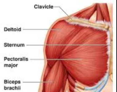
what are the main muscles in the chest
the pectoris majiour, the deltiod, serratus anterior and the pectoris minor
the pectoris majour has 3 heads this means it has 3 oragins what are they
what is the location of the cephaic vein
runs between the pectorals majour and the deltoid
describe the edges of the pectoralis minor
serrated irrugular edges
why does the pectoralis minor have serrated edges
its attahed to the 2,3,4th ribs
what are the functions of the muscles in the lowe abdomen
protect the visera, assist in breathing, coghing and throwing up
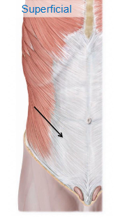
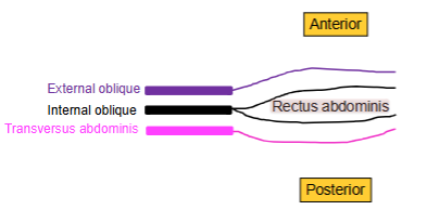
what are the muscles in the lower abdomen
the external oblique, internal oblique, transverse abdomans, rectus abdomanis and pyramidalis
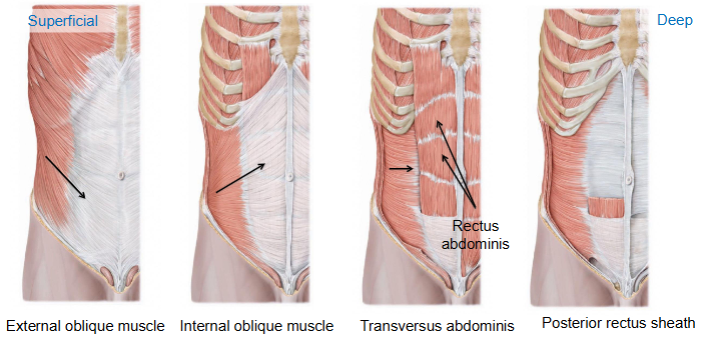
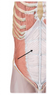
what muscle is shown in this image
the external obique
whats the orogins of the external oblique
a zig zag line from ribs 5 - 12
where does the external oblique insert
inferially on the illiac crest and the xiphoid prosses
which way do the fiers go in the external oblique
downwards (hands in pockets)
what occours when both external oblique muscles contract vs only 1 contracting
if both contract the trunk flextes if only 1 contracts it will lateraly flex the trunk
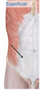
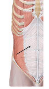
what muscle does this image show
the internal obique
describe the position of the internal oblique in relation to the external oblique
posterior to the external oblique
where does the internal oblique originate
on ribs 9-12
where does the linternal oblique insert
the midline at the linia alba also at the pubic crest
what directions do the fibers of the internal oblique run
superiolay and medialy - hands on tits
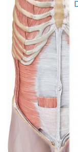
what mscle is shown in this image
the transvers abdominis
where is the transverse abdomanis located related to the internal and external obique
located posterially to the internal and external oblique
whats the insertion of the transvers abo=domalis
on the linia alba and the pubic crest
whats the orogin of the transverse abdomalis
the iliac crest
which way do the fibers run in the transverse abdomanlis
transversaly
what is an aperneurosis
tendons in the form of a sheet at the end of the muscle
which 3 muscles form an apernerosis acros the rectus abdmanils
internal and external oblique and transvers abdomanilis
what is the aperneurosis across the rectus abdomalis called
the rectus sheath
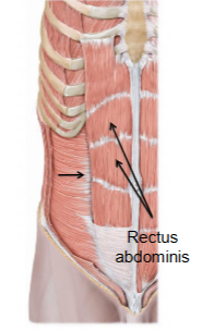
hw are the rectus abdominis organised
muscles sperated by white lines
what are the diffrent white lines called
linia alb, semi lunaris and tendenous intersections
what are the linia alba, semi lunaris and tendinous intersections formed by
the aperneurosis of the abdominal muscles
whats the orogin of the rectus abdominis
pubic symphesis
what the insertion of the rectus abdominis
costal margin and xipoid prosses
what is the piramidalis muscle
tiny muscles that arnt always present
where are the piramdalis attached
inferialy at midline arrached to the linar alba
what encloses the pyramidalis and recutas abdomalis
the tendenous rectus sheath
how is the rectus abdominus coverd at the upper ¾
coverd anterioraly by the external and internal oblique tendns and posterially by the internal and transvers muscle tendons
how is thr rectus abdominus covers on the lower ¼
coverd anterialy by the external, internal and transvers muscle tendons and only covers posterially by the transverse fascia
what does the inginal rejoin contain
the inginal ligiment
what the location of the inginal ligenent
streches from the anterior superior illiac spine to the pubic tuburcule
whats the location of the inginal canal
inthe anterior abdominal wall
what runs through the inguinal region in males/females
the spermatic cord / round ligement of the uterus
what is the anterior wall of the inguinal canal formed by
aperneurosis of the external oblique
what the posterior wall of the inguinal canal formed by
the transversalis fascia
whats the roof of the inguinal canal formed by
the muscular aperneursis of the external and internal oblique, the transversalis abdomina and fascia
what is the floor of the inginal canal formed by
the inginal ligenent
what are the testes coverd by
the tunica vaginalis
what are the 2 layers of the tunica vaginalis
parital layer and viseral layer
when the testes decent what layers go with them
the peritonium, transvers fascia, internal obique muscle and external oblique muscle
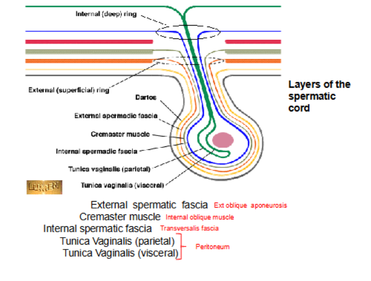
whats the only muscle that contributes to the spermatic cord
the internal oblique
what does the internal oblique muscles name change to
the cremaster muscle
what does the name of the transvers fascia change to
internal spermatic fascia
what does the name of the peitonium change to
tunica vagnalis