cxr follow up tutorial session
 Call Kai
 Learn
Learn Practice Test
 Spaced Repetition
 Match
Match Flashcards
 Knowt Play
1/19

There's no tags or description
Looks like no tags are added yet.
Name | Mastery | Learn | Test | Matching | Spaced | Call with Kai | Chat |
|---|
No analytics yet
Send a link to your students to track their progress
20 Terms
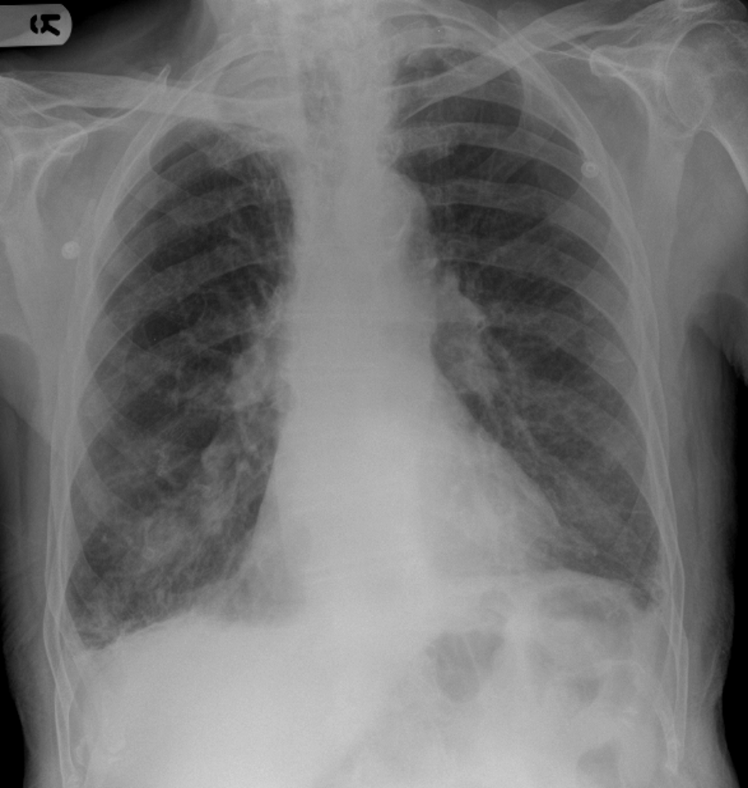
Postero-anterior chest X-ray
Abnormal
There is widening of the superior mediastinum and volume loss overall in the right lung.
There is increased density behind the medial end of the right clavicle extending to the right lung apex.
There is also ipsilateral blunting of the right costophrenic angle indicating a small, right sided pleural effusion.
The appearances are suspicious of malignancy.
Otherwise, the left hemithorax and lung markings are normal, and the cardiothoracic ratio is within normal limits.
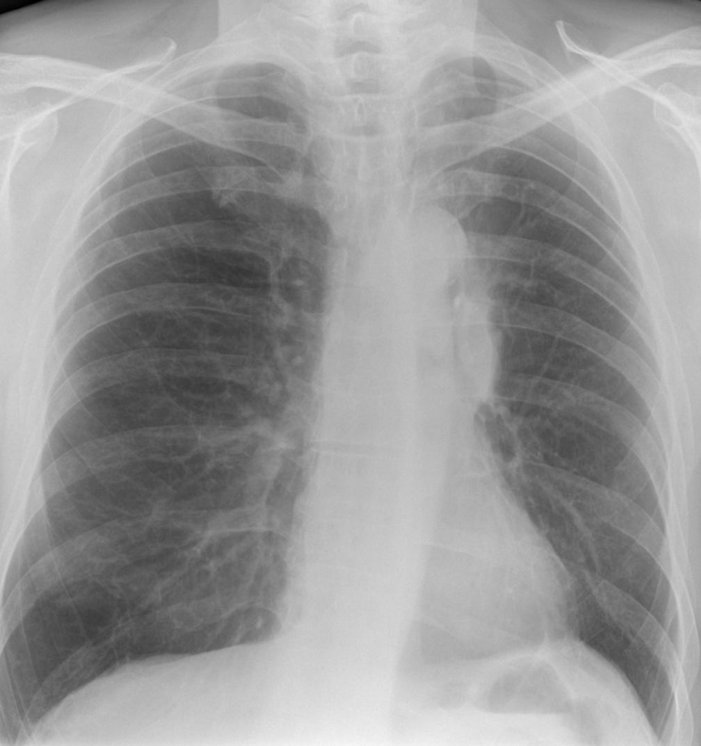
Postero-anterior chest X-ray
Abnormal
Initial inspection suggests widening of the superior mediastinum, however, there is asymmetry of the hilar region, with the left hilum appearing larger, denser and more conspicuous than the right.
The left hilum also appears to be raised in comparison with the right.
The left hilar density appears spiculated, and also appears to trach downwards, along the path of the left main bronchus.
Highly suggestive of a bronchial / hilar mass.
The left hemidiaphragm is raised, but heart size is within normal limits.
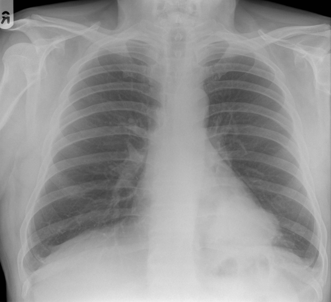
Postero-anterior chest X-ray
Abnormal
A good inspiratory effort revealing normal lung markings.
Heart size is normal, but there is an irregular left cardiac border, and the cardiac shadow appears denser on the left side.
There appears to be a large, well-defined, left sided, retro cardiac mass, situated above the left hemidiaphragm, disrupting the medial diaphragmatic silhouette, and urgent referral to the lung cancer MDT is recommended.
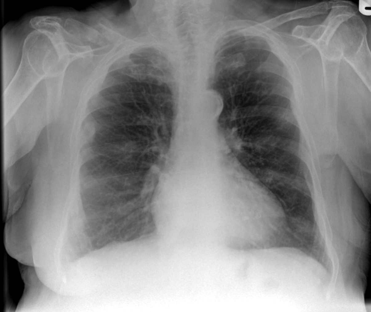
Postero-anterior chest X-ray
Abnormal
There is bilateral volume loss, and the lung markings appear more prominent.
It is difficult to assess cardiothoracic ratio with poor ventilation.
However, there is a mid shaft fracture of the right clavicle and also what appears to be a healing rib fracture in the left, posterior 4th rib.
The left breast shadow is missing – it is likely that this patient has undergone a previous, left sided mastectomy for breast carcinoma, and in the absence of trauma, the fractures are probably related to metastatic disease.
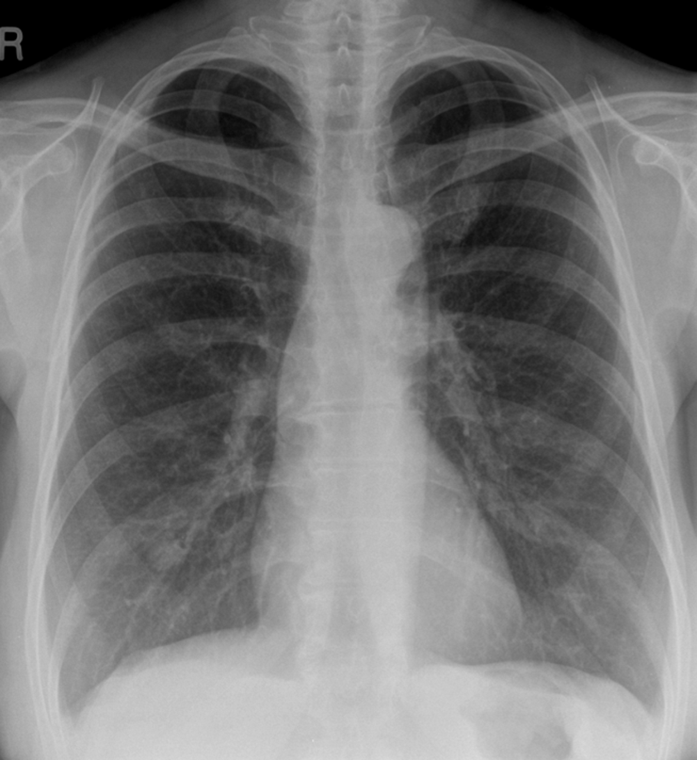
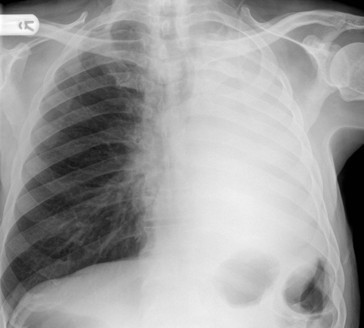
Postero-anterior chest X-ray
Abnormal
There is complete opacification of left hemithorax, and traction of the trachea and mediastinal structures to the left side.
The right lung shows signs of compensatory hyperinflation, with mild flattening of the hemidiaphragm and loss of the right cardiac imprint.
The appearances are consistent with atelectasis of the left lung, although there is no apparent cause of the collapse. An urgent MDT referral is recommended.
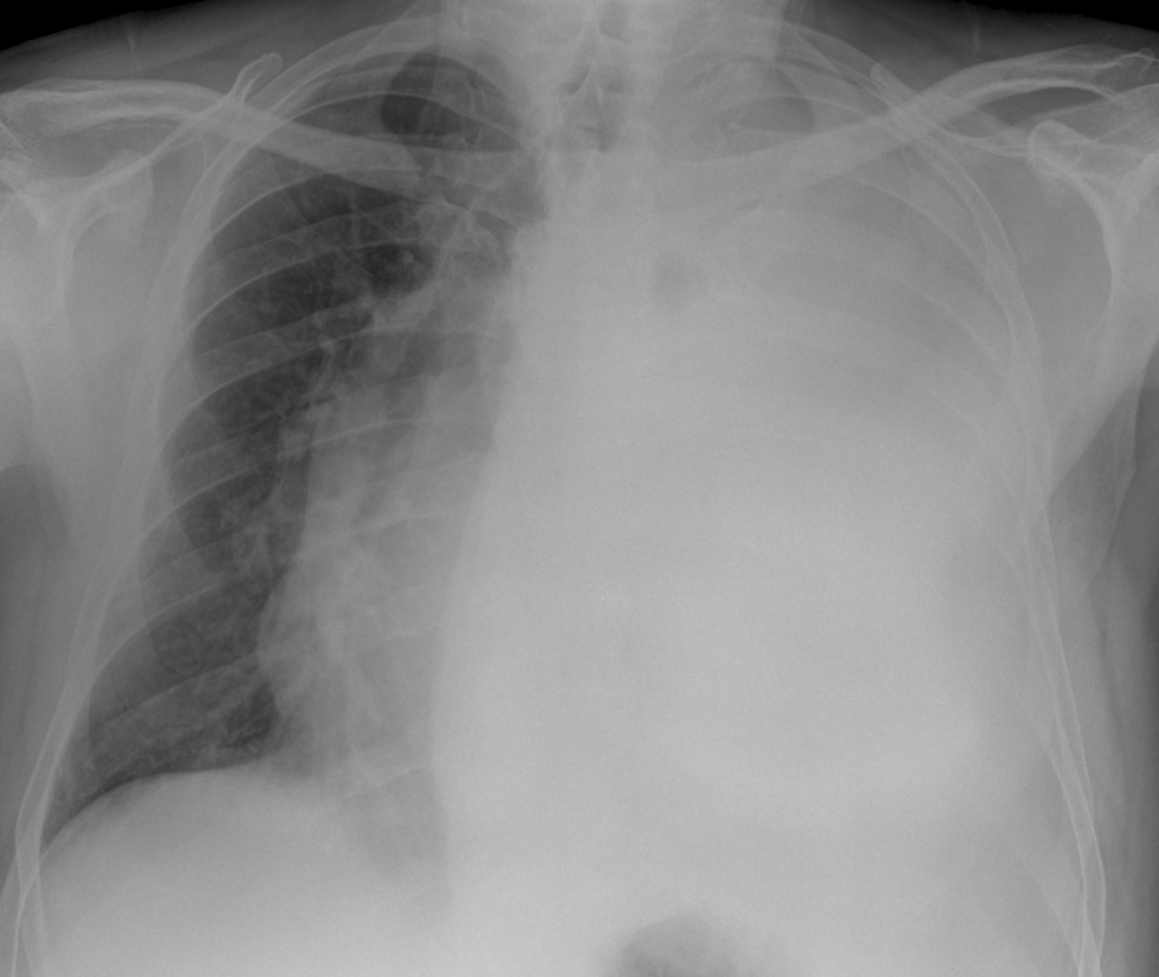
Postero-anterior chest X-ray
Abnormal
There is complete opacification of left hemithorax, and subsequent displacement of the trachea, heart and mediastinal structures to the contralateral (right) side.
The right lung shows signs of volume loss due to the compressive forces. The appearances are consistent with a massive, left sided pleural effusion exerting mass effect.
Although there is no apparent cause of the effusion an urgent MDT referral is recommended.
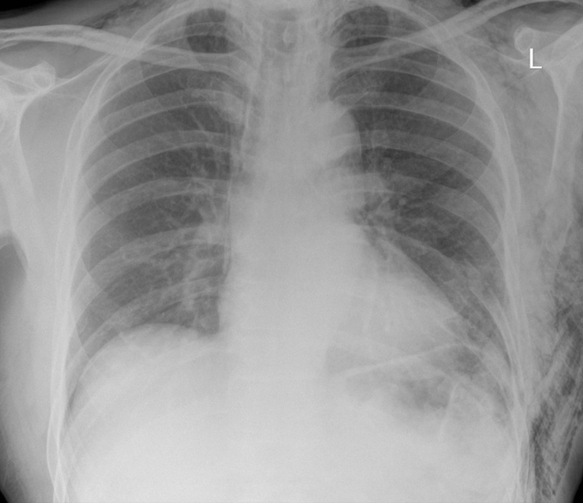
Postero-anterior chest X-ray
Abnormal
There is volume loss bilaterally , making it difficult to assess heart size.
There are multiple striating lucencies seen in and around the left hemithorax and the soft tissue planes, and these appear to be associated with posterior, left sided rib fractures (ribs 7 – 8).
The appearances are consistent with a traumatic pneumothorax and associated surgical emphysema.
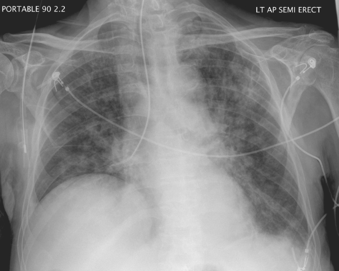
Antero-posterior semi-erect chest X-ray
Abnormal
There is a mis-placed nasogastric tube evident in the right mid/lower zone. The tube initially appears in in the midline of the oesophagus, but fails to cross the carina, instead tracking down the right main bronchus.
The tip resides in the right lower zone instead of passing below the diaphragm and into the stomach.
It must be removed immediately! There is also bilateral, hazy airways shadowing seen.
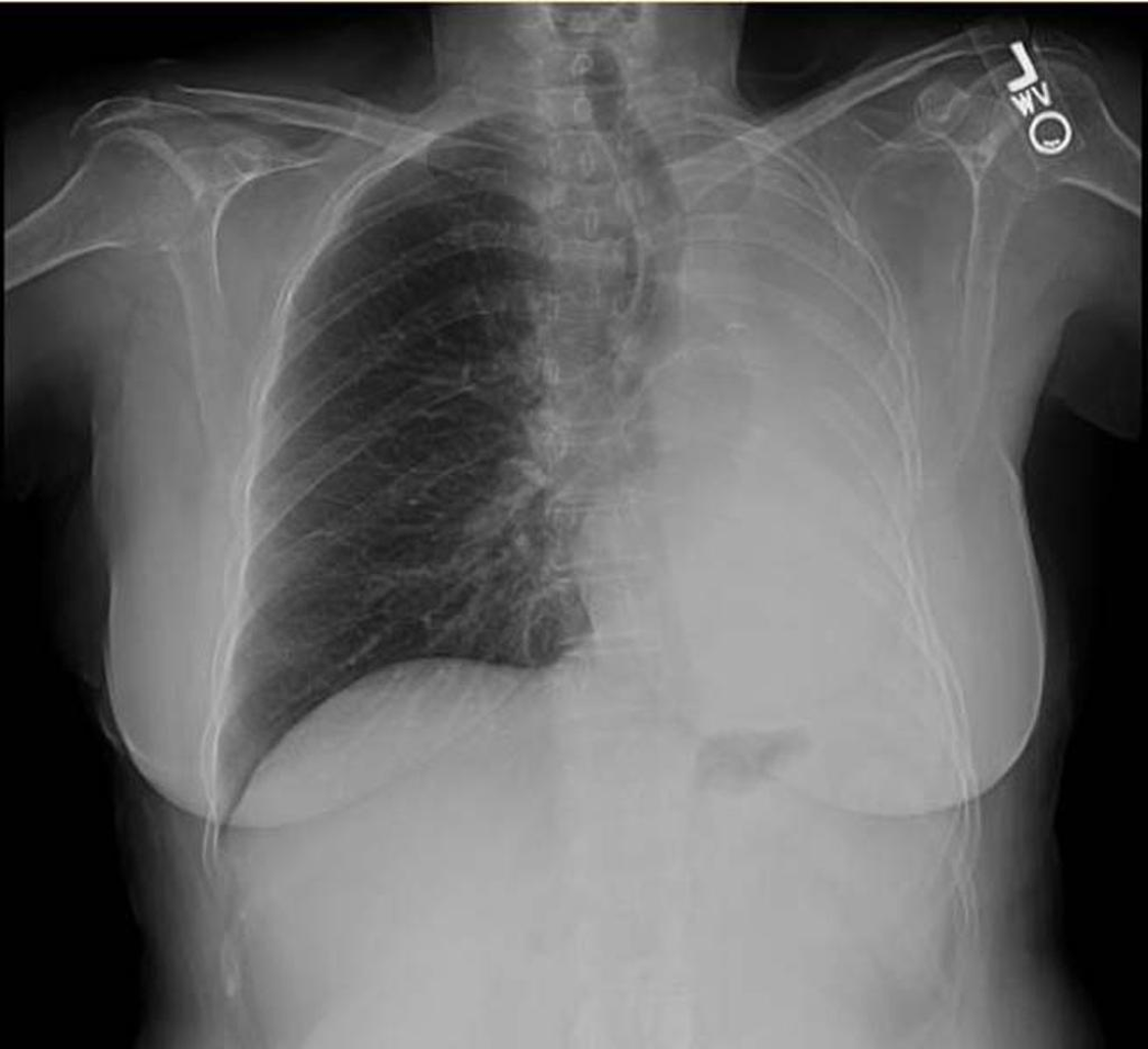
ANtero posterior chest xray
Normal
There is opacification of the left hemithorax and compensatory hyperinflation of the right lung.
Normal right lung markings can be seen.
The trachea, heart and mediastinal structures appear to be pulled towards the left side.
There are iatrogenic artefacts within the left hemithorax, most likely to be surgical clips, indicating that this patient has undergone a previous, left sided, pneumonectomy.
Normal post-pneumonectomy chest X-ray.
A 6-year old female with a history of dyspnoea and cough for 1 week. Query cause?
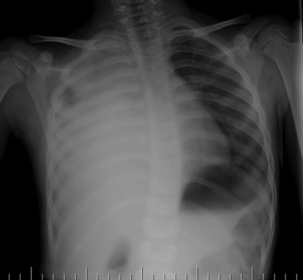
Abnormal
Opacified rt hemithorax – small lucent area right upper zone, pocket of residual air (air bronchogram)
Trachea and mediastinum remain central
This is a case of right-sided consolidation, which is one of the stages of pneumonia.
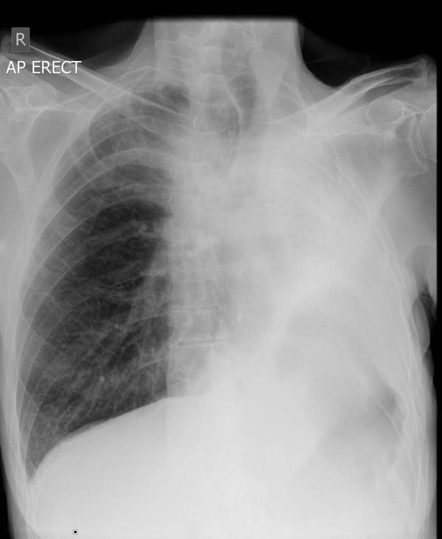
A 60 year old male, complaining of shortness of breath. His clinical history also stated that he had undergone previous thoracic surgery.
Antero posterior erect chest xray
Abnormal
Homogenous opacification of the left hemithorax with an absence of lung markings
Trachea and mediastinal deviation towards the left hemithorax
Surgical clips around the left hilum
Compensatory hyperinflation of the right lung field related to left pneumonectomy.
A 65-year-old male with severe shortness of breath. Recent weight loss.
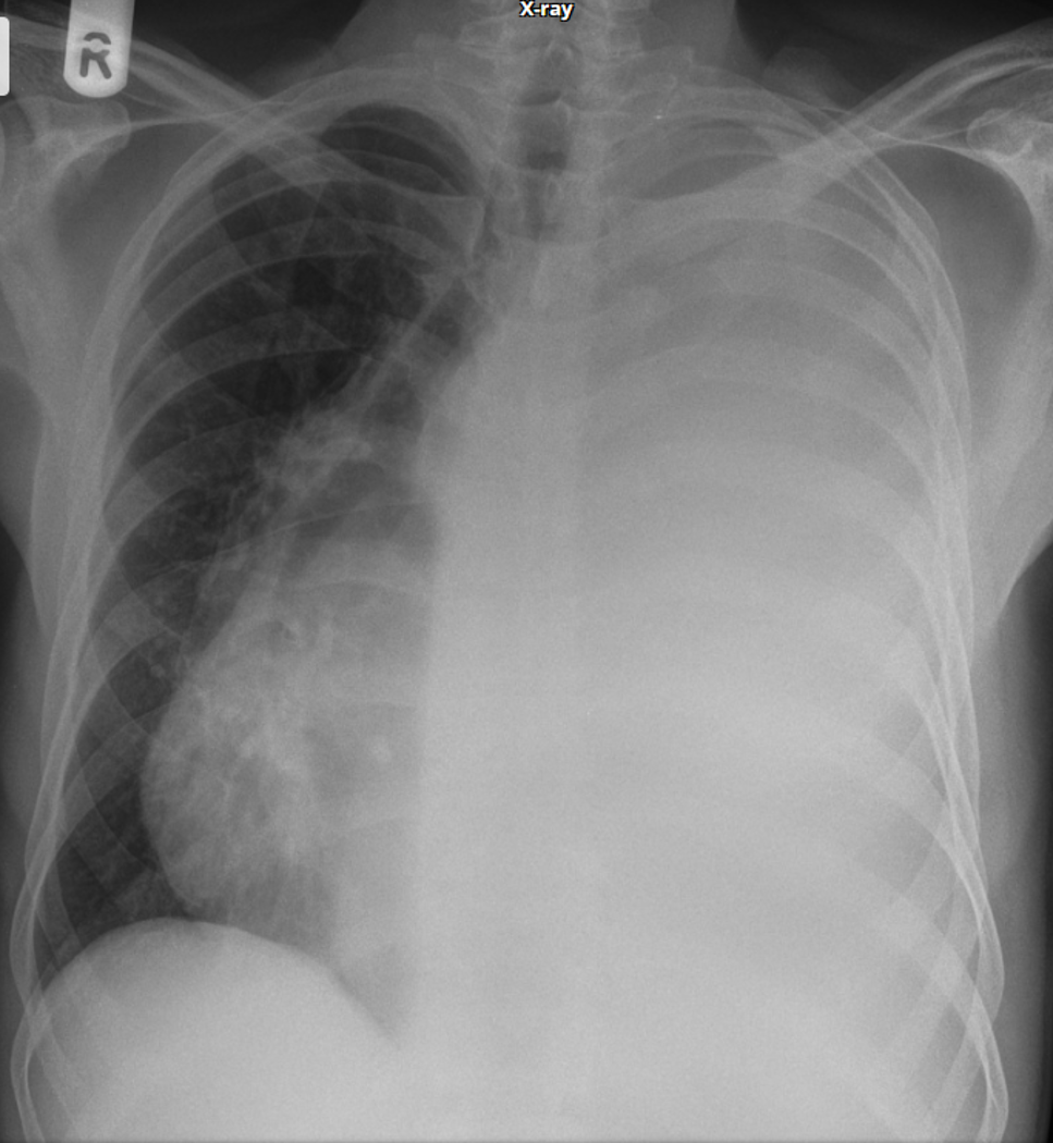
Diffuse, left sided opacification of the hemithorax, with an absence of lung markings.
Marked mediastinal and tracheal shift to the contralateral side.
The tracheal/mediastinal shift is the clue – huge effusion (relating to possible malignancy), which would be the working hypothesis until a CT examination was undertaken
A 15-year-old female with a TWO month history of dry cough, shortness of breath and weight loss (but no history of fever).
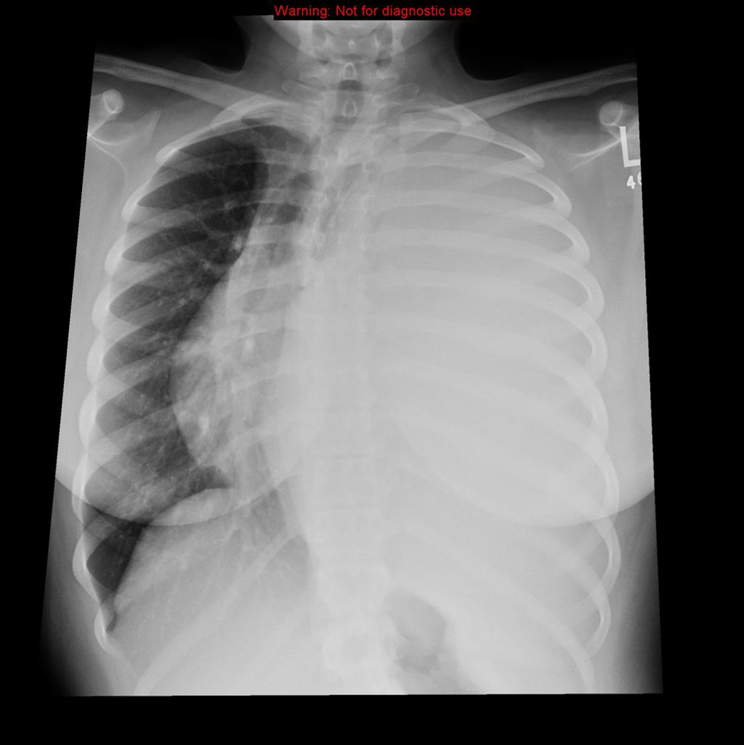
Complete opacification of the left hemithorax with absence of lung markings.
Contralateral deviation of trachea and mediastinum, with compression (volume loss) of right lung.
The appearances are consistent with a massive pleural effusion, BUT such a large pleural effusion is atypical in a child.
note-
Patient underwent CT and was discovered to have a large intrathoracic mass – subsequent histology showed this to be a Ewing’s Sarcoma.
Anteroposterior Chest X-ray
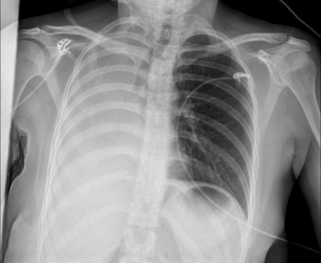
Abnormal
Total opacification of the right hemithorax and an absence of lung markings.
There is traction of the trachea and mediastinal structures to the ipsilateral side, while the left lung is inflated and demonstrates normal lung markings.
The appearances are consistent with a complete right sided atelectasis.
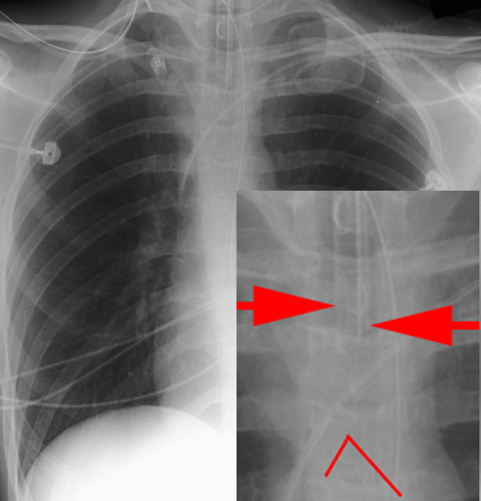
AP portable chest x-ray (and close-up) of a correctly placed endotracheal tube (arrows) and location of carina (^).
ET tubes are fixed at the mouth, so neck position affects the location of the ET tube tip.
Neck extension pulls the tube superiorly, while neck flexion pushes the tube inferiorly.
Neck rotation may also displace the tube.
On a CXR, with the neck in the neutral position, a distance of 5-7 cm above the carina is generally considered acceptable for adults.
In this position it is unlikely that the tube could be pushed beyond the carina or pulled towards the vocal apparatus.
Adult male, intubated on ICU, has poor oxygenation.
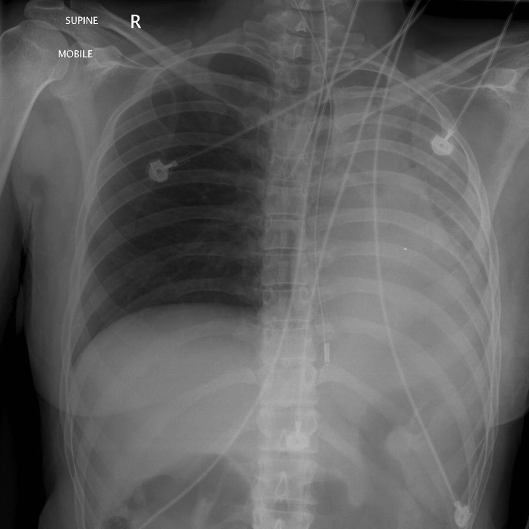
The tip of the ETT extends into the right main bronchus, resulting in endobrochial intubation with complete opacification of the left hemithorax.
This is due predominantly to collapse (atelectasis) of the entire left lung.
Note the mediastinal shift to the left!
A 50-year-old female, central venous catheter inserted for apheresis.
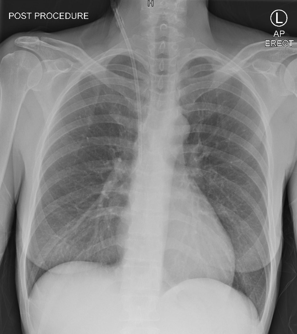
Catheter seen tracking vertically, down the right internal jugular vein, and the tip residing correctly in the distal SVC.
No pneumothorax seen.
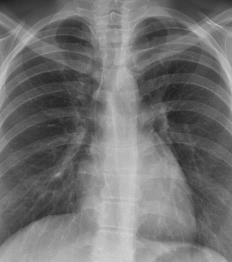
Central venous Catheter in right atrium
•This peripherally inserted central catheter (PICC) needed to be inserted with its tip at the level of the cavo-atrial junction
•The Cava-atrial junction is about the height of two vertebral bodies below the carina
•The PICC line has been inserted too far, with its tip in the right atrium (RA)
Note: Some large haemodialysis catheters are designed to be placed with their tip in the RA
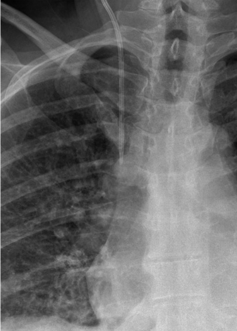
Right internal jugular vein catheter / line
•The catheter is orientated vertically
•The tip is projected over the anatomical location of the SVC - approximately 1.5 cm above the level of the carina (red arrow)
•This is an ideal position for right-sided catheters for fluid administration and venous pressure monitoring, but not for long-term chemotherapy or dialysis - no pneumothorax seen.