Lesson 19: Digestive Anatomy
 Call Kai
 Learn
Learn Practice Test
 Spaced Repetition
 Match
Match Flashcards
 Knowt Play
1/87

There's no tags or description
Looks like no tags are added yet.
Name | Mastery | Learn | Test | Matching | Spaced | Call with Kai | Chat |
|---|
No analytics yet
Send a link to your students to track their progress
88 Terms
What makes up the digestive system?
The GI, liver, pancreas and gallbladder
What is the GI?
Series of hollow organs that are connected to each other from mouth to anus
Which organs make up the GI in the order they are connected?
Mouth, Esophagus, stomach, small instestine, large intestine and anus
What does the digestive system do?
Turning food into the nutrients and energy you need to survive.
Package solid waste, stool
Why is digestion important?
Body needs nutrients from food you eat and drink
Energy growth and repairing cells
Carbohydrates, proteins, fats, vitamins, minerals, water
What are functional segments of the digestive system?
- Mouth
- Pharynx
- Esophagus
- Stomach
- Small Intestine
- Large Intestine
What are accessory structures of the digestive system?
- Liver
- Gallbladder
- Pancreas
What are the functions of the digestive system?
Ingestion: introduction of food into the mouth.
Secretion: release of water, acid, buffering substances and enzymes into the lumen of the digestive tract.
Mixing and propulsion of food through the digestive tract.
Digestion: mechanical and chemical breakdown of food.
Absorption: passage of digested products from the digestive tract into the blood and lymph.
Defecation: elimination of faeces from the gastrointestinal tract.
What are the functions of: Mouth, Pharynx and esophagus, Stomach, Small intestine, large intestine and rectum and anus
• Mouth: bite, chew, swallow.
• Pharynx and esophagus: transport.
• Stomach: mechanical disruption and chemical digestion; absorption of water & alcohol.
• Small intestine: chemical and mechanical digestion, and absorption.
• Large intestine: absorb electrolytes & vitamins (B and K).
• Rectum and anus: defecation.
What is the abdominal cavity?
It is the largest hollow space of the body.
Its upper boundary is the diaphragm
Its lower bondary is the upper plane of the pelvic cavity
Vertically enclosed by vertebral column and abdominal and lther muscles

Abdominal quadrants

What is the Peritoneum?
The peritoneum is the largest serous membrane of the body.
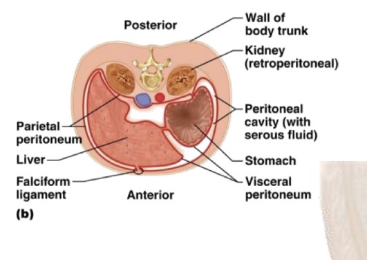
What forms the peritoneum?
• The parietal peritoneum lines the wall of the abdominal cavity.
• The visceral peritoneum covers some of the organs.
• The space between the parietal and visceral portions of the peritoneum is called the peritoneal cavity and contains serous fluid.

How are organs, such as kidneys and panreas, that lie on the posterior abdominal wall behind the peritoneum called?
Retroperitoneal
Which large folds between the viscera does the peritoneum form?
• Greater Omentum
• Falciform ligament
• Lesser Omentum
• Mesentery
• MesocoloN

How are the folds distributed and what do they contain?
These folds join the organs to each other and to the wall of the abdominal cavity, and contain blood vessels, lymphatics, and nerves that lead to the abdominal organs.
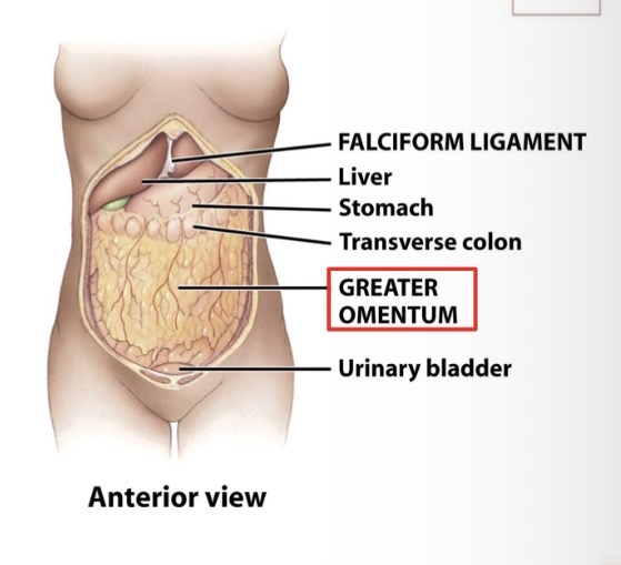
What is the Greater omentum and how many layers does it have?
It is the thickest layer of peritoneum
Has 2 layers that fold between them

How does the greater omentum extend?
Extends downward, anteriorly to small intestine.
Then, ascends again inserting in transverse colon.
What is the function of the lesser omentum?
From serosa of stomach and duodenum, connects these 2 organs with the liver.

What is the function of the ligament ligament?
Connects liver to the anterior abdominal wall and diaphragm.

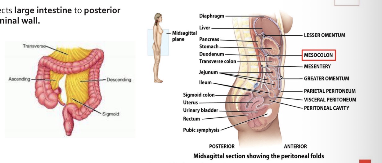
What is the function of the mesentary?
Attaches small intestine to posterior abdominal wall.
In mesentery there are blood vessels and nerves that supply intestine.
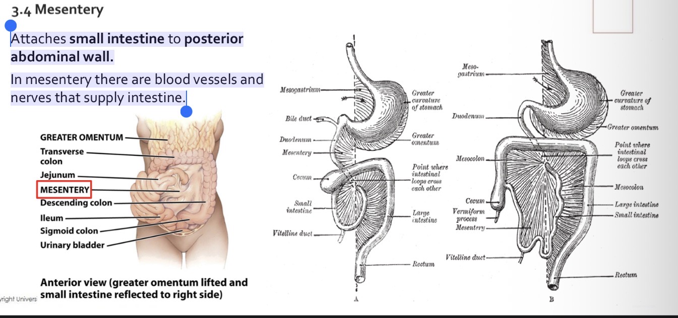
What is the function of the mesocolon?
Connects large intestine to posterior abdominal wall.
What forms the oral cavity?
- Cheeks (buccinator muscle).
- Hard and Soft palate.
- Uvula.
- Lips.
- Tongue.
- Gums.
- Teeth.
- Salivary glands.

What is the verstibule?
The vestibule of the oral cavity is bounded externally by the cheeks and lips and internally by the gums and teeth.

What forms roof and floor of the mouth?
The soft palate, hard palate, and uvula form the roof of the mouth and the tongue forms the floor.
What are the 2 muscular folds that start from the base of the uvula?
Two muscular folds start from the base of the uvula:
• anteriorly, the palatoglossal arch, which extends towards the edge of the base of the tongue;
• posteriorly, the palatopharyngeal arch, which extends to the edge of the pharynx.
What do the salivary glands release into the oral cavity?
Saliva
What are the types of salivary glands?
Minor and major salivary glands?
Where do we find minor salivary glands?
In the mucosa of the mouth and tongue are the minor salivary glands: labial, buccal and palatine glands on the lips, cheeks and palate
Where do we find major salivary glands?
3 pairs of parotid, submandibular and sublingual glands
Where are the parotid, submandibular and sublingual glands located?
• The parotid glands are located below and in front of the ears, between the skin and the masseter muscle.
• The submandibular or submaxillary glands are located on the floor of the mouth.
• The sublingual glands are located below the tongue and above the submandibular glands.

What are teeth?
Teeth are accessory digestive organs located in the alveolar processes of each jaw.
What covers alveolar processes and wha lines alveolar cavities?
The alveolar processes are covered by the gums and the alveolar cavities are lined by the periodontal ligament or membrane.
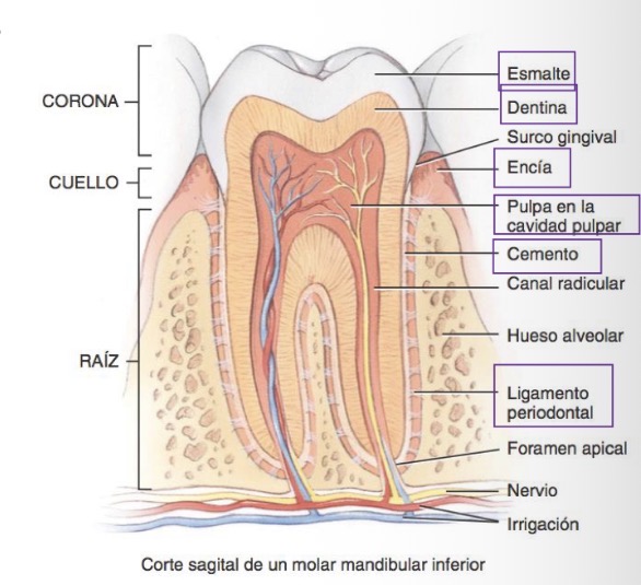
What are the three regions of a tooth?
The crown, the root and the neck

What forms the majority of the tooth and what covers it?
What covers the root?
Dentin majority of the tooth and is covered with enamel at the crown
The root is covered by cementum
What does the pulp cavity contain?
The pulp cavity contains the pulp where the blood vessels, nerves and lymphatic vessels are located.
What are the two dentitons or 2 sets of teeth called?
deciduous and permanent.
How many deciduous and permanent teeth do we have?
20 deciduous and 32 permanent
Deciduous or baby teeth structure
• The incisors and canines have only one root.
• Canines have a rounded end called a cusp.
• The first and second molars have 4 cusps.
• The upper molars (maxillary) have 3 roots; the lower molars (mandibular) have 2.

Differences between deciduous and permanent teeth?
• The deciduous molars are replaced by the first and second premolars (bicuspids), which have two cusps and one root (the upper first premolars have two roots)
• Permanent molars, which erupt after the premolars, do not
replace any deciduous teeth and appear as the jaw grows:
- first molars
- second molars
- third molars (wisdom teeth)
What are the 3 phases of swallowing?
Swallowing occurs in 3 phases:
1) The voluntary phase: the bolus passes from the mouth to the oropharynx
2) the pharyngeal phase (involuntary): the bolus passes through the pharynx into the esophagus,
3) the esophageal phase (involuntary): the bolus passes from the esophagus to the stomach.
What is the Esophagus and where is it located?
Muscular tube, 25cm
• Anterior to vertebrae
• Posterior to trachea
• Posterior to the heart

Where does it pass through the esophagus and what does it connect?
Pierces the diaphragm at esophageal hiatus.
Connects the pharynx to the stomach.
What structures does the esophagus have at the ends?
At the ends it has the upper esophageal sphincter (UES), made up of skeletal muscle, and the lower esophageal sphincter (LES), made up of smooth muscle.
What is the stomach and where is it located?
It is a J-shaped enlargement of the GI tract that begins at the bottom of the esophagus and ends at the pyloric sphincter.

What is the
It serves as a mixing and holding area for food
begins the chemical digestion of proteins
continues the digestion of carbohydrates
converting a bolus to a liquid called chyme
What are the 4 main regions of the stomach?
1) the cardias,
2) the fundus,
3) the body and
4) the pylorus

What is the function of the cardias?
The cardias surrounds the upper opening of the stomach.

What is the function of the Fundus?
The fundus is the rounded portion above and to the left of the cardia

What is the function of the body?
The body is the central portion of the stomach

What are the parts of the pyloric region and what are their functions?
The pyloric region is divided into three parts:
The pyloric antrum connects to the body of the stomach.
the pyloric canal
the pylorus, which communicates with the duodenum through the pyloric sphincter.

What are the curvatures of the stomach called?
The concave inner edge of the stomach is the lesser curvature,
and the convex outer edge is the greater curvature.

What are the functions of the small instestine?
The major events of digestion and absorption occur in the
small intestine

How is the small intestine divided?
It is divided into the duodenum, jejunum, and ileum

How does the small intestine extend?
The small intestine extends from the pyloric sphincter to the ileocecal sphincter.
What kind of structures does it have?
It has circular folds (plicae circularies): permanent ridges in the mucosa that increase surface area.
What kind of structures does does have on the surface area?
Villi and microvilli also increase absorption surface area.
The duodenum
- is the shortest segment
- is retroperitoneal
- begins at the pyloric sphincter of the stomach
It measures 25 cm and is shaped like a C tube.
The jejunum
measures about 1 meter
The ileum
- It measures around 2 meters
- It joins the large intestine through the sphincter or ileocecal valve.
How does the large intestine extend?
The large intestine extends from the ileocecal sphincter to the anus.

What is the diameter of the large intestine?
1.5 m. by 6.5 cm. diameter.
What are the subdivisions of the large intestine?
Its subdivisions include the: cecum, colon, rectum, and anal
canal.
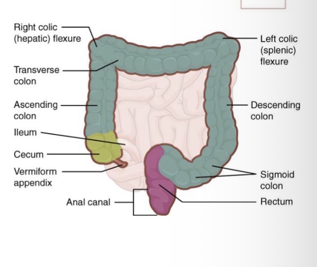
Which parts are retroperitoneal and which ones are intaperitoneal?
Ascending, descending colon and rectum are retroperitoneal.
Transverse and sigmoid colon are intraperitoneal.
How is the large intestine attached to the abdominal wall?
Attached to posterior abdominal wall die to the mesocolon
How is the colon divided?
Into ascending, transverse, descending and sigmoid

The ascending colon
ascends the right side of the abdomen, reaches the undersurface of the liver, and turns to the left to form the right colonic (hepatic) angle.
How does the ascending colon continue?
It continues as the transverse colon and curves below the
lower border of the spleen, where it forms the left colonic
(splenic) angle.
How does the transverse colon continue?
It then descends as the descending colon and continues as
the sigmoid colon and finally the rectum.
What is formed by the last 2-3 cm of the rectum?
The anal canal
How is the external opening of the anal canal called and what structures does it have?
At the external opening of the anal canal, the anus, there is an
internal anal sphincter of smooth muscle (involuntary) and an external anal sphincter of skeletal muscle (voluntary)
The liver
The liver is the largest gland in the body and weighs about 1.4 kg in adults.
Where is the liver located?
It is located below the diaphragm, in the right hypochondrium and part of the epigastrium, in the abdominopelvic cavity.
What covers the liver?
Visceral peritoneum
Structure of the liver
It is divided into 2 main lobes (a large right lobe and a smaller left lobe)

What separates both lobes of the liver
falciform ligament (a part of the peritoneum).

What is at the free edge of the falciform ligament?
The round ligament
What are the left and right coronary liagemnts?
The left and right coronary ligaments are narrow
extensions of the parietal peritoneum, running from
the liver to the diaphragm.

What is the gallbladder?
The gallbladder is a sac located in a depression on the posterior surface of the right lobe of the liver, called cystic fossa
What are the parts of the gallbladder?
Body, neck and fundus

What is the function of the gallbladder?
Receives bile from liver, assembles and stores it. Then it is released through the cystic duct
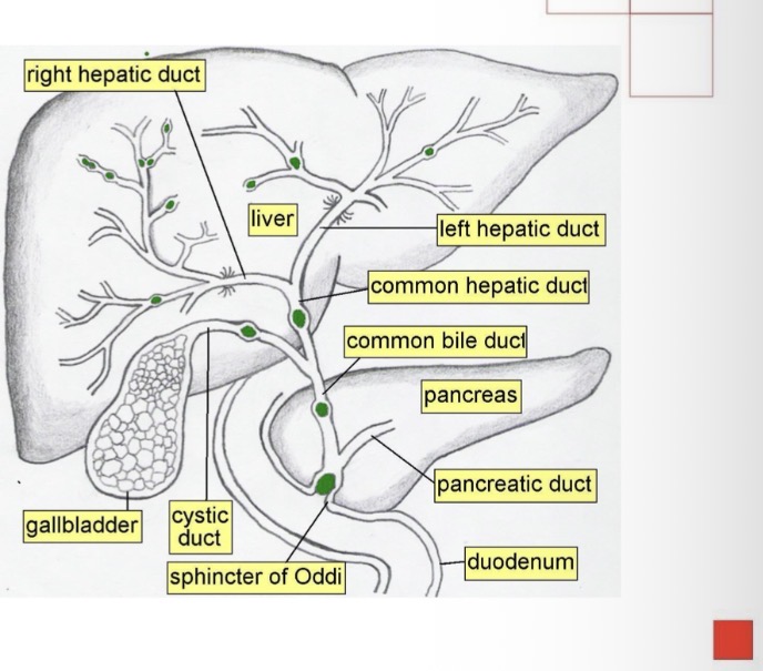
Bile Ducts of gall bladder
Left and right hepatic ducts merge to form
→ common hepatic duct that exits liver and joins
→ cystic duct (from gall bladder) forming
→ common bile duct
→ joins with pancreatic duct and enters duodenum at major duodenal papilla.

What are pancreas and where are they located?
The pancreas is a gland that measures around 12-15 cm in length and 2.5 cm in width and is located behind the greater curvature of the stomach.

What are the parts of the panchreas and how are they connected to the duodenum?
Head, body and tail
connected to the duodenum by two ducts: the pancreatic duct (duct of
Wirsung) and the accessory duct (of Santorini).
What does the pancreatic duct join?
The pancreatic duct joins the common bile duct (or hepatocystic) and enters the duodenum as a common duct called the hepatopancreatic ampulla (ampulla of Vater) that has a sphincter (sphincter of Oddi).
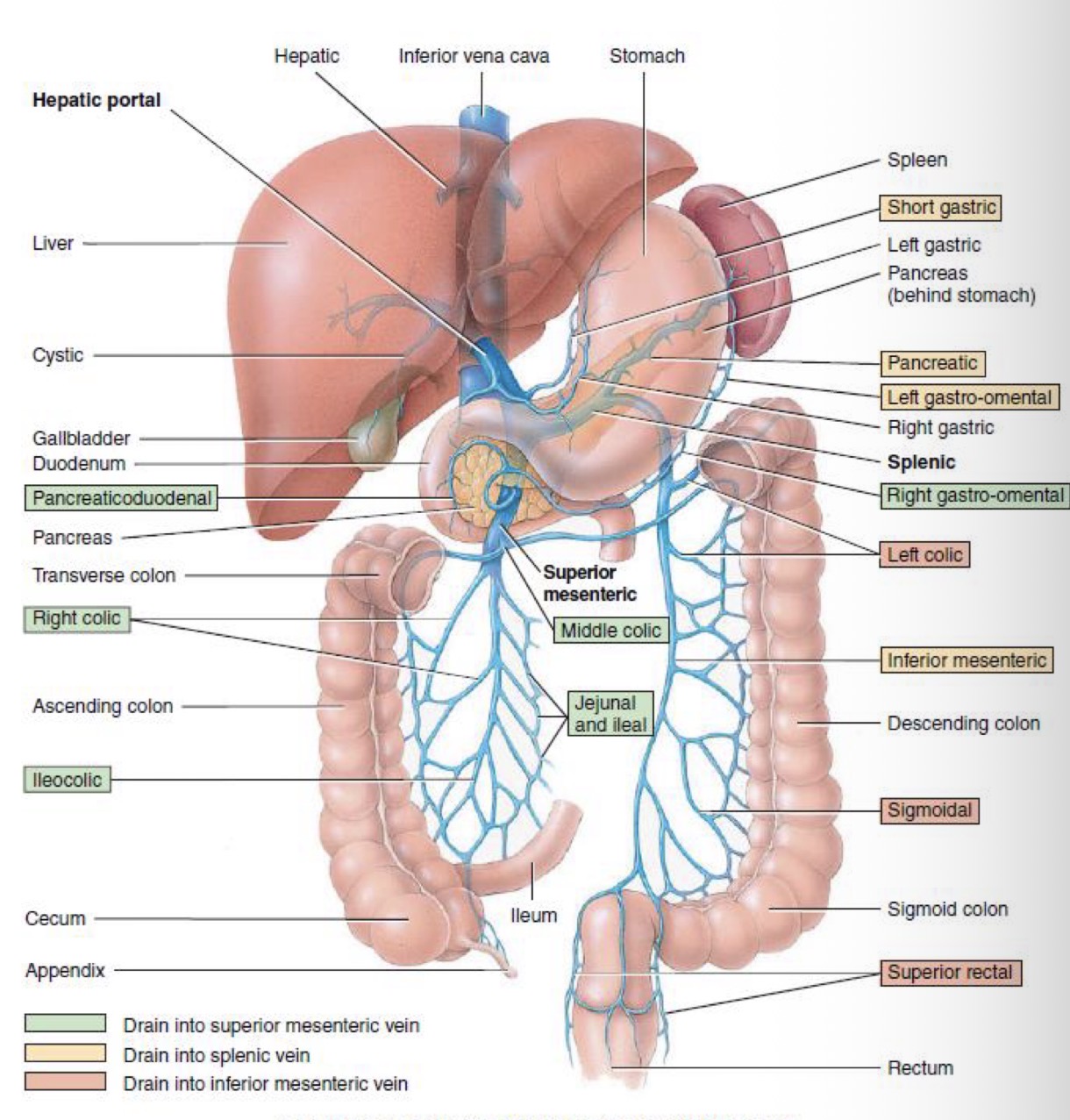
What is responsible for the vascularisation of the organs of the digestive tract and spleen to liver?
The portal venous system

Where does the liver receive blood?
1)The hepatic artery, obtains oxygenated blood
2) The portal vein, receives deoxygenated blood, which contains nutrients, drugs, and possibly microorganisms and toxins absorbed by the digestive tract

What happens with products of hepatocytes and nutrients required by other cells?
Products made by hepatocytes and nutrients required by other cells are released back into the blood, which drains into the central vein and then flows into the hepatic vein.