Chapter #3 Positioning (Part 2)
 Call Kai
 Learn
Learn Practice Test
 Spaced Repetition
 Match
Match Flashcards
 Knowt Play
1/59

There's no tags or description
Looks like no tags are added yet.
Name | Mastery | Learn | Test | Matching | Spaced | Call with Kai | Chat |
|---|
No analytics yet
Send a link to your students to track their progress
60 Terms
What are the general procedural guidelines for chest radiography?
Patient preparation
Patient position
Collimation
SID
ID markers
Radiation protection
Patient instructions.
What should be removed during patient preparation?
Artifacts from the anatomy of interest, such as
long earrings
necklaces
clothing artifacts.
How should ambulatory patients generally be positioned?
Upright or seated erect.
What may be used when a nonambulatory patient cannot sit upright and air-fluid levels are important?
A decubitus position.

What is the recommended minimum SID for chest radiography?
183 cm (72 inches).
Why is a 72-inch SID used for chest radiography?
To decrease heart magnification
Increase spatial resolution.
Where should the right or left side marker be placed?
In the collimated exposure field,
Clear of the anatomy of interest.
Should digital annotation be used instead of a physical side marker?
No.
When are chest exposures usually made?
After the second deep inspiration.
How is the patient positioned for a PA chest?
Upright, either standing or seated.

What does an upright PA chest demonstrate regarding air/fluid?
It demonstrates air or fluid levels.
How is the patient's body positioned for a PA chest?
Facing the vertical grid with the MSP centered perpendicular to the IR.
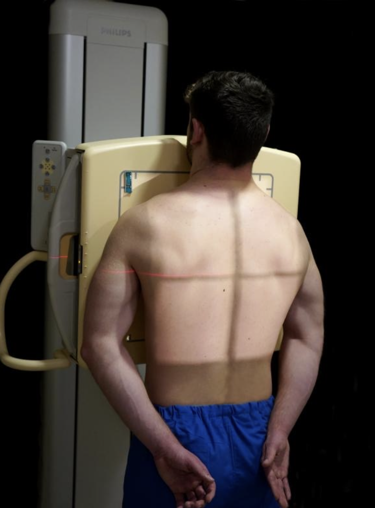
What should the patient do with their shoulders for a PA chest?
Depress them into the same transverse plane and rotate them forward to touch the grid.
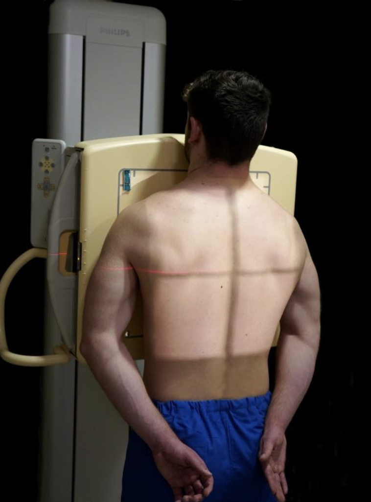
Where does the CR enter for a PA chest?
At the MSP at the level of T7.
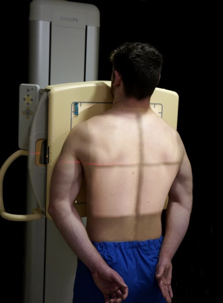
When is the PA chest exposure made?
At the end of the second deep inspiration.

What should be included on a PA chest?
Entire lung fields from the apices to the costophrenic angles.
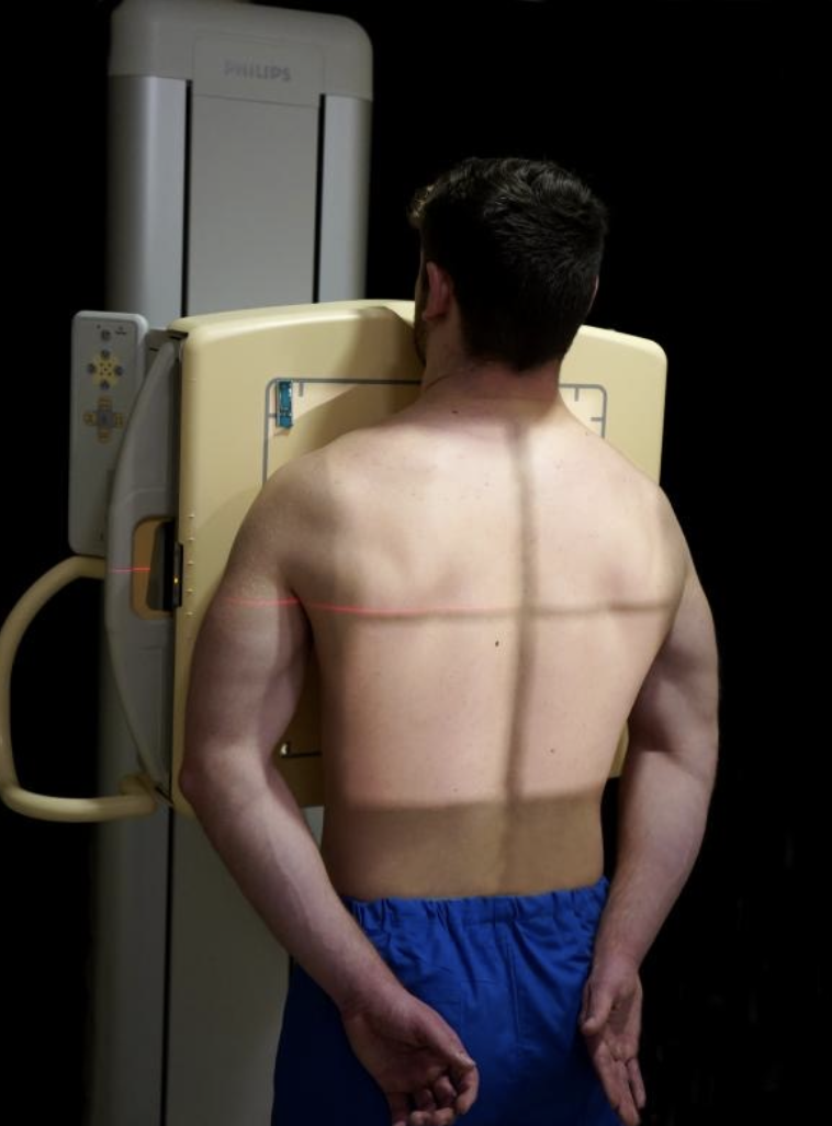
Where should the scapulae be on a properly positioned PA chest?
Projected outside the lung fields.

How many posterior ribs should be visible above the diaphragm?
10.
When is an AP chest commonly used?
When the patient is too ill to sit or stand.

Where does the CR enter for an AP chest?
8 cm (3 inches) below the jugular notch.
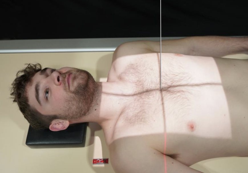
When is an AP chest exposure made?
After the second full inspiration.

How does the heart appear on an AP chest?
Magnified.

How do the lung fields appear on an AP chest compared with PA?
Shorter.
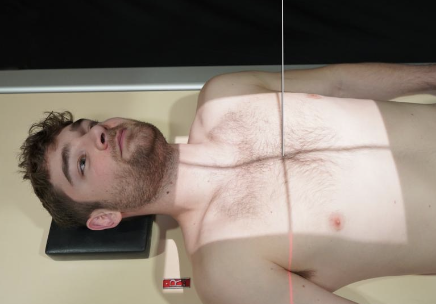
How do the clavicles appear on an AP chest?
Projected higher and more horizontally.
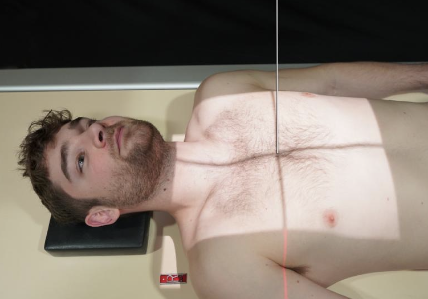

PA =
standard upright chest
AP =
often sick/nonambulatory patient + magnified heart

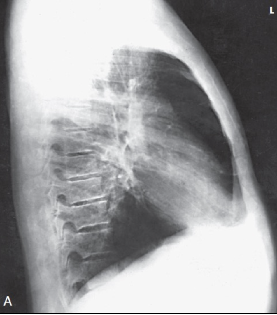
Which lateral position is routinely used for the chest?
Left lateral.
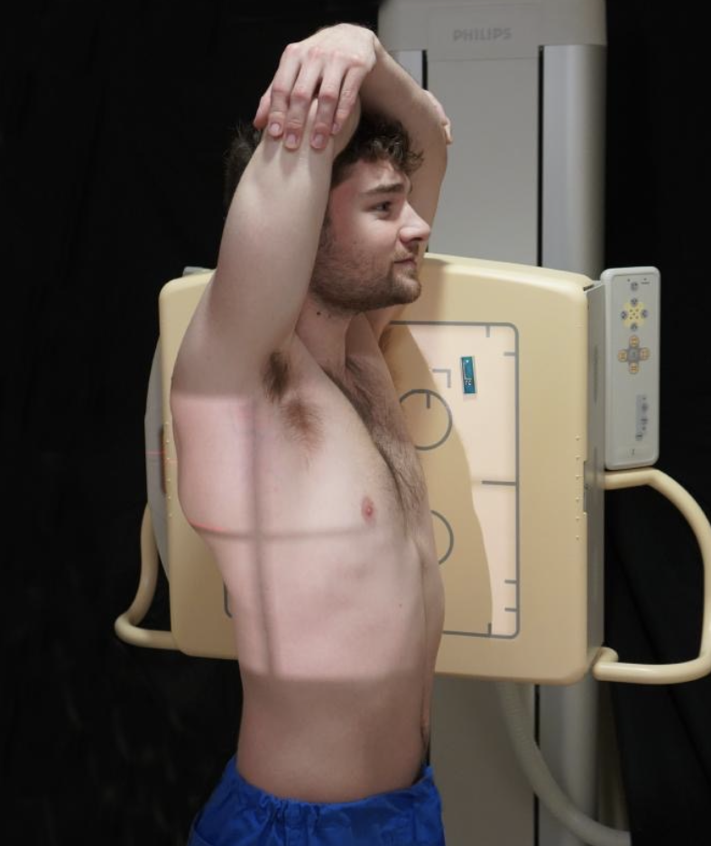
Why is the left lateral routinely used?
To minimize magnification of the heart.
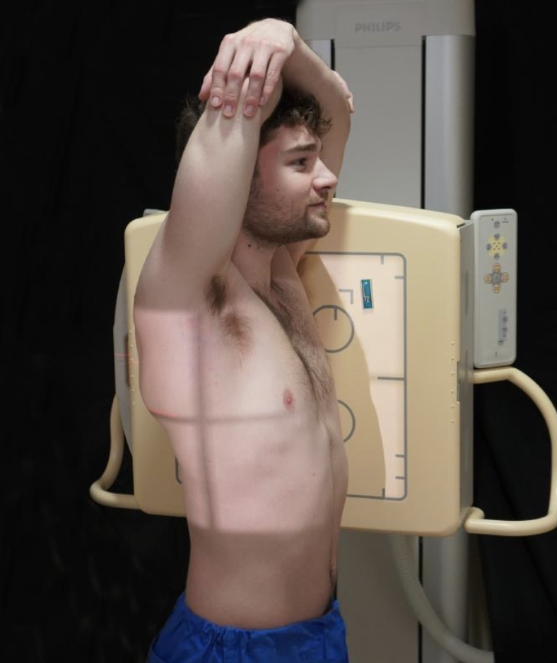
Which side is placed closer to the IR?
The side being demonstrated.

How is the patient positioned for a true lateral chest?
MSP parallel to IR
MCP perpendicular to IR.
Where are the arms positioned?
Extended over the head
Elbows flexed
Forearms resting on the head.

Where does the CR enter for a lateral chest?
At the MCP at the level of T7.

When is the lateral chest exposure made?
At the end of the second deep inspiration.

What should be demonstrated on a lateral chest?
Costophrenic angles
Lower lung apices
Hilum centered and sternum in profile.
What is another name for the AP axial chest position?
Lordotic position (Lindblom).

How does the patient position their body for the lordotic chest?
Upright, facing the tube
Leaning backward until the shoulders rest against the grid.

What is the coronal plane of the thorax positioned at?
15–20° from vertical.
Where does the CR enter for the lordotic position?
MSP at the midsternum
8–10 cm (3–4 inches) below the jugular notch.
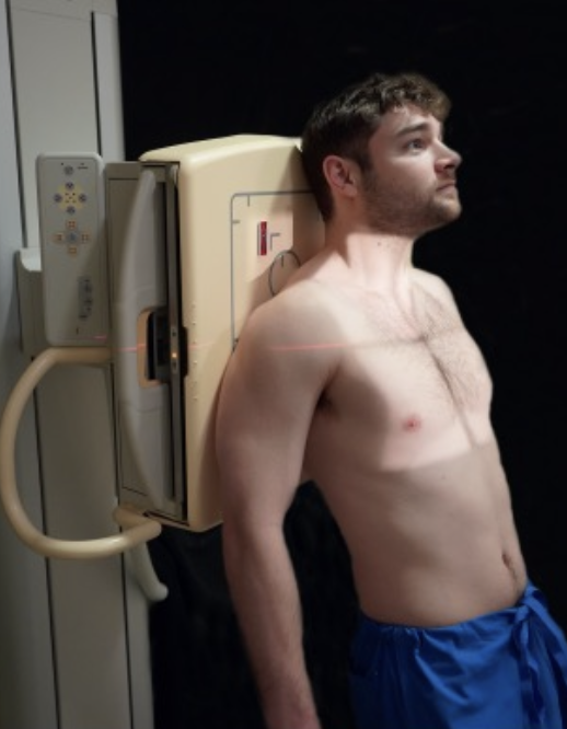
What does the lordotic position demonstrate especially well?
Lung apices
Interlobar effusions.

Where should the clavicles appear?
Superior to the lung apices.
LORDOTIC =
APICES
What are the two types of AP/PA decubitus positions?
Right or left lateral decubitus.

What position demonstrates fluid?
Patient lies on the affected side.

What position demonstrates free air?
Patient lies on the unaffected side.
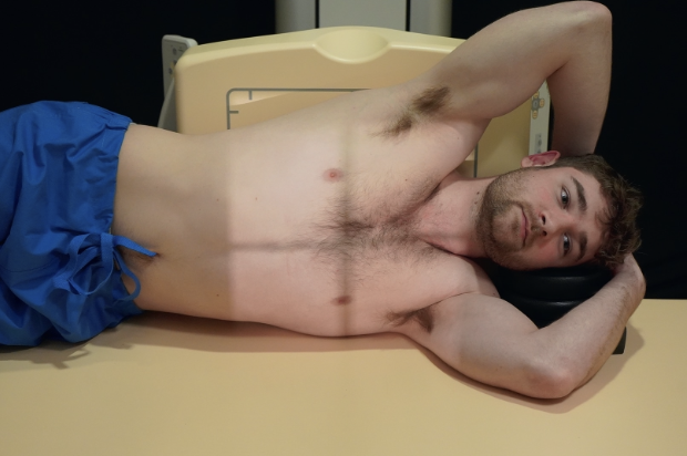
Why does the patient remain in the decubitus position for 5 minutes?
To allow fluid to settle or air to rise.
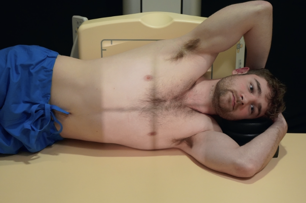
What is the key rule for decubitus positioning?
Fluid = affected side down;
Free air = unaffected side down.
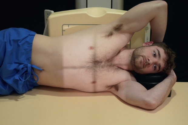
What is the CR for an AP/PA lateral decubitus?
Horizontal and perpendicular to the center of the IR.

What is the respiration instruction for AP/PA lateral decubitus?
Exposure on the second full inspiration.
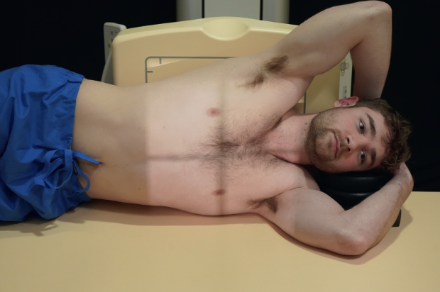
What positions are used for lateral ventral/dorsal decubitus?
Prone or supine.
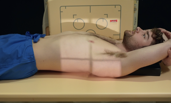
How long does the patient remain in position?
5 minutes.
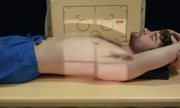
Why does the patient remain in position for 5 minutes?
To allow fluid to settle or air to rise.
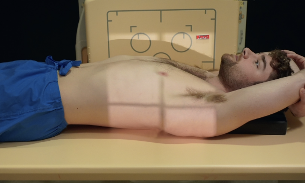
Where is the IR centered?
At T7.

What is the CR direction?
Horizontal and perpendicular to the IR.
What is aspiration pneumonia?
Pneumonia caused by aspiration of foreign particles.
What is pulmonary edema?
Replacement of air with fluid in the lung interstitium.
What is pneumothorax?
Accumulation of air in the pleural cavity resulting in collapse of the lung.
What is pleural effusion?
Collection of fluid in the pleural cavity.
What is COPD?
A chronic condition of persistent obstruction of bronchial airflow.
What is bronchitis?
Inflammation of the bronchi.
What is atelectasis?
Collapse of all or part of the lung.